@aestheticsgroup
@aestheticsjournaluk
Aesthetics
aestheticsjournal.com
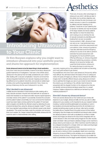
There are, of course, other advantages to ultrasound use which I will discuss later in this article, but my primary objective was to help minimise the risk of occlusion and hasten recovery in case of a complication by utilising vascular mapping on my patients. I would aim to scan patients prior to injection to identify and map the location of the large vessels and rescan patients after injection to check the blood flow, and in doing so try to minimise the risk of vascular occlusions by identifying and treating them as soon as possible. Having worked in A&E, ultrasound was not new to me. I had performed many nerve blocks, central line placements and cannulations under ultrasound guidance. I had also attended several emergency ultrasound courses to be able to perform extended focused assessment with sonography for trauma (eFAST) scans. Offering Ultherapy, an ultrasound-guided Dr Kim Booysen explains why you might want to lifting and tightening procedure, similarly introduce ultrasound into your aesthetic practice allowed me to become familiar with and shares her approach for implementation superficial facial anatomy ultrasound. So, taking the leap to scanning and Facial ultrasound seems to be the latest thing in facial aesthetics. vascularly mapping all my new patients did not seem as daunting, Every time I read new journal articles, it seems to be about ultrasoundand I was quite excited to get started. guided treatments or investigations. I had seen a few articles about For practitioners who have never used ultrasound, the journey will be ultrasound in the past but had not really considered its use in clinic.1,2 more difficult. You will need to learn the basics of how an ultrasound After dealing with a vascular complication, I became convinced that works, the types of images you will see, how to interpret the different ultrasound-guided treatments and vascular mapping would make a images, learn to identify different tissues using ultrasound and learn big difference to my patients’ downtime, and provide an added layer how to optimise the images you see on your screen to best interpret of safety when performing injectable treatments.1 Here, I share my the anatomy you are looking at. You will also need to become journey of discovery into the use of ultrasound in my clinic. acquainted with the doppler function on the ultrasound so that you can identify normal and abnormal blood vessel flow in a vessel. Why I decided to use ultrasound However, I believe ultrasound is a skill that can be learnt by most I initially became interested in facial ultrasound after dealing with a medical practitioners.4,6 tricky vascular occlusion. Anyone who has dealt with an occlusion is aware that resolution is not instant and often requires repeat injections How did I choose my ultrasound device? and daily reviews of the patient.3 I successfully reversed the occlusion There are several ultrasounds on the market and your choice will and the patient healed uneventfully, but I was convinced the treatment depend on your budget, ultrasound skills and how you plan to utilise could have been faster and less painful for the patient. If I had been ultrasound in your practice, so it’s important to do your research before able to doppler and identify the affected vessel by detecting the area investing. I wanted something lightweight and portable as I work in of abnormal blood flow using the ultrasound doppler mode,1,4,5 I could two locations and provide teaching around the UK and Ireland, so I then have injected hyaluronidase into the affected vessel under direct needed something easy to pack up and go. I also had to consider visualisation. This would have likely made the resolution and recovery the battery life as I needed there to be good amount of time before time much quicker. So, I began researching affordable ultrasounds that recharging. How the images are transferred and stored is also a factor; could be used in a facial aesthetic clinic setting. the ultrasound device I went with is visualised on an iPad or mobile device and images are stored to the cloud which integrated with my current devices in clinic and made saving images for the clinic records easier and faster. I also purchased a desktop charger, so the machine is always close by and ready to scan. Other machines need the battery to Epidermis and Dermis Subcutaneous Fat be unclipped and placed in a charger which can make scanning longer Frontalis Muscle Frontal Bone and take up more of the appointment time. In my experience, you don’t need the most expensive machine, but you do want a minimum 4-5cm visualisation depth and a scanning frequency of around 20MHz as this will allow you to visualise all the necessary facial structures. The higher the frequency the smaller the Figure 1: Visualising the depth of the forehead to improve injectable placement depth of penetration and the bigger spatial resolution. Facial structures
Introducing Ultrasound to Your Clinic
Reproduced from Aesthetics | Volume 8/Issue 9 - August 2021