


3D LASER SINTERING ADDITIV E MA NU FACTURING CUSTO MI ZE D IMPLANTS SYSTEM

3D LASER SINTERING ADDITIV E MA NU FACTURING CUSTO MI ZE D IMPLANTS SYSTEM


ALSO INTEGRATED
THE

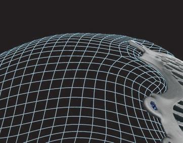
A.B. Dental’s new Customized Implants system exemplifies the pinnacle of technological initiative in our field, combining the latest 3D laser printing technology with ABGuidedService, A.B. Dental's exclusive patented computerized planning system that enables doctors to plan dental and facial restoration treatments with extraordinary precision.
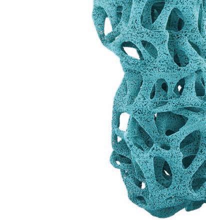
The existing methods of manufacturing implants didn't allow for the control at the Nano level and the correct surface energy that we were looking for. We were determined to create something from scratch –to build an implant according to our precise design. And we weren't willing to compromise.
The Customized Implants system provides an effective, personalized restoration solution for especially complex clinical cases where conventional implants are not a suitable option. These high-end implants are individually designed and manufactured for patients who require mandibular and/or maxillofacial reconstruction due to severe disfigurement or massive bone loss caused by cancer, major bone trauma, or other medical conditions.
Our search brought us to 3D printing, a new manufacturing technique that had never before been used to build dental implants of this kind. For the first time, we could manufacture a form from titanium dust in a sequential layering process that allows for unprecedented precision.

Novel osteogenic Ti-6Al-4V device for restoration of dental function in patients with large bone deficiencies: design, development and implementation
D.J. Cohen, A. Cheng, A. Kahn, M. A viram, A. J. Whitehead, S. L. Hyzy, R. M. Clohessy, B. D. Boyan & Z. SchwartzPublished: Scientific Reports, 08 February 2016
Abstract
Custom devices supporting bone regeneration and implant placement are needed for edentulous patients with large mandibular deficiencies where endosteal implantation is not possible. We developed a novel subperiosteal titanium-aluminum-vanadium bone onlay device produced by additive manufacturing (AM) and post-fabrication osteogenic micro-/nano-scale surface texture modification.
Human osteoblasts produced osteogenic and angiogenic factors when grown on laser-sintered nano/microtextured surfaces compared to smooth surfaces. Surfaceprocessed constructs caused higher bone-to-implant

Cellular
contact, vertical bone growth into disk pores (microCT and histomorphometry), and mechanical pull-out force at 5 and 10 w on rat calvaria compared to non-surfacemodified constructs, even when pre-treating the bone to stimulate osteogenesis. Surface-modified wrap-implants placed around rabbit tibias osseointegrated by 6 w. Finally, patient-specific constructs designed to support dental implants produced via AM and surface-processing were implanted on edentulous mandibular bone. 3 and 8 month post-operative images showed new bone formation and osseointegration of the device and indicated stability of the dental implants.

Cellular response to

Published: Advances in Dental Research 2016, Vol. 28(1) 10–17
Abstract
Changes in dental implant materials, structural design, and surface properties can all affect biological response. While bulk properties are important for mechanical stability of the implant, surface design ultimately contributes to osseointegration.
This article reviews the surface parameters of dental implant materials that contribute to improved cell response and osseointegration. We focus on how surface design affects mesenchymal cell response and differentiation into the osteoblast lineage.
Surface roughness has been largely studied at the microscale, but recent studies have highlighted the importance of hierarchical micron/submicron/Nano surface roughness, as well as surface roughness in combination with surface wettability. Integrins are transmembrane receptors that recognize
changes in the surface and mediate downstream signaling pathways. Specifically, the noncanonical Wnt5a pathway has been implicated in osteoblastic differentiation of cells on titanium implant surfaces.
However, much remains to be elucidated. Only recently have studies been conducted on the differences in biological response to implants based on sex, age, and clinical factors; these all point toward differences that advocate for patient-specific implant design. Finally, challenges in implant surface characterization must be addressed to optimize and compare data across studies.
An understanding of both the science and the biology of the materials is crucial for developing novel dental implant materials and surface modifications for improved osseointegration.


T10-FS Handle fixation screw
T10-FS,30 Fixation Screw Driver insert 30 mm

FS-2,7


Ib-LPI
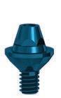
Cover screw for customized implant

T5-FS-3,20 Contra angle driver for fixation screw
T5-FS-3,25 Contra angle driver for fixation screw


Healing cap for customized implant

Healing cap for customized implant

P25-LPI,1

P35-LPI,2
P25-a,b/20

P25 Metal cover

Locator Extended Range Male processing package (Yellowm - extra soft)

P25-a,b/20 Pink - soft

P12-LPI,T/L
Temporary Flat Connection Abutment for customized implant

D1-LPI
Analog for customized implant

P12a-LPI Flat connection abutment for customized implant

P25-a,b/20 Purple - strong

P25-a,b/20 Transparent - standard

P12-LP1 Flat connection abutment for customized implant

D2-P12-LPI,15
Impression Transfer for Flat Connection Abutment for customized implant

P12p Flat connection Plastic sleeve for customized implant

D2al-LPI Long fixing screw for customized implant
P16-LPI,1
Straight adaptor for customized implant

P16-LPI,2
Straight adaptor for customized implant

P16-LPI,3
Straight adaptor for customized implant

Plastic sleeve for angular adaptor

P14-bR
Cobalt chrome sleeve for angular adaptor

P14-bT
Titanium sleeve for angular adaptor

P0-P14,5
Healing cap for P14

P0-P14,7
Healing cap for P14
D1-P14
Analog for P14

D2-P14 Transfer for P14

P14,sc
Scanning base for angular adaptor

P14-bRs
Cobalt chrome short sleeve

P14,bTs
Scanning base for angular adaptor D2-P14a

DBM-DEMINERALIZED BONE MATRIX READY TO USE NO REHYDRATION OR MIXING
Bone Graft - putty texture. Consists of 93% demineralized bone and 7% Hyaluronic acid. Available in a variety of volumes.


LPI - A subperiosteal implant for resorbed jaws.
Dr. Adrian Kahn
Age: 61 | Gender: female | Bilateral mandibular edentulism | Desire for fixed restoration

Panoramic view - severe posterior alveolar ridges atrophy
Prof. Zvi Schwartz

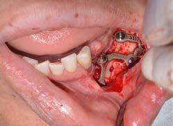
Right mandibular customized implant - screw fixation


Customized subperiosteal implant was created on the right mandible

A customized Ti6Al4V implant was designed using software with orange representing implant posts and green representing stabilizing screws

Bone allograft covering the bone surface

Implant in position, covered by bone allograft

A follow-up panoramic X-ray was taken to evaluate osteointegration and bone-to-implant contact after three months
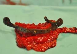
Customized subperiosteal implant was created for a situation where there is not enough bone for a conventional implant

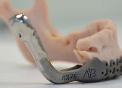
Customized implants were designed using 3D planning
Patient specific implant

Small holes were drilled into the patient’s jaw prior to implant placement to ensure exposure to stem cells and progenitor cells


Bone allograft placed over the implant A follow-up panoramic X-ray was taken to evaluate osteointegration and bone to-implant contact after three months
LPI - Laser Precision Implant: A subperiosteal implant for resorbed jaws. Customized subperiosteal implant was created for a situation where there is not enough bone for a conventional implant

3D Computerized Planning, severe posterior alveolar ridges atrophy

Healing caps 6 weeks post-op

A guide to drill the endosseous depth

Post-op pano xray

After drilling with the guide

AB scanning abutments for intra-oral scanning
U implant fitting accurately into the bone and on the bone surface

Final restoration
LPI - Laser Precision Implant: A subperiosteal implant for resorbed jaws. Customized subperiosteal implant was created for a situation where there is not enough bone for a conventional implant

3D planning on CT of left and right maxilla

Bone to cover the implant surface for additional augmentation

Custom implants left and right on 3D printed model of maxilla

Post-op pano xray

Right implant fitting accurately with screw fixation

3 months later, with multiunit abutments

Left implant with cover screws on the abutments

Patient’s denture adapted to be temporary screw retained restoration


Sinus lift bone augmentation
A New 3D “Custom Made Roofing” Technique for Maxillary Sinus Lift Augmentation
Dr. M. Shteif / Department of Oral, Maxillofacial Surgery, “Carmel” medical center, Haifa, Israel
A 45-year-old woman was referred after failure of sinus augmentation with oro-antral communication for reconstruction of her posterior maxilla.

Surgical guide for bonny slot


7 months’ post op CT


TSR (titanium sinus roofing) insertion

Bone substitute and implants insertion

Immediate post op CT (with implants)


Immediate post op CT


7 months’ post op CT (with implants)

10 months’ post op CT (with implants)
A New 3D “Custom Made Roofing” Technique for Maxillary Sinus Lift Augmentation
Dr. M. Shteif / Department of Oral, Maxillofacial Surgery, “Carmel” medical center, Haifa, Israel
A 45-year-old woman was referred after failure of sinus augmentation with oro-antral communication for reconstruction of her posterior maxilla.


Panoramic view of the Implants Final restoration


Orbital Floor Reconstruction
Prof. Imad Abu el-Naaj D.M.D / Head, Department of Oral & Maxillofacial Surgery, The Baruch Padeh medical center
Dr. Avi Toeg / Oral & Maxillofacial senior staff specialist, The Baruch Padeh medical center
A 24-year-old patient suffered from a blowout fracture of his left orbital floor, following facial trauma. He required reconstruction of the floor. To prevent increased or reduced volume of the orbit, which would result in enophtalmus or exophthalmos.
The conventional treatment to restore the orbital floor using autogenic bone, titanium or vicryl mesh, or other materials. There is difficulty in exactly restoring the shape of the orbit due to the complex anatomy and the danger of damage to the optic nerve.
The implant was designed using the shape of the orbit of the other eye which was intact.
The surgery was made with general anesthetic, and was completed in 20 minutes, using the custom-made implant with screw fixation. Conventional surgery would last more than an hour.
The low dose post-op CT shows excellent adaptation, and the patient has full function and aesthetics two years later.


Oral and maxillofacial restoration treatments
TMJ Condyle Replacement
Prof. Imad Abu el-Naaj D.M.D / Head, Department of Oral & Maxillofacial Surgery, The Baruch Padeh medical center
Dr. Avi Toeg / Oral & Maxillofacial senior staff specialist, The Baruch Padeh medical center
A 64-year-old patient with metastatic tumor to his right mandibular ramus. Resection of the tumor required removal of the condyle, resulting in loss of the Temporomandibular Joint. The joint disc was intact.
The tumor was resected with the help of a 3D Template surgical guide, which enabled cutting the mandible at exactly the planned position.
The custom implant was placed using specific screws. The surgery was completed in 2 hours, which was far less than anticipated and less than the conventional treatment with much better recovery. Post op CT showed good position of the condyle in the eminence fossa.
The patient was able to chew 2 days after the surgery, good mouth opening, excellent function with no pain on chewing.
After more than one year follow up the patient is functioning with the TMJ prosthesis with no complications.


Dr. Vadim Reiser\ Director of Oral and Maxillofacial Surgery Department of the Tel Aviv Sourasky Medical Center, Dr. Aharon Amir, Assuta Hospital
Patient Specific Implant to reconstruct maxilla, due to Central Giant Cell Granuloma on the right side.
This 24-year-old patient was diagnosed at an early age with an aggressive benign Central Giant Cell Granuloma on the right side. The right maxilla was removed, extending to 26 area on the left.
The patient had poor aesthetics and function, and speech difficulty.

3D Planning of Reconstruction
Implant and Bone Shape from Fibula

Cutting Guide for Leg Fibula Donor Site

Implant and Bone


Reconstructed maxilla based on a 3D image before surgery

Surgical Process 3D Designed Bone Cutting Guide to remove a precise shape of bone

Implant and Bone

The image was superimposed on the new CT.


Custom Implant on 3D Model
Bone with Blood Supply and Reconstruction Implant
Post-Op Panoramic Xray

6 months’ after reconstruction: Panoramic View after Implant Placement
Facial Reconstruction
Prof. Samer Srouji DMD, Ph.D / Chief of Oral And Maxillofacial, Oral Medicine Institute, Head Of “Bone Regeneration” Lab, Galilee Medical Center, Naharia

3D Planning of Reconstruction Implant and Bone Shape from Fibula

Cutting Guide for Leg Fibula Donor Site

The shape of the reconstructed maxilla was based on a 3D image of the patient before her first surgery.

Surgical Process 3D Designed Bone Cutting Guide to remove a precise shape of bone

The image was superimposed on the new CT.

Custom Implant on 3D Model

Published: Volume 27, Number 8, November 2016
Abstract
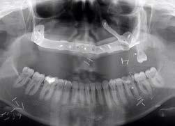
We describe the planning and surgery as well as pitfalls and management of patients exhibiting near total mandibular avulsion injury rehabilitated using threedimensional (3D) laser printing of a titanium lower jaw. Laser-sintering involves zapping layers of powdered metal to recreate a 3D implantable skeletal defect. The process involves using either mirror imaging of the unaffected side or using archival image database of healthy individuals. A 25-year-old man presented with a gunshot injury that left him with a near total avulsed mandible.
The patient received state-of-the-art treatment using a
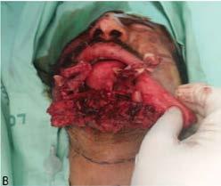


laser 3D printed mandible which was connected to the muscles of mastication for functionality.
The inner side of the titanium jaw was filled with the patient’s comminuted fractured bones in addition to harvested iliac crest bone graft that was covered with the patient’s remaining periosteal tissue.
The implantation of a near total mandible using 3D laser printing is a fast and predictable process that in selected patients can result in aesthetically as well as functionally excellent results. The authors believe that the future of craniofacial reconstruction will employ these methods for facial bony reconstruction.


Published by Elsevier Ltd., 9 January 2017
Abstract
Reconstruction of the craniofacial complex is extremely challenging due to the unique anatomy, presence of vital structures and the diversity of defects. In craniofacial reconstruction, restoration of aesthetics and function is the primary goal.
Auto-grafts are the gold standard for craniofacial skeletal reconstruction, however they possess several disadvantages. These disadvantages led to the research of alloplastic materials.
Development of computer assisted design and computer assisted manufacturing systems allows for precise preoperative planning and designing of patient specific implants.
Materials and methods: A case of facial bone reconstruction is presented exhibiting major deficiency in the mandible. Reconstruction was performed using a custom-made titanium implant. This implant was shaped as a crib, thus allowing for artificial and autogenous bone


graft augmentation for future implant placement.
Results: The mandibular defect was reconstructed using titanium for strength and function and resulted in proper mouth opening, function and aesthetics. Another innovation was the addition of dental implants which were designed as part of the patient specific implant thus allowing for future implant supported dental rehabilitation not necessarily requiring support of the bone graft.
Conclusions: Individual computer assisted design & computer assisted manufacturing systems for preexisting facial defects can become an alternative to auto-grafts. Results are promising and exhibit excellent aesthetic and functional outcomes, while reducing operating time and avoiding donor site morbidity. This procedure provides a simple way to reconstruct a complex three-dimensional structure with precision which is difficult achieving with standard methods.


