Sankoff et al
Rapid Assessment of CVP
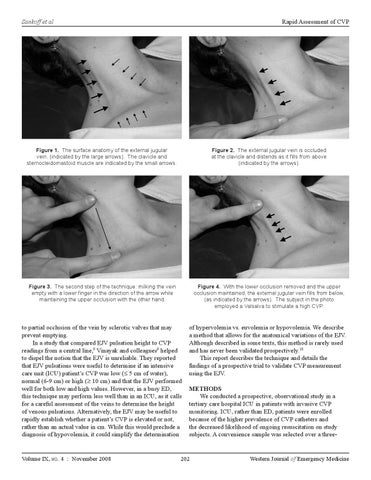
Figure 1. The surface anatomy of the external jugular vein, (indicated by the large arrows). The clavicle and sternocleidomastoid muscle are indicated by the small arrows.
Figure 2. The external jugular vein is occluded at the clavicle and distends as it fills from above (indicated by the arrows).
Figure 3. The second step of the technique: milking the vein empty with a lower finger in the direction of the arrow while maintaining the upper occlusion with the other hand.
Figure 4. With the lower occlusion removed and the upper occlusion maintained, the external jugular vein fills from below, (as indicated by the arrows). The subject in the photo employed a Valsalva to stimulate a high CVP.
to partial occlusion of the vein by sclerotic valves that may prevent emptying. In a study that compared EJV pulsation height to CVP readings from a central line,9 Vinayak and colleagues9 helped to dispel the notion that the EJV is unreliable. They reported that EJV pulsations were useful to determine if an intensive care unit (ICU) patient’s CVP was low (≤ 5 cm of water), normal (6-9 cm) or high (≥ 10 cm) and that the EJV performed well for both low and high values. However, in a busy ED, this technique may perform less well than in an ICU, as it calls for a careful assessment of the veins to determine the height of venous pulsations. Alternatively, the EJV may be useful to rapidly establish whether a patient’s CVP is elevated or not, rather than an actual value in cm. While this would preclude a diagnosis of hypovolemia, it could simplify the determination
of hypervolemia vs. euvolemia or hypovolemia. We describe a method that allows for the anatomical variations of the EJV. Although described in some texts, this method is rarely used and has never been validated prospectively.10 This report describes the technique and details the findings of a prospective trial to validate CVP measurement using the EJV.
Volume IX, no. 4 : November 2008
METHODS We conducted a prospective, observational study in a tertiary care hospital ICU in patients with invasive CVP monitoring. ICU, rather than ED, patients were enrolled because of the higher prevalence of CVP catheters and the decreased likelihood of ongoing resuscitation on study subjects. A convenience sample was selected over a three-
202
Western Journal of Emergency Medicine