Images In Emergency Medicine
A Case of Syncope Kristin H. Dwyer, MD, MPH* Joshua S. Rempell, MD MPH†
*Brown University, Warren Alpert School of Medicine, Department of Emergency Medicine, Providence, Rhode Island † Cooper Medical School of Rowan University, Cooper University Hospital, Department of Emergency Medicine, Camden, New Jersey
Section Editor: Rick A. McPheeters, DO Submission history: Submitted January 26, 2017; Revision received April 7, 2017; Accepted April 19, 2017 Electronically published October 3, 2017 Full text available through open access at http://escholarship.org/uc/uciem_cpcem DOI: 10.5811/cpcem.2017.4.33725
[Clin Pract Cases Emerg Med. 2017;1(4):427–429.]
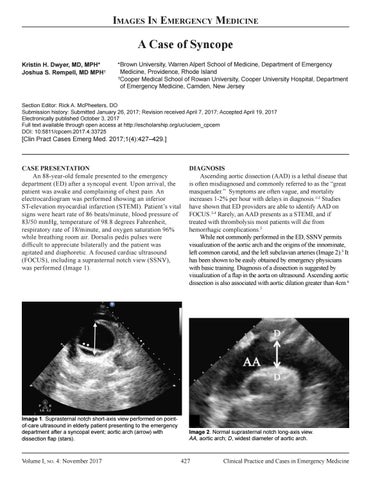
CASE PRESENTATION An 88-year-old female presented to the emergency department (ED) after a syncopal event. Upon arrival, the patient was awake and complaining of chest pain. An electrocardiogram was performed showing an inferior ST-elevation myocardial infarction (STEMI). Patient’s vital signs were heart rate of 86 beats/minute, blood pressure of 83/50 mmHg, temperature of 98.8 degrees Fahrenheit, respiratory rate of 18/minute, and oxygen saturation 96% while breathing room air. Dorsalis pedis pulses were difficult to appreciate bilaterally and the patient was agitated and diaphoretic. A focused cardiac ultrasound (FOCUS), including a suprasternal notch view (SSNV), was performed (Image 1).
Image 1. Suprasternal notch short-axis view performed on pointof-care ultrasound in elderly patient presenting to the emergency department after a syncopal event; aortic arch (arrow) with dissection flap (stars).
Volume I, no. 4: November 2017
DIAGNOSIS Ascending aortic dissection (AAD) is a lethal disease that is often misdiagnosed and commonly referred to as the “great masquerader.” Symptoms are often vague, and mortality increases 1-2% per hour with delays in diagnosis.1-2 Studies have shown that ED providers are able to identify AAD on FOCUS.3-4 Rarely, an AAD presents as a STEMI, and if treated with thrombolysis most patients will die from hemorrhagic complications.5 While not commonly performed in the ED, SSNV permits visualization of the aortic arch and the origins of the innominate, left common carotid, and the left subclavian arteries (Image 2).3 It has been shown to be easily obtained by emergency physicians with basic training. Diagnosis of a dissection is suggested by visualization of a flap in the aorta on ultrasound. Ascending aortic dissection is also associated with aortic dilation greater than 4cm.6
Image 2. Normal suprasternal notch long-axis view. AA, aortic arch; D, widest diameter of aortic arch.
427
Clinical Practice and Cases in Emergency Medicine