4 minute read
This is what they do Four


This is what they do
From collaborating with patients to research at cellular level: Brain Center Rudolf Magnus has a wide variety of scientists. They are all searching for the best answers to their questions, as are these four motivated researchers.
Advertisement
By Aafke Kok Images: Bram Belloni
Frank Meye Translational neurosciences
Brain changes and binge eating
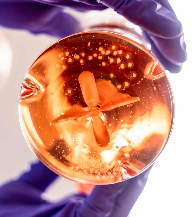
In response to stress, both people and animals consume a great deal of sugar and fat. ‘Binge eating is a problem for people with obesity, eating disorders or people with impulsive behavior such as ADHD,’ says Frank Meye. The need for ‘bad’ food is accompanied by changes in communication between different areas of the brain involved in the processing of rewards. This is what Meye is studying. He is using mice that have been subjected to social stress and, therefore, display binge-eating behavior. Meye examines the mice’s brain tissue. Using electrodes, he measures the electrical signal with which nerve cells communicate. The more they communicate, the stronger the connection. Meye studies the differences in the strength of this connection between stressed mice and control mice. For example, he looks at connections with dopamine neurons, an important group of cells in the reward circuit of the brain.
Meye can then control specific nerve cells in living mice and study the mice’s eating behavior. He does this through optogenetics or chemogenetics, with which he can turn nerve cells ‘on’ or ‘off’ using light or special viruses, respectively. This is how he is trying to uncover the link between changes in the brain and binge-eating behavior. Ultimately, he hopes to understand how to prevent these changes in the brain, in order to reduce binge eating.
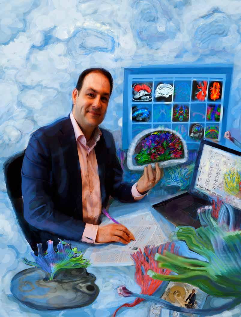
Natalia Petridou Imaging technology
Understanding fMRI

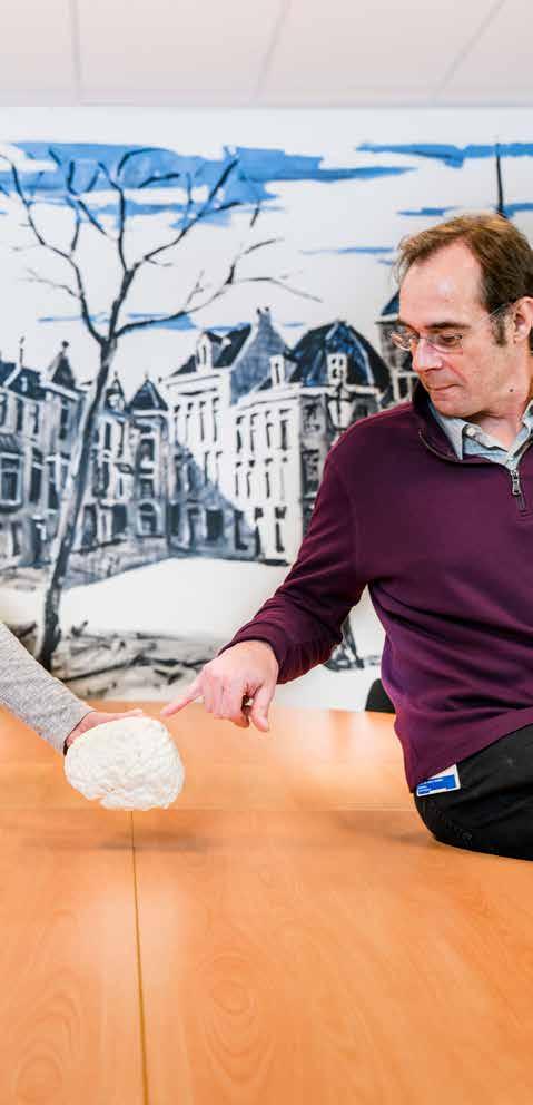
Marjolijn Ketelaar Rehabilitation medicine
True patient collaboration
For the study of cerebral palsy (CP), a congenital disruption of the brain, Marjolijn Ketelaar invited twelve teenagers with CP to join her study as ambassadors. One example of their participation was thinking about research questions – patients often have a very different view of what is really important in the study of their disease. Among other things, Ketelaar and her team studied problems young people with CP have within the educational setting. In addition to scientific publications, this also led to products that she created with the teenagers, like a poster for teachers explaining CP, to eliminate misunderstandings about the disease. This is true patient participation, not just a tick on researchers’ checklists. ‘True collaboration with patients, from beginning to end, leads to much better research,’ says Ketelaar. She also tries to help her colleagues understand the importance of patient participation. For example, she contributed to the development of a tool that researchers can use to discuss desired participation at every step during studies. She also organized a conference with not one, but two chairs at every session: a researcher or doctor, and a patient or the parent of a patient. This keeps the focus on the question ‘for whom are we doing this?’ In order to understand the functioning of the brain, many researchers use fMRI scanners. In the images that they create, the active areas of the brain light up. This technology is also frequently used in the clinic. ‘But fMRI measures brain activity in an indirect way,’ says Natalia Petridou. What you are really measuring are the characteristics of blood flow in parts of the brain. The idea is that this flow of blood is connected to activation of nerve cells. After all, they need oxygen from the blood to do their job. ‘But we don’t know how the link between blood and nerve cells works exactly,’ says Petridou. She hopes to find out. She is building models to predict how certain parts of the brain work. She then tests these models. For this, she uses a 7-Tesla MRI scanner, a scanner with an extremely strong magnetic field. Petridou is developing methods to use this scanner to make highly detailed images of the brain. In addition, she uses special cameras to study minute changes in the bloodstream. She also takes measurements using electrodes in the brain. Ultimately, Petridou hopes to understand the link between blood and nerve cells, and to use her findings to improve fMRI use.
Jan Veldink Neurogenetics

Gene therapy
As a doctor, Jan Veldink sees ALS patients every week. ‘Their life expectancy is currently three to five years, and with the medication we now have this can be extended up to six months,’ says Veldink. He hopes to change this with his research. The goal of Project MinE, set up through crowdfunding, is to map out the DNA of 22,500 patients. The counter is currently at 10,000. By comparing DNA, researchers have already found five spots in the DNA that are involved in the occurrence of ALS. This may be a line of approach for new treatment, such as gene therapy. In gene therapy, drugs focus on the protein in a gene identified by the study, or they block these genes directly. But ALS is not caused by genetic anomalies alone. That is why Veldink also studies the outer shell of DNA, which is affected by life style factors such as diet and stress. On the basis of the outer DNA shell, Veldink can divide ALS patients into new subgroups, incorporating the interaction with lifestyle. The starting points for new therapies are promising, but because these therapies serve a small subgroup of patients, it is difficult to find companies for further development. Veldink remains hopeful, ‘mainly because the increasing interest in ALS in recent years.’