9 minute read
Diagnostic Imaging Insights Accelerated by Artificial Intelligence


Mark Hitchman
Mark Hitchman, Managing Director of Canon Medical Systems UK, highlights the exciting AI innovations in diagnostic imaging and discusses the need for home-grown data to power advancements in the future
Advertisement
Arti cial Intelligence (AI) in healthcare is big news. Nearly every day there is a new study or research result announced, powering the potential advances in how we diagnose, treat and prevent healthcare conditions in the future. It stirs the imagination and is an exciting exploration to be part of, but it also has to be a manageable process, realistic and scalable.
With every positive news story on the advances of AI in healthcare, there is also the counterbalance that questions when we will realistically be able to use these advancements in clinical practice. Our response is that AI is already here. It's being used in many hospitals and clinics up and down the UK, perhaps without the radiographers even realising, as it works seamlessly behind the scenes.
There is no plan to shock and awe the healthcare community, it has been through enough in the last few years. So our approach is to calmly and carefully introduce the advancements embedded within our imaging systems and visualisation software via familiar user interfaces and training plans. Diagnostic imaging evolution is happening every day and it is making a di erence to clinical con dence and work ow e ciency.
AI helps with decision support, it’s not about replacingclinicians The reality of today’s AI in action is about solving many of the acute problems in the modern healthcare environment. It is about providing the tools to help clinicians make con dent decisions faster when faced with growing backlogs. It is also about simplifying work ows that optimise sta ng and equipment resource deployment that ultimately deliver time e ciency gains that can be passed onto better patient care. Whether this is being able to see an extra 3-5 patients in the worklist per day to keep on top of waiting lists, or to provide extra time for radiographers to spend with patients needing reassurance and comfort during a stressful examination, the bene ts are clear.
AI in imaging is also about reducing the stress and exhaustion on health professionals. Replacing radiologists or oncologists with AI is not an option, the human element is needed more than ever. But AI can provide an e ective decision support tool to help triage reporting cases and speed up decision making.
The journey of AI in imaging began with the introduction of AI-assisted imaging rstly through CT and now MRI via a Deep Learning reconstruction AI algorithm that di erentiates ‘noise’ from true signal to clean up images resulting in high-quality scans free from distortion. This helps to preserve edges,improve textures and maintain details to assist with clearer clinical interpretation. It also reduces the need for image retakes and is at a much lower patient radiation dose than o ered before (Fig. 1, 2 & 3). This technology is now in place in dozens of hospitals across the UK, and from conversations with the imaging community, it is clear that the bene ts to patients, processes and people are helping daily diagnosticservice life.
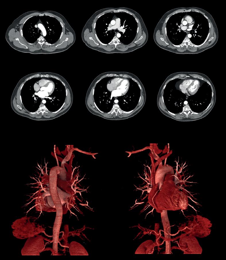
Fig 2. Image obtained using AI-assisted CT demonstrating excellent visualisation of detail in small vessels enabled by lownoise and high-resolution characteristics. The patient arrived in the Emergency Department hypertensive with atypical chest pain. Helical Gated Chest CTA was conducted to rule out acute coronary syndrome and/or Aortic dissection.
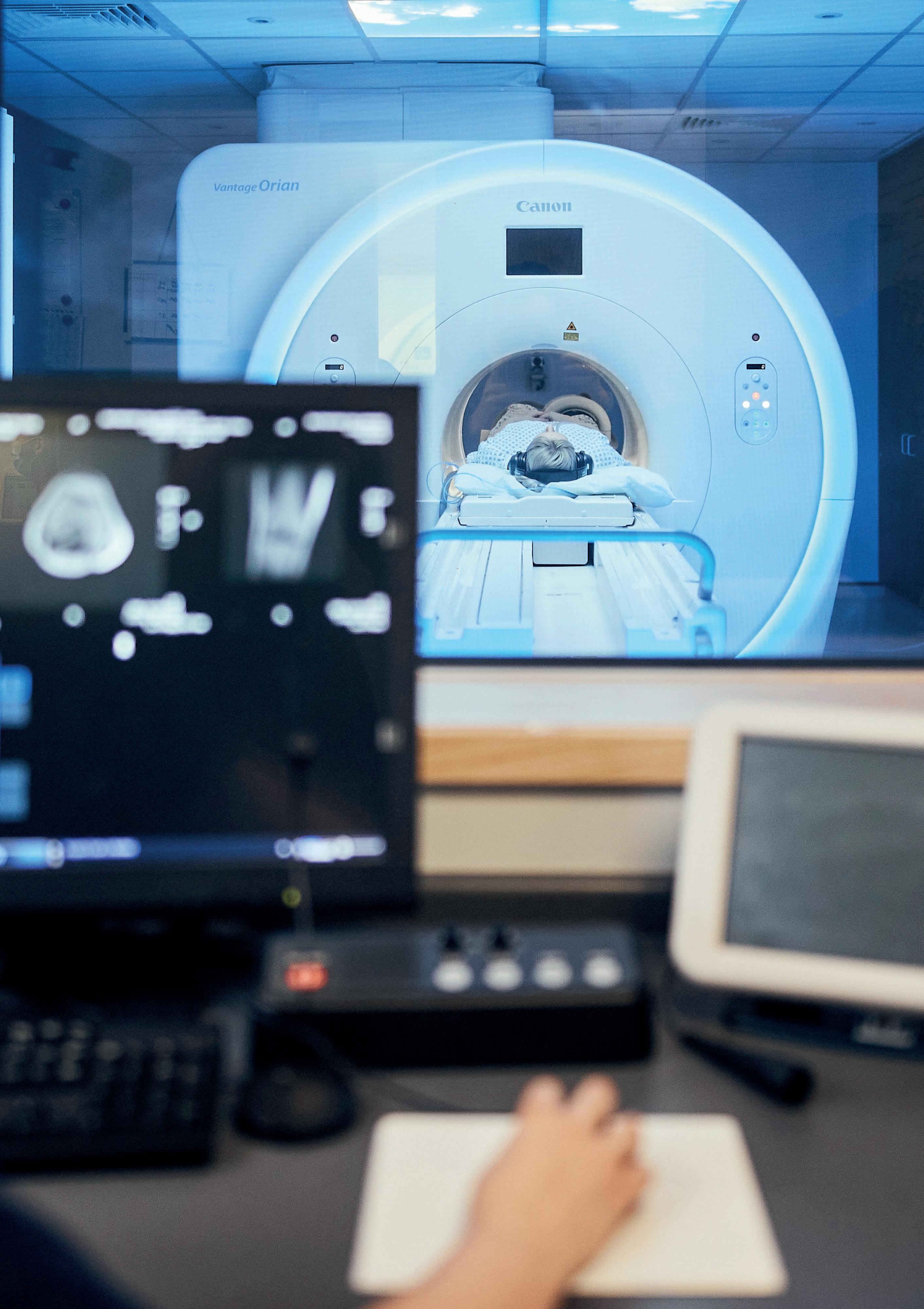
At Wycombe Hospital, part of Buckinghamshire Healthcare NHS Trust, the Deputy Lead MRI Radiographer gave feedback on the AI-assisted MRI (Fig. 4) stating that, “the Advanced intelligent Clear-IQ Engine Deep Learning reconstruction technology “produces great images that are detailed and low in noise. This reduces scan times and produces valuable diagnostic images rst time”.
Improved diagnostic accuracy is also cited as one of the bene ts at NHS Lothian’s Royal Hospital for Children and Young People in Edinburgh using an AI-assisted CT. Along with making complex paediatric examinations easier, it speeds up scans and reduces dose for young patients. The CT Radiographer told us that, “Dose reduction for paediatric patients is amazing. The 16cm detector can achieve a volume scan in 0.5 seconds on a head which is really helpful when examining young patients – we no longer need anaesthetics or strategies to try and keep them still for as long. The fast speed really helps us perform the procedure quicker and is better for the small person concerned”.
Similar feedback has been received about the Advanced intelligent Clear-IQ Engine powered CT from a major acute teaching hospital in Scotland, the Royal In rmary of Edinburgh. The CT/MRI Superintendent stated, “As radiographers we can become blasé about the imaging equipment we use on a daily basis, but the arrival of our new CTs reminded us of the amazing innovation going into medical imaging today. Our cardiologists have been blown away with how quick a cardiac CT is acquired using the wide detector, as well as the image quality achieved at such low doses”. AI-assisted diagnosis of key diseases But AI has far more potential to demonstrate in imaging. More recently, we have started rolling out AI and automation applications for imaging that drill into speci c conditions.
Take, for example, strokes. These occur once every ve minutes in the UK. It is the fourth single leading cause of death and responsible for 35,000 deaths annually. Great results have been achieved in bringing these gures down over the years due to expanding research, medical innovations and awareness campaigns but ‘time is brain’ has always been a ubiquitous phrase and it is still relevant today. The quicker clinicians can identify and treat a patient with a stroke, the better the outcome from death or long-term neurological damage.
The automation of stroke diagnosis using AI in diagnostic imaging has the potential to streamline stroke-related work ow by automatically consolidating results into a single summary and alerting for abnormalities. It can help to swiftly analyse and categorise images to detect signs of ischemic and haemorrhagic stroke in minutes. This has the potential to provide access to information that can speed up the administration of life-saving treatment.
Gathering up data to teach AI and cement itsplaceindaily clinical practice Great strides are also being made on the visionary future of AI, the stu that medical futurists dream of. Behind every deep-learning innovation there is data,
lots of it. This is what builds the algorithms and feeds the machines with knowledge.
This is a great use, recycling you could say, of the information our healthcare establishments have been collecting for years. Only a few years ago it was estimated that 97% of the 50 petabytes of data produced by hospitals per year went unused. Thebig data sources include patient medicalrecords, CT or MRI scan images, pharmacy records, laboratory results and allthe other sub-sectors of the healthcare ecosystem.
It is the decanting of this relevant healthcare data safely, which is pseudonymised to ensure protection of patients’ personal details, that is key to accelerating the pace of
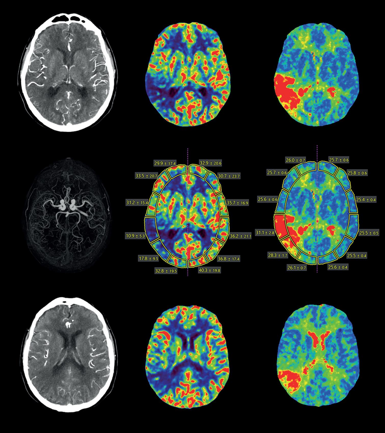
Fig 3. Brain perfusion right occlusion using AI-assisted CT: Low-dose whole brain dynamic 4D CTA DSA and perfusion, enabled by wide-area CT and Advanced intelligent Clear-IQ Engine (AiCE) DLR. The 4D CTA shows a blood ow interruption of the right MCA-M3 segment. Perfusion maps display reduced cerebral blood ow and increased MTT in M3 territory corresponding to the vascular abnormality.
Fig 4. AI-assisted MRI producing images that are detailed and low in noise. This reduces scan times and produces valuable diagnostic images rst time.
turning great AI ideas into daily clinical practice reality. Each AI application needs up to 100,000 data sets or even more to learn from along with a development process involving human clinical evaluation.
We are very proud to have a home-grown hub of AI research and development in Canon Medical Research Europe in Edinburgh. This group of software engineers and architects collaborates with 15 partners in the UK across academia, the NHS and industry via its Safe-Haven Arti cial Intelligence Platform (SHAIP). This means, in essence, that the data being used to develop future AI innovations is gathered from UK speci c data sources, making the development accurate and speci c to our patient populations, at the same time as working closely with colleagues in Japan, China, Europe and the USA.
The team is responsible for building a set of services and web applications that include useful tools for clinicians to select and annotate patient data for machine learning, together with infrastructure for data scientists to develop, train and validate algorithms within the hospital environment. Once an algorithm has been created using SHAIP, it can be deployed into a Clinical Cockpit to allow e ortless demonstration to clinicians.
Many projects including stroke and Covid are fully underway and wider work in progress includes oncology, congenital heart disease, ultrasound, interventional angiography and diagnostic support. AI is here today and scaling ready for a smarter tomorrow AI is only as good as the data it is built from, which means that when trying to get solutions to market to start helping patients and speeding up the way healthcare is delivered post-Covid, collaboration is key. Federated learning, that is, not moving patient data outside the rewalls of the institutions they reside in, is a growing methodology and requires the involvement of lots of proactive parties.
We all play a role in the acceptance and belief in AI. From welcoming its place within the health environment to driving the creation of new ideas and tech-partnering to make concepts a reality. It is the here and now to deliver improved imaging insights and its future is certain to power the reshaping of tomorrow’s clinical services. //
References i Brain Research UK, https://www.brainresearchuk.org.uk/ neurological-conditions/stroke accessed January 2022. ii AUTOStroke, https://eu.medical.canon/products/healthcare_it/ auto-stroke-solution.html


