EYES SELECTION
Fat-rich diets and liver health Dietary management is crucial in caring for people with non-alcoholic fatty liver disease (NAFLD), now known as metabolically associated steatotic liver disease (MASLD). NAFLD is a chronic liver condition ranging from simple steatosis to steatohepatitis (NASH) with or without significant fibrosis. With rapidly increasing rates of incidence, the estimated global prevalence of NAFLD is around 30% of the population, with progression to cirrhosis in around 1−2%.1
A new name
NAFLD encompasses various fatty liver diseases that are not caused by excessive alcohol consumption, viral hepatitis, drug-induced liver disease or monogenic conditions. Recently, a Delphi consensus led to a new nomenclature: steatotic liver disease (SLD).2 SLD includes: • MASLD − metabolically associated SLD • MetALD − a combination of MASLD with secondary causes • Cryptogenic SLD − SLD without metabolic features or due to other causes. This article will use the term MASLD to replace NAFLD, aiming to identify the same group of patients.
Identifying MASLD
MASLD is defined as evidence of steatosis on imaging or biopsy, along with at least one of several cardiometabolic risk factors. These include a body mass index (BMI) ≥25kg/m² or waist measurement above specified thresholds adjusted for sex and race, elevated fasting glucose levels or diabetes diagnosis, high blood pressure or antihypertensive treatment, elevated plasma triglycerides or lipid-lowering treatment, and low levels of high density lipoprotein−cholesterol.2 MASLD is, as such, linked to the insulin resistance syndrome, also called metabolic syndrome. Indeed, the condition is linked to chronic excess caloric consumption and lack of physical activity. It is known that the Western diet, rich in carbohydrates and saturated fats, is a major contributor to the development of steatosis.3 Evidently, this can be directly attributed to the caloric excess that these foods represent. But what is exactly the ‘Western diet’? It is a modern dietary pattern, highly characterised by pre-packaged foods, refined grains, red and processed meat, sugar-sweetened beverages, industrially produced animal products, high-fat dairy products and fried products (see Figure). If broken down in structural components, it is a diet low in vegetables, and high in sugar, sodium and especially saturated fats.
Saturated fats in the diet
Fats can be divided into three groups: transfatty acids, unsaturated fats and saturated fats. Saturated fats have a reinforcing effect on liver fat accumulation. One particular study examined the addition of muffins high in saturated fats versus those high in unsaturated fats to the diet of participants. Weight gain was similar in both groups, but liver fat content increased significantly in the group receiving high saturated fats.4 Similar results
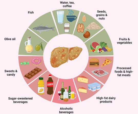
Better dietary options for good liver health (green versus red panels).
were seen in a trial with overfeeding using saturated fats versus unsaturated fats versus simple sugars over three weeks.5 Thus, since the importance of reducing saturated fats in the diet is established, foods rich in palm oil (such as processed foods), butter and other high fat dairy products, and high fat meats (such as sausages) must be avoided, as well as cakes, ice creams and other sweets.3 The Mediterranean diet, rich in nuts, seeds, vegetables, olive oil, fish and wholegrains high in fibre, is highly recommended, since it is a good source of essential unsaturated fats (n-3 fatty acids) but lacks saturated fats. Extra attention must be made to sugar-sweetened beverages, since they are not energy-dense, but also contain refined sugars such as sucrose, fructose or high-fructose corn syrup, that are easily converted into fats.6
Other factors to consider
Obviously, MASLD is more than just the consumption of too many saturated fats, and multiple risk factors and molecular pathways are at play. MASLD is, in essence, a condition of excess intrahepatic triglyceride accumulation, which stems from the relative imbalance of lipid inflow and lipid removal in the liver. Lipid influx is derived from a number or sources, of which dietary intake is estimated at around 15%. Other sources are adipose tissue lipolysis and de novo lipogenesis. So, if only 15% is dietary, why is it so important? Because nutritional lipids are the only depot that we can actively control. We should also note that the molecular effects of several nutrients on organ receptors are still largely unknown, while the composition of our food is getting increasingly difficult
to monitor, creating an important paradox. Different dietary patterns and nutrients may promote hepatic lipid accumulation by acting on a variety of hepatic nuclear receptors. Most is known about the liver X receptor and peroxisome proliferator-activated receptors, or PPARs. These PPARs are currently one of the most promising targets in pharmaceutical attempts to treat hepatic steatosis. Despite this, we are still guessing how most of our processed foods might contribute to hepatic steatosis.
In conclusion
Nutrition and diet have a significant impact on MASLD (formerly NAFLD). A balanced diet with a reduced caloric intake, an emphasis on essential fats and avoidance of saturated fats and added sugars is essential for managing and potentially reversing MASLD, coupled with lifestyle modifications, including regular physical activity and weight management. It is crucial for individuals with MASLD to seek professional guidance from healthcare experts to tailor an appropriate diet plan and monitor liver health effectively. Jonathan Mertens Belgium REFERENCES 1. Francque 2023 Reviews in Endocrine & Metabolic Disorders 24 885−899 https://doi.org/10.1007/s11154-023-09820-6. 2. Rinella et al. 2023 Journal of Hepatology https://doi.org/10.1016/j.jhep.2023.06.003. 3. Francque et al. 2021 JHEP Reports 3 100322 https://doi.org/10.1016/j.jhepr.2021.100322. 4. Rosqvist et al. 2019 Journal of Clinical Endocrinology & Metabolism 104 6207−6219 https://doi.org/10.1210/jc.2019-00160. 5. Luukkonen et al. 2018 Diabetes Care 41 1732−1739 http://doi.org/10.2337/dc18-0071. 6. Jensen et al. 2018 Journal of Hepatology 68 1063−1075 https://doi.org/10.1016/j.jhep.2018.01.019.
7