9 minute read
Surgical Colic


By Lauren Thurgood
Cert IV VN, Dip LM, RVN
AVN and AVNAT Continuing Professional Development
PATIENT DETAILS: Species:Equine Breed: Thoroughbred Age: 12 years old Sex: Gelding Colour: Brown Weight:520 kg
HISTORY AND EXAMINATION A gelding was assessed on farm by one of our ambulatory veterinarians for the acute onset of severe colic symptoms. The gelding was uncomfortable, showing marked abdominal distention but permitted a basic examination. His heartrate was slightly increased at 52, which is above the ideal reference range of 28–48 beats per minute. His respiration rate of 16 and a rectal temperature of 37.6 degrees Celsius were within normal limits for his breed and age group. To permit further examination, intravenous analgesia was provided with 10 ml of flunixin meglumine (1 mg/kg) and 2 ml of xylazine (0.4 mg/kg). His mucous membranes were pink and moist with a capillary refill time of less than 2 seconds, indicating adequate perfusion. Gastrointestinal auscultation was reduced in all four quadrants, but with tympanic sounds throughout. The gelding indicated increasing pain despite analgesia and was reluctant to stand. This uncontrollable pain indicated the need for further diagnostics with the possibility of surgical intervention. The gelding was to be referred to the hospital for further evaluation by our surgeons and internists. For the aid of transportation, 0.5 ml of butorphanol (0.1 mg/kg) was administered intravenously. On arrival at the hospital the gelding’s condition had deteriorated and he was recumbent in the trailer. Cardinal signs were consistent with the onfarm assessment, but physically he was showing severe pain. A rectal examination performed by the afterhours surgeon found moderate gas distention and displacement of the large colon. THE PLAN These findings, in combination with the uncontrollable pain exhibited by the gelding, indicated the need for surgical correction. With consent for surgical intervention, the team prepared the patient and the theatre for an emergency exploratory laparotomy. PRE-ANAESTHETIC BLOOD PANEL A blood sample was collected from the left jugular vein to run a pre-anaesthetic panel. The results showed a mild hyperlactatemia, as blood lactate levels were at 2.6 mmol/L, with a normal range below 2 mmol/L in adults. The packed cell volume or PCV of 32% was on the low end of normal, with the reference range at 32–48%. The total protein result of 42 g/L, a mild hypoproteinemia was apparent, as the suggested range is 57–80 g/L (Zoetis LLC, 2022). These values can be suggestive of compromised blood circulation and can be representative of an early intestinal strangulation. INDUCTION AND PREPARATION The right jugular vein was clipped and prepped using a chlorhexidine and methylated spirit preparation. The nurse aseptically placed a 14-gauge Extended Use MILACATH®, attached a heparinised extension set and sutured into position. Antimicrobials were given prophylactically, 38 ml of procaine penicillin (22 IU/kg) was given into the muscle, and 34 ml of gentamicin (6.6 mg/kg) was given intravenously. As premedication, 3.1 ml of xylazine (0.6 mg/kg) and 5.2 ml of methadone (0.1 mg/kg) were calculated. With 2 ml of xylazine and the full dose of methadone administered intravenously, his mouth was rinsed to clear any food material and a nasogastric tube passed to ensure no accumulation of fluid in the stomach. The remaining 1.1 ml of xylazine was given in the induction box and he was allowed to settle. The patient was induced with 11.4 ml of ketamine and 4.2 ml of midazolam at the listed rates. The patient was intubated with a 26 mm ET tube and connected to a rebreathing circuit with mechanical ventilation. Isoflurane was used as the anaesthetic agent with 8L of oxygen as the carrier.
PATIENT PREPARATION The surgery site preparation begins in the induction box as the clip starts from the base of the sternum and caudally to include the entire area around the sheath. The area was scrubbed with a chlorhexidine-based detergent. To transfer the patient from induction to the surgery room, hobbles were attached around the
pasterns and connected to an electric hoist from the ceiling. Together the team transported the gelding onto the surgical bed and positioned in dorsal recumbency. While anaesthetised, the patient received Hartmann’s at 5 L/hr for the first hour, reducing to 3 L/hr afterwards. To ensure adequate fluid therapy was provided, urine output was monitored by the placement of an indwelling 28 French Foley catheter. The sheath was sutured closed to ensure a sterile preparation of the surgical site could be achieved. The surgical site was prepared with a 4% chlorhexidine solution, methylated spirits, and sterile gauze swabs were used to prepare the surgical site. The chlorhexidine was used in a circular motion starting over the incision site and working towards the periphery. The chlorhexidine was removed with the spirit-soaked swabs using the same principle. The surgical scrub was repeated three times to ensure an aseptic technique. SURGICAL INSTRUMENTATION Prior to the gelding moving into the surgery room, the instrument table was prepared. The surgical table provided basic instrumentation for the surgeon to gown and glove, drape the patient, and open the abdomen via a ventral midline incision. The laparotomy revealed a right dorsal displacement of the large colon, although with further exploration there also seemed to be a strangulating lesion. A portion of the small intestine was herniated within the epiploic foramen, which caused the blood supply and motility to become compromised. A small intestinal resection was performed, removing 3 feet of damaged bowel. To complete the resection, further instrumentation was needed by the surgeon. The following instrumentation was provided to the surgical assistant: • an extensive instrument kit • two doyen intestinal clamps • two medium Penrose drains • a Caiman® seal and cut device • additional abdominal sponges • warmed saline • Poole suction tube • new gloves. The surgeon exteriorised the strangulation to understand the extent of the comprised small intestine and how much would need to be removed to allow for healthy margins. With the damaged bowel identified, the surgeon marked the two outer edges of the resection with the Penrose drains. The surgeon utilised the Caiman to seal and cut the vessels
Surgical Colic
Continued from previous page
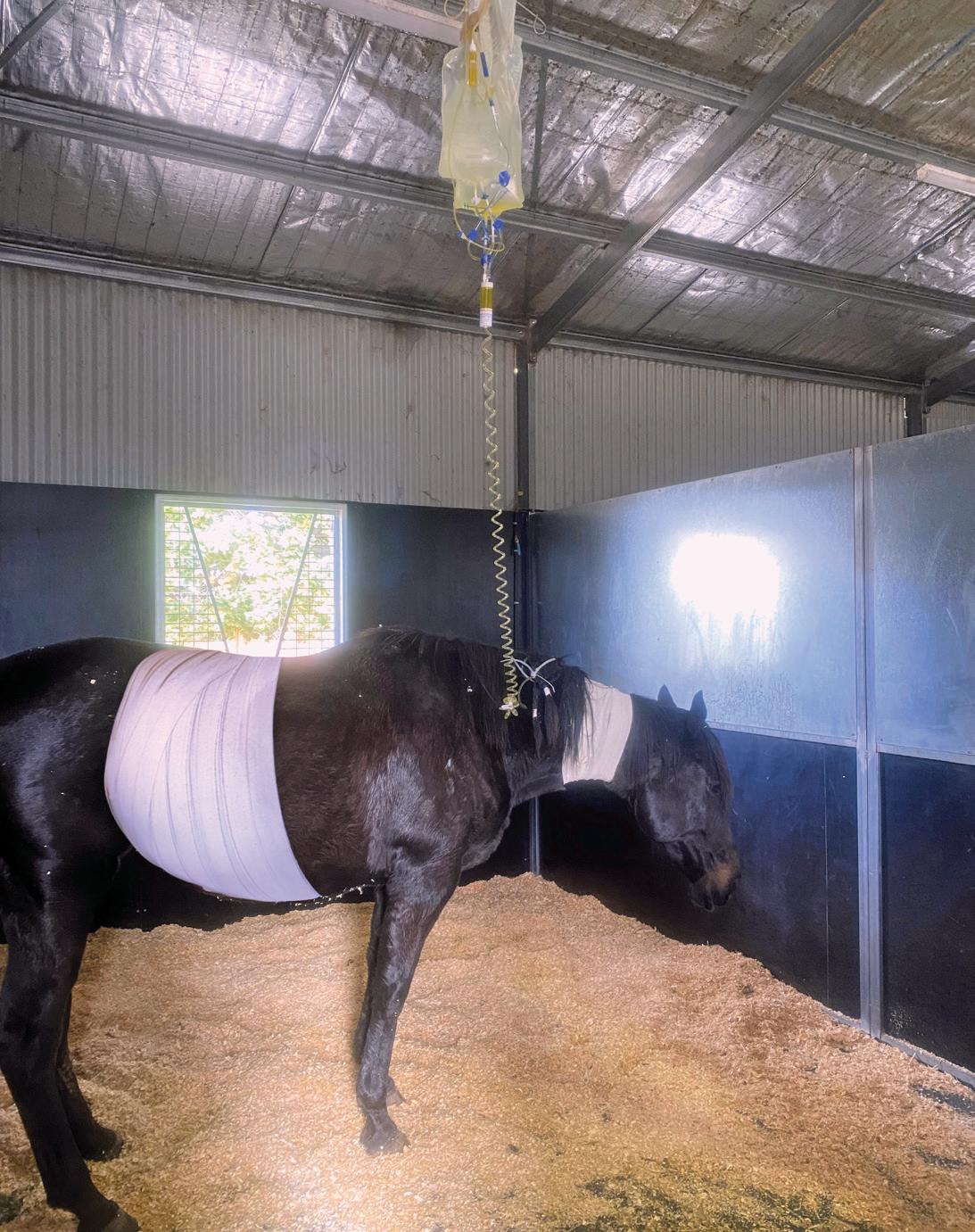
to ensure a clean removal of the damaged bowel. The abdomen was lavaged using 10 L of warmed sterile saline to remove any contamination and withdrew under suction using the Poole suction tube. The surgeon continued to investigate the abdomen, ensuring the placement of bowel was correct, and prepared for closure. A 5 Safil on a cutting needle was used to close the linea, with a 2/0 Monosyn for subcutaneous and skin closure. A stent was sutured over the top of the incision with a non-absorbable suture and a Hypafix dressing placed over the top. The surgery team transferred the patient into recovery where he was provided with flow-by oxygen at 15 L/ min and monitored closely on camera. The gelding sat into sternal and stood on his first attempt. POSTOPERATIVE CARE An abdominal bandage was placed in recovery to ensure the incision remained clean and dry prior to moving to the ICU. The nursing team removed the Hypafix dressing in a sterile manner, ensuring the stent was intact. An additional sterile bandage was placed along the incision and covered with a layer of cohesive bandage and a layer of Elastoplast over the top. As he was transferred to ICU, he was placed on intravenous fluid therapy (IVFT), with Hartmann’s at a rate of 1.5 L/hr through a gravity fed administration set allowing 4–5 drops per second. Analgesia was provided with lignocaine as a continuous rate of infusion (CRI), administered through a fluid pump at 78 ml/hour. Lignocaine CRIs require an initial loading dose at 1.3 mg/kg, to be given intravenously over a 15-minute period. The lignocaine CRI required careful patient monitoring to ensure no signs of toxicity or neurological side effects presented. Flunixin meglumine was continued postoperatively at 1.1 mg/kg for the first 12 hours and reduced to 0.5 mg/kg twice daily. Antimicrobials were continued postoperatively at the same rates given at the time of surgery. At 48 hours post-surgery, the gelding had passed cowpat-like faeces and was urinating well. His appetite and demeanour strengthened with an increasing interest in gradual feeding and surrounding activity. IVFT was reduced to 1 L/hr, with the lignocaine CRI discontinued, but continuation of
flunixin intravenously twice daily. To monitor fluid therapy, PCV and TP were monitored showing a PCV of 40% and total protein of 60 g/L, which indicated we were providing adequate fluid therapy. At 72 hours post-surgery, mild intermittent colic symptoms presented, with him becoming agitated and flank watching. A nasogastric tube was passed, and 3 L of gastric reflux was obtained. Additional flunixin was administered and fluid therapy increased to compensate the loss. Six hours after the additional flunixin, the gelding demonstrated mild colic signs and a further 2 L of gastric reflux. He was then refluxed every 2 hours, consistently producing upwards of 1 L each time. The owner was contacted to discuss the likelihood of a small intestinal obstruction. His poor prognosis was noted, and the owner unfortunately decided to subject the gelding to euthanasia. Despite best efforts with medical management, it was evident that a small intestinal obstruction was present. This was most likely at the anastomosis site leading to the accumulation of fluid proximal to this (Rubio Martinez and Hendrickson, 2021).

CASE DISCUSSION With the slow introduction of feed, IVFT and analgesia as medical support, postoperative ileus still became evident. Cases that present financial limitations are challenging, as the patient may have recovered with continued medical management or further surgical investigation, but this was not an option for our client. Afterhours emergency admissions of such a large animal take a team of highly trained and skilled members, and this case highlights the utilisation of multiple departments in the equine industry. The cases aren’t always successful, but they can offer learning experiences for what we can do better or what we can change next time.
References
Coumbe KM. Equine Veterinary Nursing. 2nd ed. Wiley-Blackwell. 2012. Rubio Martinez L & Hendrickson D. Complications in equine surgery. 1st ed. Wiley & Sons inc., 2021:332–337. Services LLC, z. Vetscan VS2 Reference Ranges. Retrieved 5 June 2022, from https://www2.zoetisus.com/content/_assets/docs/Diagnostics/technicalpapers/VETSCAN-VS2-Reference-Ranges-VTS-00038.pdf Zoetis LLC. HM5 Reference Ranges. Retrieved 5 June 2022, from https://www2. zoetisus.com/content/_assets/docs/Diagnostics/technical-papers/HM5Reference-Ranges-iPad-VTS-00426.pdf