8 minute read
Feeding tubes – placement and maintenance


by Anita Parkin
RVN, AVN, Dip (Surg & ECC), VTS (Anes & Analgesia), CVPP, TAE or ‘pilling’ balls of food used to be suggested; however, this method can be exceptionally stressful to the patient and will foster the development of food aversion. Enteral nutrition is the most appropriate choice of providing protein and calories. It is simple, well tolerated and the most cost-effective.
INDICATIONS FOR NUTRITIONAL SUPPORT Significant anorexia (> 3 days, > 1-day neonates). Significant weight loss (> 10%, > 5% neonates). Increased nutritional losses (diarrhoea, vomiting, renal disease, wounds and burns). Increased nutritional requirements (fever/infection, trauma/surgery, cancer, burns). Anticipated loss of appetite (animal not expected to eat for 3 days). Bypass of specific parts of alimentary canal (head injury, surgical site, pancreatitis). It has been well established that nutritional support in critically ill
Anita presented on this topic at the 2022 VNCA Conference and is this year’s VNCA Vet Nurse of the Year
Nutrition is a very important component of patient management, especially in the critical care setting. There is significant information indicating that animals may be hypermetabolic during many disease states. For this reason, understanding and meeting nutritional requirements cannot be overlooked when managing a critically ill or severely traumatised animal. Other factors, such as immune system function and maintaining a healthy gastrointestinal mucosal barrier, may also play a role in managing diseased dogs and cats. Enteral feeding is indicated in patients who cannot ingest adequate calories but have sufficient gastrointestinal function to allow digestion and absorption of feeding solutions delivered into the gastrointestinal tract via an enteral feeding device. The most important stimulus for mucosal cell proliferation is the direct presence of nutrients in the intestinal lumen. For the most part, the old adage ‘If the gut works, use it’ applies in most situations. Practical measures to improve food intake include the use of highly odorous foods, warming the foods prior to feeding and stimulating eating by positive reinforcement with petting and stroking behaviour. Assisted feeding, appetite stimulation and tube feeding (orogastric) methods can all be used. Assisted feeding by gently syringing a liquid food into the corner of the patient’s mouth
– placement and maintenance
patients will decrease morbidity and mortality, improve tolerance to invasive procedures, shorten hospitalisation periods, decrease incidence of infections, enable earlier ambulation, hasten wound healing, and reduce complications. DIET SELECTION The type of formula to feed the patient will depend on the selected route of feeding, the functional status of the gastrointestinal tract and the patient’s nutrient requirements. Other factors such as cost, availability and ease of use may also be important. Patients that are fed via naso-oesophageal or jejunostomy feeding tubes are limited to receiving liquid enteral formulas. Most commercially available liquid diets have a caloric density of approximately 1 kcal per ml. Commercial blended pet food diets should be used for feeding into the stomach via esophagostomy or gastrostomy tubes. In select cases, the feeding of a liquid enteral formulation may be indicated (naso-oesophageal or jejunostomy tube feeding). There are a number of complete and balanced veterinary enteral formulations that contain adequate amounts of protein, taurine, and micronutrients, precluding the need for supplementation in most situations. Feeding should be delayed for 24 hours after placing a gastrostomy tube to allow gastric motility to return and to allow formation of a fibrin seal.
CALCULATION OF NUTRITIONAL REQUIREMENTS An estimate of an animal’s nutrient requirements is needed to determine the minimum amount of food necessary to sustain critical physiologic processes. The resting energy requirement (RER) is the animal’s energy requirement at rest in a thermoneutral environment and in a postabsorptive state. A linear formula can be applied to determine the RER of dogs and cats weighing at least 2 kg but less than 45 kg. Alternatively, one can utilise an allometric formula that can be applied to dogs and cats of all body weights. Linear formula: RER (kcal/day) = (30 x BWkg) + 70 Allometric formula: RER (kcal/day) = 70 (BWkg)0.75
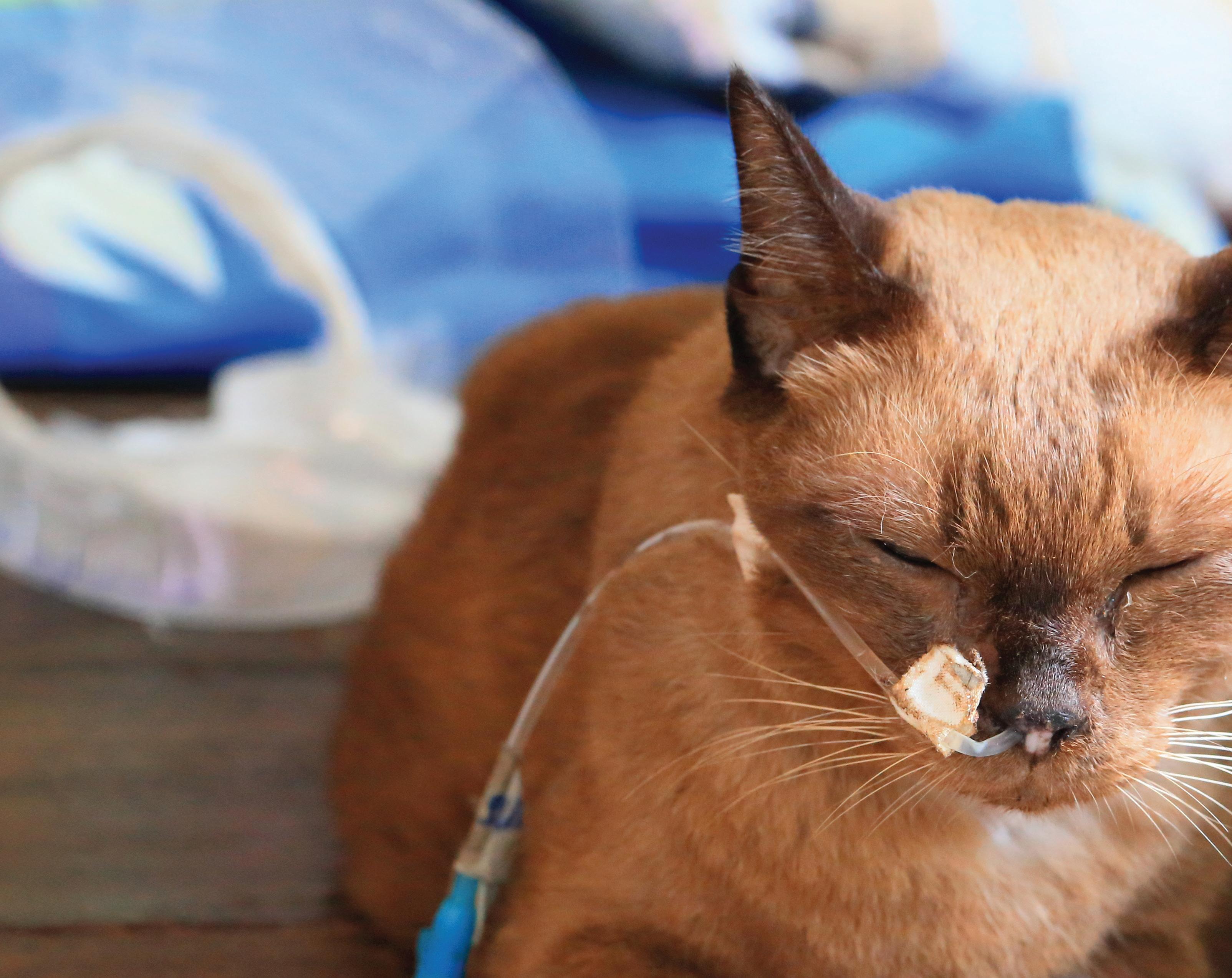
Photo: ©shutterstock/SUJITRA CHAOWDEE Hospitalised patients should be fed a third of their calculated RER initially, increasing to their full RER over 3 days, realising that their actual energy requirement is likely to change over the course of the disease process through recovery. Close observation of changes in body weight, physical examination findings (decreased subcutaneous fat stores, muscle wasting, and presence of oedema or ascites), and ongoing losses (diarrhoea, vomiting, exudative wounds), will help determine whether to increase or decrease the patient’s caloric intake towards the illness energy requirement (IER) or RER, respectively. IER (kcal/day) in dogs = 1.25-1.5 x RER: in cats 1.10-1.25 x RER Feeding can be instituted immediately following oesophagostomy tube placement once the animal has fully recovered from anaesthesia. Diet can be administered as bolus feedings or continuous infusion when feeding via oesophagostomy and gastrostomy tube. Capacities for cats and dogs are 5 to 10 ml/kg body weight during initial food reintroduction. Maximum capacities as high as 45 to 90 ml/kg body weight have been measured in cats and dogs when fully realimented. Most often, meeting the patient’s RER can be done in volumes far less than these maximums. Salivating, gulping, retching and even vomiting may occur when too much food has been infused or when the infusion rate is too fast. With bolus feeding, the required daily volume of food should be divided into four to six feeds. Patients are usually fed approximately 33% of their caloric requirement on the first day of feeding, with a gradual increase of 33% of the caloric requirement per day. Most patients can reach their energy requirement by the third or fourth day of feeding. The food should be warmed to room temperature and

Feeding tubes – placement and maintenance
Continued from previous page
fed slowly through the tube to prevent vomiting. Flushing of the tube with 10 to 15 ml of lukewarm to warm water helps prevent clogging. Before each feeding, aspirate the tube with an empty syringe to check for residual food left in the stomach from the previous feeding. If more than half the last feeding is removed from the stomach, skip the feeding and recheck the residual volume at the next feeding. NASO-OESOPHAGEAL/ NASOGASTRIC Naso-oesophageal/nasogastric intubation is an easy, effective, and efficient means of providing enteral nutritional support. The availability of small bore, soft polyvinyl and silastic feeding tubes, low viscosity, nutritionally complete liquid diet formulations and patient tolerance of tube placement has made naso-oesophageal/ nasogastric tube placement a popular avenue for feeding malnourished patients. Naso-oesophageal/ nasogastric tube placement is indicated in any patient with proteincalorie malnutrition that will not undergo oral, pharyngeal, oesophageal, gastric or biliary tract surgery. Tube management: Place a column of water in the tube and cap it when not in use; this prevents intake of air, reflux of oesophageal contents, and occlusion of the tube by diet. Tube occlusion by diet can frequently be unblocked by flushing the tube with a little carbonated soft drink, leaving it in situ for 10–15 minutes then flushing with a little pressure. Naso-oesophageal/ nasogastric tubes can be left in place for several weeks, are well tolerated, easily removed, the patient can drink and swallow around the tube, and repeated orogastric intubation is prevented. Complications: Tracheal intubation, inadvertent dislodgement (use Elizabethan collars), dislodgement through sneezing, vomiting or regurgitation, rhinitis, unilateral dacryocystitis tube occlusion are uncommonly encountered. Reflux oesophagitis can result from improper tube placement (i.e. through the lower oesophageal sphincter) or oesophageal irritation from the tube itself.
OESOPHAGOSTOMY Indications: Oesophagostomy tube feeding is indicated in anorexic patients with disorders of the oral cavity or pharynx or anorexic patients with a functional gastrointestinal tract distal to the oesophagus. Contraindications: Oesophagostomy tube placement is contraindicated in patients with a primary or secondary oesophageal disorder (for example, oesophageal stricture after oesophageal foreign body removal or oesophageal surgery, oesophagitis, megaoesophagus). Advantages: Advantages of oesophagostomy tube feeding include ease of tube placement, tubes are well tolerated by the patient, large bore feeding tubes (8 Fr or greater) can be used allowing the use of blended diets, tube care and feeding is easily performed by the client, patients can eat and drink around the tube, and tube removal can be performed any time after placement. Oesophageal tube placement eliminates coughing, laryngospasm, or aspiration occasionally associated with pharyngostomy tubes. Disadvantage: The major disadvantage of the oesophagostomy tube is the need for general anaesthesia during placement. Complications: Complications associated with oesophagostomy tube placement include early removal by the patient or vomiting. No significant long-term complications have been reported (for example, oesophagitis, oesophageal stricture, oesophageal diverticulum or subcutaneous cervical cellulitis). As with naso-oesophageal tubes reflux oesophagitis can result from improper tube placement (i.e. through the lower oesophageal sphincter) or oesophageal irritation from the tube itself. TUBE OBSTRUCTION Obstruction of the feeding tube is one of the most common complications of enteral feeding. Most obstructions are secondary to coagulation of formula, although obstruction by tablet fragments, tube kinking, and precipitation of incompatible medications can also result in tube obstruction. Naso-oesophageal tubes are prone to obstruction because of their small diameters and obstruction also occurs up to three times more frequently in patients fed by continuous vs bolus feedings. Sucralfate and antacids have been reported to precipitate with enteral formulas and cause tube obstruction. Several ‘remedies’ have been advocated to relieve tube obstruction. Warm water injected with gentle pressure and suction will relieve most obstructions. For more unyielding obstructions, carbonated water is instilled into the tube and allowed to sit for up to one hour before applying gentle pressure and suction. Tube obstructions can be minimised by flushing the feeding tube with warm water before and after administering medications or enteral feedings. Tablets should be crushed and dissolved in water prior to administration through the feeding tube if no alternative form of medication is available.
TAKE HOME POINTS • ‘If the gut works, use it’. • Warm food before feeding – even if using the tubes. • Keep a column of water in the tube between feeds. • Use carbonated drink to unblock the tube if required. • Introduce food slowly (even via the tubes).


