2023 University of Michigan Kellogg Eye Center Annual Report

Page 28

IF WE CAN SEE MOLECULAR CHANGES WHILE THEY

ARE HAPPENING, INSTEAD OF JUST SEEING AN

EYE BLEED, WE CAN

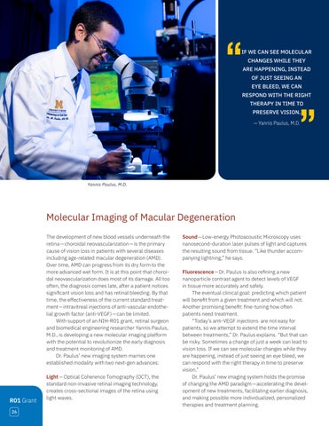
RESPOND WITH THE RIGHT THERAPY IN TIME TO PRESERVE VISION. — Yannis Paulus, M.D.

Yannis Paulus, M.D.

Molecular Imaging of Macular Degeneration The development of new blood vessels underneath the retina— choroidal neovascularization—is the primary cause of vision loss in patients with several diseases including age-related macular degeneration (AMD). Over time, AMD can progress from its dry form to the more advanced wet form. It is at this point that choroidal neovascularization does most of its damage. All too often, the diagnosis comes late, after a patient notices significant vision loss and has retinal bleeding. By that time, the effectiveness of the current standard treatment— intravitreal injections of anti-vascular endothelial growth factor (anti-VEGF)— can be limited. With support of an NIH-R01 grant, retinal surgeon and biomedical engineering researcher Yannis Paulus, M.D., is developing a new molecular imaging platform with the potential to revolutionize the early diagnosis and treatment monitoring of AMD. Dr. Paulus’ new imaging system marries one established modality with two next-gen advances:

R01 Grant 26

Light — Optical Coherence Tomography (OCT), the standard non-invasive retinal imaging technology, creates cross-sectional images of the retina using light waves.

Sound—Low-energy Photoacoustic Microscopy uses nanosecond-duration laser pulses of light and captures the resulting sound from tissue. “Like thunder accompanying lightning,” he says. Fluorescence—Dr. Paulus is also refining a new nanoparticle contrast agent to detect levels of VEGF in tissue more accurately and safely. The eventual clinical goal: predicting which patient will benefit from a given treatment and which will not. Another promising benefit: fine-tuning how often patients need treatment. “Today’s anti-VEGF injections are not easy for patients, so we attempt to extend the time interval between treatments,” Dr. Paulus explains. “But that can be risky. Sometimes a change of just a week can lead to vision loss. If we can see molecular changes while they are happening, instead of just seeing an eye bleed, we can respond with the right therapy in time to preserve vision.” Dr. Paulus’ new imaging system holds the promise of changing the AMD paradigm—accelerating the development of new treatments, facilitating earlier diagnosis, and making possible more individualized, personalized therapies and treatment planning.


Turn static files into dynamic content formats.

Create a flipbook

Articles inside

Partnership between U-M Med School, Business School, and the Kellogg Eye Center Drives Latest Kenya

4min
pages 38-39

The Edna H. Perkiss Research Professorship in Ophthalmology and Visual Sciences

3min
page 37

Honoring the Visionary Leadership of Paul P. Lee, M.D., J.D.

3min
page 36

The Alan Sugar, M.D., Research Professorship in Ophthalmology

3min
page 35

Mark W. Johnson, M.D., Honored with Heed-Gutman Award

2min
page 34

Protecting Retinal Neurons from Diabetes

2min
page 34

Mining Big Data for Novel Glaucoma Genes

3min
page 33

Beyond the Electronic Health Record

5min
pages 32-33

Applauding a Good Catch

2min
page 31

Microneedles for Sustained Retinal Drug Delivery

2min
page 30

Alumni Highlights

4min
pages 29-30

Lecture in Professionalism and Ethics

1min
page 29

Molecular Imaging of Macular Degeneration

2min
page 28

Institutional Grants Anchor Research Infrastructure, Training

5min
pages 26-27

2023-2024 Heed Fellows

5min
pages 24-25

Pre-Med Awarded NIH Research Supplement

2min
page 23

Kellogg PGY4 Sole Resident on ACGME Residency Program Review Committee

2min
page 22

Kellogg Post-Doc Receives Prestigious NIH Grant

2min
page 21

An Out-of-This-World Perspective on Residency from one of Forbes’ Thirty-Under-Thirty

3min
page 20

Expanding Personalized Treatment and Clinical Research in Uveitis

3min
page 19

KCRC Assists in Michigan Medicine Research with Consequences for Eyes

3min
page 18

Editing Genes to Treat Corneal Dystrophies

3min
page 17

Using Artificial Intelligence to Improve IOL Formulas

3min
page 16

Selfless Service Beyond Kellogg’s Walls

1min
page 15

The Genes That Drive Eye Size

2min
page 15

Image-Guided Medical Robotics Comes to Kellogg

3min
page 14

How Inflammation Triggers Photoreceptor Regeneration

2min
page 13

The Molecular Physiology of the Blood-Retinal Barrier

3min
page 12

Prioritizing Patient Wellness—and Our Own

3min
page 11

Michigan's 15th President Joins the Department

3min
page 10

Patent Issued for Photo-Mediated Ultrasound Therapy

1min
page 9

Unlocking the Therapeutic Potential of Tears

2min
page 9

Oculoplastics: Building on an Extraordinary Legacy

3min
page 8

Assessing Age-Related Vision Impairment

3min
page 7

For IRD Patients, Tailored Interventions Address Impaired Vision and Related Distress

3min
page 6

A Rare Syndrome, A Team Approach

4min
pages 4-5

2023 University of Michigan Kellogg Eye Center Annual Report

3min
page 3
Issuu converts static files into: digital portfolios, online yearbooks, online catalogs, digital photo albums and more. Sign up and create your flipbook.
2023 University of Michigan Kellogg Eye Center Annual Report by U-M Kellogg Eye Center - Issuu