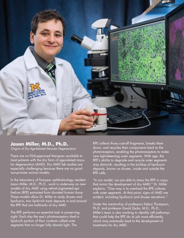
Jason Miller, M.D., Ph.D.
Origins of Dry Age-Related Macular Degeneration
There are no FDA-approved therapies available to treat patients with the dry form of age-related macular degeneration (AMD). Dry AMD lab studies are especially challenging because there are no good non-primate animal models. In the laboratory of first-year ophthalmology resident Jason Miller, M.D., Ph.D., work is underway on new models of dry AMD using retinal pigmented epithelium (RPE) extracted from donated human tissue. These models allow Dr. Miller to study drusen and lipofuscin, two lipid-rich trash deposits in and around the RPE that are hallmarks of dry AMD. The RPE performs an essential task in preserving sight. Each day the eye’s photoreceptors shed a lipid-rich portion of their contents called outer segments that no longer fully absorb light. The
RPE collects those cast-off fragments, breaks them down, and recycles their components back to the photoreceptors, enabling the photoreceptors to make new light-detecting outer segments. With age, the RPE’s ability to degrade and recycle outer segments may diminish, resulting in the buildup of lipofuscin deposits, known as drusen, inside and outside the RPE cells. “In our model, we are able to stress the RPE in ways that mimic the development of dry AMD,” Dr. Miller explains. “One way is to overload the RPE cultures with outer segments. At that point, signs of AMD are evident, including lipofuscin and drusen secretions.” Under the mentorship of professors Debra Thompson, Ph.D. and professor David Zacks, M.D., Ph.D., Miller’s team is also working to identify cell pathways that could help the RPE do its job more efficiently, which may eventually lead to the development of treatments for dry AMD.
