26 Restorative Dentistry Adhesion to Mild Fluorosed Enamel
a
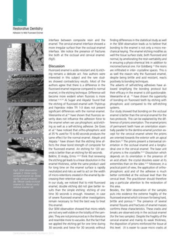
Fig 3
interface between composite resin and the enamel. The cervical enamel interface showed a more irregular surface than the occlusal enamel interface. We notice the presence of fractures line both at the occlusal and cervical enamel. (fig3).
Discussion
b
c
d
Fig3: SEM observation: sample 2: Distal cavity: occlusal enamel (a). Distal cavity: cervical enamel (b). Mesial cavity:occlusal enamel (c). Mesial cavity: cervical enamel (d).
Fluorosed enamel is acido-resistant and its etching remains a delicate act. Few authors were interested in this subject and the rare studies showed contradictory results. Most of the authors agree that there is a difference in the fluorosed enamel response compared to normal enamel, in the etching technique. Difference will become more evident when fluorosis is more intense.13,16,19 Al Sugair and Akpata1 found that the etching of fluorosed enamel with Thylstrup and Fejerskov index TFI 1-3 does not present significant differences with the normal enamel. Weerainhe et al.16 have shown that fluorosis severity does not influence the adhesion force to the enamel when we use phosphoric acid etching as well as a self-etching adhesive. De Goes et al.6 have noticed that ortho-phosphoric acid at 35 % used for 15 to 60 seconds produces the same effect for the normal enamel. Ateyah and Akpata 2 have shown that the etching time affects the shear bond strength of composite for the fluorosed enamel. An etching for 120 seconds is better than an etching for 60 seconds. Belkhir, El Araby, Ermis 4,8,9 think that renewing the etching gel leads to a linear dissolution in the enamel thickness, while the same product used for long time on the enamel surface is rapidly neutralized and risks as well to act on the width of micro-retentions created in the enamel by decreasing their retentive value. In our study, we noticed that to mild fluorosed enamel, double etching did not give better results than the simple etching: etching of one time 30 seconds is enough. However, in case of severe fluorosed enamel other investigations remain necessary to find the best way to treat this enamel. Our SEM observation showed that micro reliefs are not very well visible on the totality of the samples. They are not pronounced as in the literature and resemble more to vacuoles. But the fact that we applied the etching gel for one time during 30 seconds and twice for 30 seconds without
Dental News, Volume XXI, Number II, 2014
finding differences in the statistical study as well in the SEM observation leads us to believe that bonding to the enamel is not only a micro mechanical keying. The enamel etching modifies as well the tissue surface state, both fluorosed and normal, by ameliorating the resin wettability and in ensuring a physic-chemical link in addition to micromechanical one. For Goldberg 10 the resins are infiltrated in inter- crystalline spaces. This is as well the reason why the fluorosed enamel, despite being brittle and acid resistant, reacts positively to bonding techniques. The adverts of self-etching adhesives have allowed simplifying the bonding protocol but their efficacy in the enamel is still questionable. Weerainhe et al. 16 have shown the superiority of bonding on fluorosed teeth by etching with phosphoric acid compared to the self-etching systems. Our study showed that bonding on the occlusal enamel is better than the cervical enamel for the two protocols. This can be explained by the difference of prisms orientation. The enamel prisms of permanent teeth have an orientation essentially parallel to the dentino-enamel junction except for the cervical enamel where the prisms are oriented towards the exterior with an apical direction.14 The prisms present a transversal orientation in the occlusal enamel and a longitudinal one in the cervical enamel. The base unit of prisms is the crystallite.10,14 Dissolution which depends on its orientation in the presence of an acid attack: the crystal dissolves easier at its extremities than on the sides.14,15 Moreover, in a practical point of view, the application of orthophosphoric acid and of the adhesive is much better controlled at the occlusal level than the cervical level. The practitioner should therefore pay a particular attention to the restoration of this area. Besides, the SEM observation of the samples puts into evidence the extreme fragility of the fluorosed enamel which is known for being hard, brittle and porous.4,5 The presence of several enamel fissures and fractures of enamel masses confirms these characteristics. These cracks and breaks are observed only in the occlusal enamel (for the two samples). Despite the fragility of the cervical enamel and mainly its weak thickness, the disposition of prisms reinforced the tissue at this level. (It is easier to cause micro cracks at