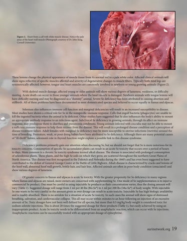
Figure 2. Heart from a calf with white muscle disease. Notice the pale areas of the heart wall muscle (Photograph courtesy of Dr. John King, Cornell University)
These lesions change the physical appearance of muscle tissue from its normal red to a pale white color. Affected clinical animals will show signs reflective of specific muscles affected and severity of degenerative changes to muscle fibers. Typically both hind legs are symmetrically affected; however, tongue and heart muscles are commonly involved in newborn or young growing animals (Figure 2). With skeletal muscle damage, affected young or older animals will show various degrees of lameness, weakness, or difficulty moving. Acute death can occur in those younger animals where the heart muscle is damaged. Newborn animals with tongue lesions will have difficulty nursing and may be diagnosed as a “dummy” animal. Severe Se deficiency has been attributed to causing abortion and stillbirth. All of these problems have been documented in most domesticated species and believed to occur equally in llamas and alpacas. Selenium also influences immune cell function and marginal deficiencies will result in an increased susceptibility to disease. Most studies have shown a critical role for Se in the nonspecific immune response. Cells that engulf bacteria (phagocytes) are unable to kill the ingested bacteria when the animal is Se deficient. Other studies have suggested that Se also influences the body’s ability to mount an appropriate antibody response to an infectious agent. Subclinical Se deficiency in growing animals, through its affect on immune response, may predispose them to diarrhea and pneumonia conditions. Young animals infected with coccidia may not be able to mount a sufficient immune response to help them recover from the disease. This will result in a prolonged disease condition and a perception of disease treatment failure. Adult females with marginal Se deficiency may be more susceptible to uterine infections (metritis) around the time of breeding. Premature, weak, or poor doing babies have been attributed to Se deficiency. Although there are many potential causes of “ill-thrift” babies, selenium’s role in thyroid function might explain a possible link to this disease syndrome. Deficiency problems primarily gain our attention when discussing Se, but we should not forget that Se is more notorious for its toxicity concerns. Consumption of specific Se-accumulator plants can result in an acute Se toxicity that occurs over a period of hours to days. More common is a chronic Se toxicity syndrome termed alkali disease. The disease is associated with prolonged consumption of seleniferous plants. These plants, and the high Se soils on which they grow, are scattered throughout the northern Great Plains of North America. This disease was first recognized in the Dakota’s and Nebraska during the 1860’s and has even been suggested to have contributed to the defeat of General George Custer at the Battle of Little Bighorn. Alkali disease is characterized by cracks and lesions of the hoof wall, abnormal hoof wall growth, brittle hair, and hair loss. Affected animals are often in poor body condition (emaciated) and show various degrees of lameness. Of greater concern to llamas and alpacas is acute Se toxicity. With the greater propensity for Se deficiency in many regions where llamas and alpacas are raised, more owners are concerned with supplementing Se. One mode of Se supplementation is to inject a commercial Se product. One must be careful in using injectable Se products as their concentration varies, thus the dosing amount will vary (Table 1). Suggested dosage will range from 1 ml per 40 lbs (Bo-Se®) to 1 ml per 200 lbs (Mu-Se®) of body weight. With injectable Se one wants to be very careful in the amount given as over dosage can result in acute toxicity. Injectable Se has high biologic availability and is readily absorbed. There is no antidote for a situation of acute Se toxicity. In such cases the animal will show signs of distressed breathing, salivation, and cardiovascular collapse. This all may occur within minutes to an hour following an injection of an excessive amount of Se. Toxic dosages have not been well defined for all species, but more than 0.5 mg/kg body weight is considered toxic for sodium selenite injections. This is about 20 times the suggested dosage for these products (Table 1), but easily achieved by using an inappropriate product. Acute Se toxicity needs to be differentiated from an anaphylactic reaction, which can occur with Se injections. Anaphylactic reactions can be successfully treated with an appropriate dosage of epinephrine.
surinetwork.org
19