TECHNICIAN UPDATE
Managing a Serious Wound By Sally Schwartz CVT
Photos courtesy of Sally Schwartz
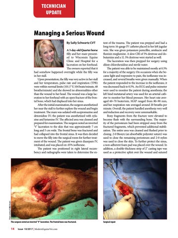
A 3-day-old Quarter horse filly and her mare presented to Wisconsin Equine Clinic and Hospital for a laceration on her forehead. The owners reported that it had somehow happened overnight while the filly was in her stall. Upon presentation, the filly was very active in her stall and her temperature, pulse rate and respiration (TPR) were within normal limits (101.1° F, 104 beats/minute, 48 breaths/minute) and she showed no abnormalities other than the wound to her head. The wound was a large laceration to her forehead with an open fracture of the frontal bone, which had displaced into her sinus. After the initial examination, the surgeon anesthetized her near the stall to further explore the wound and begin treatment. The mare was sedated with acepromazine and detomidine IV; the patient was anesthetized with xylazine and ketamine IV. The affected area was cleaned and prepared for examination. The surgeon noted an inverted ‘V’ laceration to the skin that was approximately 7 cm long and 5 cm wide. The frontal bone was fractured and had collapsed into the frontal sinus. It was then decided to move the filly into the surgical room for further treatment of the wound. The patient was given diazepam IV, intubated, and was placed on .05% isoflurane. The patient was positioned in right lateral recumbency and radiographs were taken to determine the ex-
tent of the trauma. The patient was prepped and had a long-term 16-gauge IV catheter placed in her left jugular vein. She was given potassium penicillin, amikacin and flunixin meglumine. A slow CRI of 5% dextrose and dobutamine and a 1L 5% dextrose were started as well. The laceration was then prepped for surgery using dilute chlorohexidine and sterile water. The patient was able to be maintained mostly at 0.5% for a majority of the surgery. On occasions when she became light and responsive to pain, the isoflurane was increased, and several breaths were given manually. When the patient responded to the increase in the isoflurane, it was decreased back to 0.5%. An ECG and pulse oximeter were used to monitor the patient during anesthesia; the left hind metatarsal artery was used for an arterial catheter to monitor her blood pressure. Her heart rate averaged 60–70 beats/min, MAP ranged from 80–90 mm, and her respiration rate averaged around 20 breaths per minute. Overall, the patient handled anesthesia very well and induction and recovery were unremarkable. Bony fragments from the fracture were elevated to become flush with the surrounding bone. The majority of the periosteum had been stripped away from the fractured fragments, which prevented additional stabilization. The entire area was cleaned and flushed prior to closing. 2-0 Biosyn (an absorbable polyester suture) was used to close the remaining periosteum and 2-0 nylon was used to close the skin. To further protect the sinus, a non-adherent foam pad was placed over the wound. In addition, a double-thickness strip of 2" casting tape was used as a protective splint over the wound and sutured
The surgeon noted an inverted "V" laceration. The frontal bone was fractured.
14
Issue 10/2017 | ModernEquineVet.com
Surgical repair