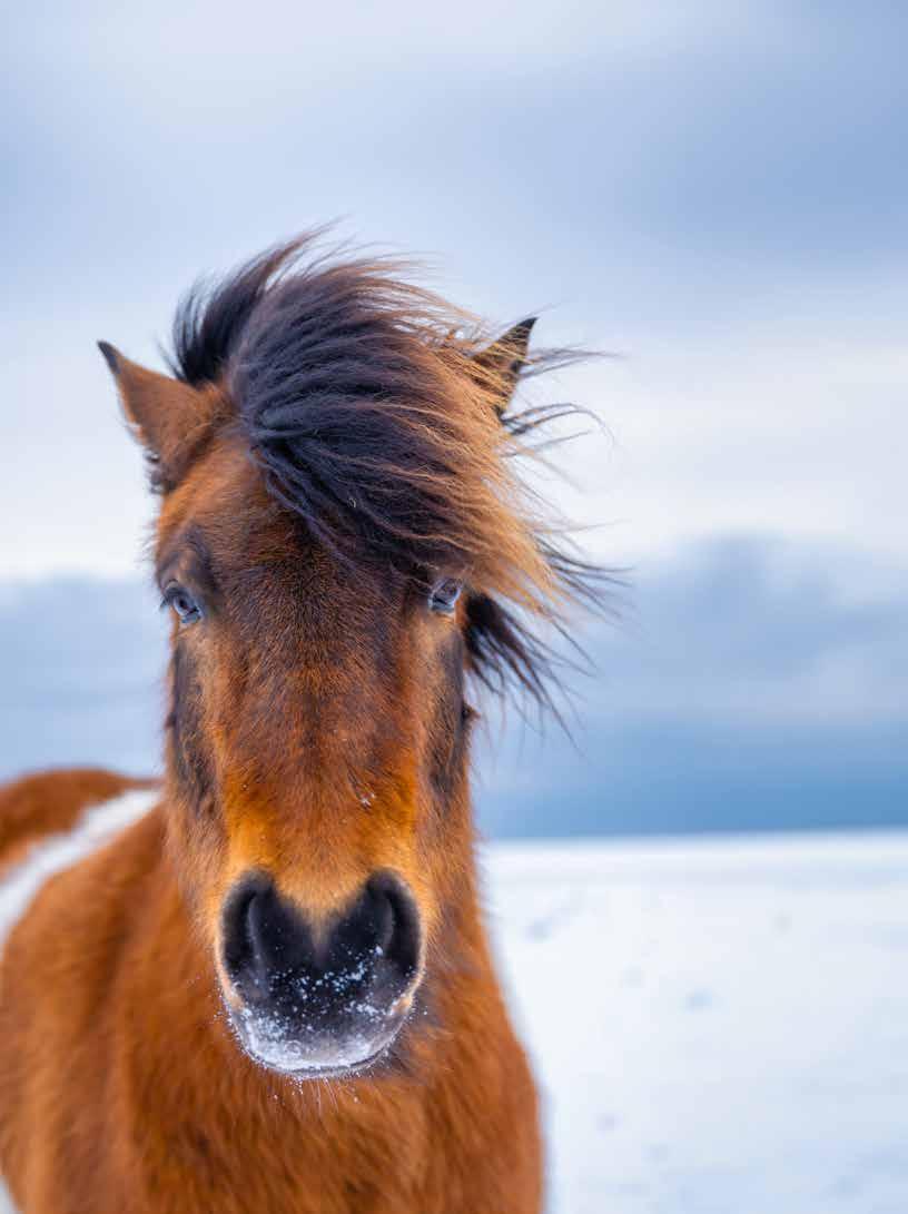
Lameness vs. Ataxia Treating Equine Asthma Treating Equine Osteoarthritis With Plasma Therapies Tech Update: Lateral Suspensory Ligament Branch Lesion Challenges of Diagnosing Neck Pain Equine Vet The Modern Vol 14 Issue 2 2024 www.modernequinevet.com
in
digital
is for general informational purposes only. PercyBo Publishing Media LLC makes no representations or warranties of any kind about the completeness, accuracy, timeliness, reliability or suitability of any of the information, including content or advertisements, contained in any of its digital content and expressly disclaims liability of any errors or omissions that may be presented within its content. PercyBo Publishing Media LLC reserves the right to alter or correct any content without any obligations. Furthermore, PercyBo disclaims any and all liability for any direct, indirect, or other damages arising from the use or misuse of the information presented in its digital content. The views expressed in its digital content are those of sources and authors and do not necessarily reflect the opinion or policy of PercyBo. The content is for
in whole or in part without
TABLE OF CONTENTS
DISCLAIMER: The content
veterinary
ALL RIGHTS
permission
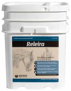
SALES: ModernEquineVet@gmail.com EDITOR: Marie Rosenthal ART DIRECTOR: Jennifer Barlow CONTRIBUTING WRITERS: Paul Basilio • Landon Grey COPY EDITOR: Patty Wall Published by PERCY BO media publishing PO Box 935 • Morrisville, PA 19067 Marie Rosenthal and Jennifer Barlow, Publishers Equine Vet The Modern ORTHOPEDICS Lameness vs Ataxia: Suspect the Unexpected 8 RESPIRATORY Equine Immunosuppression Linked to Dexamethasone Treatment for Asthma ............................................................................. 12 VET STATS Should Reporting Strangles Be a National Mandate? 13 TECHNICIAN UPDATE Thoroughbred Filly With Lateral Suspensory Ligament Branch Lesion 14 NEWS NOTES Equine Devices Successful at Creating Good Biological Concentrations for Regenerative Medicine ................................................. 17 Neck Pain: Clinical Exam is The Foundation COVER STORY 4 Cover: Shutterstock/biletskiyevgeniy.com ADVERTISERS Arenus Animal Health/Sore No-More 3 American Regent/Adequan 7 Arenus Animal Health/Aleira-Releira 9 Shanks Veterinary Equipment 10
LEGAL
this
issue
professionals.
RESERVED. Reproduction
is prohibited.

Clinical Exam
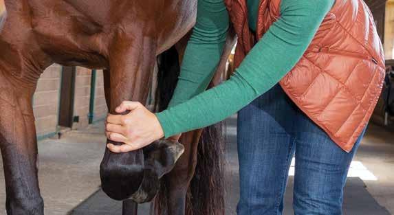
With a laundry list of presenting signs that are not always clear, even after imaging, diagnosing neck pain is a challenge.
“Therefore, even today with all the advanced imaging and modalities that we have available to us, the clinical examination still becomes the foundation by which we need to look at these horses and decide how we tackle the diagnostic algorithm as we go forward,” said Elizabeth Davidson, DVM, DACVS, DACVSMR, an associate professor of sports medicine, at the University of Pennsylvania’s New Bolton Center.
Palpate the entire neck looking for evidence of focal asymmetry, diffuse muscle atrophy, focal muscle atrophy and other issues, remembering that subtle abnormalities may be present. For instance, sweating
can suggest trauma within that area.
“Don't forget to move the mane and forelock out of the way,” Dr. Davidson said at the 69th AAEP Annual Convention, held in San Diego.
“I'm sure everyone has their own way of palpating the neck and that is fine,” Dr. Davidson said. “We should palpate the neck all the time. Gather information about the neck, seeing if there's a particular area that's more sensitive than another area.”
She suggested palpating the right and left side simultaneously, which makes it easier to compare the two.
As many veterinarians do, Dr. Davidson uses food to entice the horse to move its neck from side to side and up and down to see its flexibility and the horse’s willingness to move the neck. In one video she showed a horse that turned his entire body to-
By Marie Rosenthal, MS
ORTHOPEDICS
4 Issue 2/2024 | ModernEquineVet.com
The Challenge of Diagnosing Neck Pain is the Foundation
ward the food when she was on its right side, rather than just moving his head, indicting the inability or unwillingness to move his head and neck laterally.
“These things may give you clues as to whether or not they can move their neck when you ask them to bend in one particular way,” she said.
One thing to remember, however, is that by asking the horse to move its neck to a particular side means the other side is being simultaneously extended (both sides move), so “it still becomes very challenging to decide which area or side is painful,” she admitted.
“When we do these exercises to assess whether or not horses have mobility in the neck, we still are in essence manipulating and causing movement of the entire top line on these horses,” which can continue
to confuse the picture.
An underappreciated part of the physical examination is body movement. Although a dynamic examination is common when limb lameness is suspected, it may not be part of the examination for suspected neck pain, but it should be, she said.
“In my opinion the walk is underappreciated; the gait can give you so much information about how the horse is trying to navigate its environment.”
As they walk look at them from behind as well as on each side. Watch its posture and how it holds its head. Is raised a little too high or too low? Is it bent ever so slightly to one side?
Turning tight circles is typically done during a neurological examination, but it can assess other abnormalities besides proprioceptive and ataxic defi-
Shutterstock/Juergen
ModernEquineVet.com | Issue 2/2024 5
Bauer Pictures
CHECK OUT
a story about Dr. Sue Dyson's equine pain ethogram in the February 2021 issue of Modern Equine Vet
SIGNS OF NECK PAIN
• Neck stiffness
• Decreased flexibility
• Abnormal head carriage, head tilt
• Resistance to rein tension
• Inability to maintain frame
• Asymmetrical cervical musculature
cits, she said. Is the neck stiff as it tries to make a circle?
“This is a maneuver that traditionally we use for neurologic assessment, which we can also use when we're trying to sort out orthopedic problems in these horses,” she said.
Note its unwillingness to move in a certain way, which can be a signal of pain. “Horses are amazingly kind,” Dr. Davidson said. “They try so hard to do what we ask them to do.”
• Abnormal posture
• Previous fall or collision
• Unilateral forelimb lameness
• Patchy sweating
• Weak or disjointed gait
• Toe drag
• Rearing or bolting
Don’t forget to watch the neck in relation to the rest of the top line.
Don’t forget to watch the neck in relation to the rest of the top line.
“The examination continues by assessing horses exercising, with and without a rider,” she said. “We also need to do go beyond the walk and look at these horses when they trot and canter.”
How low does it carry its head and neck? Does it have a hard time maintaining its stride? Does it need more encouragement? Does it rear or bolt, swish its tail? These all can be signs of pain.
Many of the horse’s historical behaviors are challenging, even dangerous to reproduce, but they are important for veterinarians to see which can help pinpoint the problem.
“We add things such as a bridle or side reins or rider to see if we can entice different types of exercising abnormalities that may give us clues about whether or not the horses indeed have problems that potentially might be located within the neck,” she said.
Sometimes the signs are subtle. And it’s important to remember Dr. Sue Dyson’s Ridden Horse Pain Ethogram that comprises a list of behaviors that are more likely to be seen in horses with musculoskeletal pain. For example, abnormal facial expressions and things
• Refusal to go forward
• Unwilling to work or general laziness
• Lack of connection between front and hind limbs
• Abrupt stops when ridden
• Neck 'stuck'
such as the movement of its ears behind vertical or flat, she suggested, might be signs of pain. A “bad horse” might not have behavior issues; it might be in pain.
“We now know that these types of abnormalities aren't just bad behaviors. They can be associated with underlying pain conditions with these horses. It's up to us to be able to identify them, recognize them, and decide where that pain might be coming from.
“A complete physical and clinical examination is so important. It is the foundation, the most important thing by which we go forward with our next step of diagnostics. And some of these maneuvers may help us sort out the abnormalities, the underlying conditions, especially if they have an underlying neck condition.”
The clinical examination is the foundation, but also just the beginning, Dr. Davidson added, pointing to other tests and imaging that may be needed to make a diagnosis.
It can be difficult to separate orthopedic conditions, such as stifle pain or bilateral limb lameness from neck conditions. “A lot of times gait abnormalities are not, at least in my experience, super specific or pathognomonic for neck problems. But we can still use these maneuvers that then allow us to decide if the neck needs to be on our list of differentials and then sort through our next diagnostics,” Dr. Davidson said.
“Horses are good natured creatures, and we have the unbelievable honor to work on them,” she said. It is up to the veterinarian to decide, “What is the information that I can gain by doing different maneuvers, doing specific things to sort out when horses can no longer do their job?” Dr. Davidson said. MeV
6 Issue 2/2024 | ModernEquineVet.com ORTHOPEDICS
There’s nothing else like it.
For more than 30 years, Adequan® i.m. (polysulfated glycosaminoglycan) has been administered millions of times1 to treat degenerative joint disease, and with good reason. From day one, it’s been the only FDA-Approved equine PSGAG joint treatment available, and the only one proven to.2, 3
Reduce inflammation
Restore synovial joint lubrication
Repair joint cartilage
Reverse the disease cycle
When you start with it early and stay with it as needed, horses may enjoy greater mobility over a lifetime.2, 4, 5
Discover if Adequan is the right choice. Visit adequan.com/Ordering-Information to find a distributor and place an order today.
BRIEF SUMMARY: Prior to use please consult the product insert, a summary of which follows: CAUTION: Federal law restricts this drug to use by or on the order of a licensed veterinarian. INDICATIONS: Adequan® i.m. is recommended for the intramuscular treatment of non-infectious degenerative and/or traumatic joint dysfunction and associated lameness of the carpal and hock joints in horses. CONTRAINDICATIONS: There are no known contraindications to the use of intramuscular Polysulfated Glycosaminoglycan. WARNINGS: Do not use in horses intended for human consumption. Not for use in humans. Keep this and all medications out of the reach of children. PRECAUTIONS: The safe use of Adequan® i.m. in horses used for breeding purposes, during pregnancy, or in lactating mares has not been evaluated. For customer care, or to obtain product information, visit www.adequan.com. To report an adverse event please contact American Regent, Inc. at 1-888-354-4857 or email pv@americanregent.com.
Please see Full Prescribing Information at www.adequan.com
www.adequan.com
1 Data on file.
2 Adequan® i.m. Package Insert, Rev 1/19.
3 Burba DJ, Collier MA, DeBault LE, Hanson-Painton O, Thompson HC, Holder CL: In vivo kinetic study on uptake and distribution of intramuscular tritium-labeled polysulfated glycosaminoglycan in equine body fluid compartments and articular cartilage in an osteochondral defect model. J Equine Vet Sci 1993; 13: 696-703.
4 Kim DY, Taylor HW, Moore RM, Paulsen DB, Cho DY. Articular chondrocyte apoptosis in equine osteoarthritis. The Veterinary Journal 2003; 166: 52-57.
5 McIlwraith CW, Frisbie DD, Kawcak CE, van Weeren PR. Joint Disease in the Horse.St. Louis, MO: Elsevier, 2016; 33-48.
All trademarks are the property of American Regent, Inc.
© 2021, American Regent, Inc.
PP-AI-US-0629 05/2021
LAMENESS VS ATAXIA:
Suspect the Unexpected
By Paul Basilio
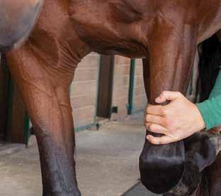
Differentiating an orthopedic problem from a neurologic one can be a difficult but common issue for lameness practitioners. It’s critical to recognize these issues early on, however, as neurologic deficits can present safety issues for clients with riding horses.
“If you have concern for a neurologic deficit in a riding horse, please make that be known to the client,” said Jackie Christakos, DVM, of the Littleton Equine Medical Center in Colorado. “Falling to the ground on a groomed surface is abnormal. If it happens multiple times, I’m suspicious that something is wrong. When they’re healthy, horses can climb up the side of a mountain on a 100-mile ride. They should not fall while on groomed footing.”
During a Burst Session at the 69th AAEP Convention in San Diego, Dr. Christakos described her process when she suspects a neurological issue in a horse that’s presented for lameness.

8 Issue 2/2024 | ModernEquineVet.com
ORTHOPEDICS
photography
Shutterstock/AnnaElizabeth
During the exam
“Horses don't usually come to me for neurologic dysfunction,” Dr. Christakos explained, “but I end up being suspicious of it when they come to me for lameness.”
While performing the lameness exam, she will look for certain clues that may suggest a neurological component.
“Horses don't usually come to me for neurologic dysfunction, but I end up being suspicious of it when they come to me for lameness.”
Dr. Jackie Christakos
“One of the first things to test is whether there is consistent, blockable limb lameness,” she said. “If it’s present, then I can do my part to take that out of the equation.”
Another clue is if the horse has a “regular” irregular gait, which does not always fit with an orthopedic diagnosis.
“If the horse struggles to hold a gait that should

come naturally to it, then that’s a clue,” she explained. “If a horse looks horrible at a walk but looks fine at a trot, then I start to get suspicious of a neurologic problem.”
Another way to differentiate an orthopedic vs neurologic horse is by checking whether the horse’s gait can be thrown off by changing the head position or by walking it down a hill.
“If the horse’s signs are still confusing at the end of the exam, you could do an NSAID [nonsteroidal antiinflammatory] trial and have the horse come back for a re-check,” Dr. Christakos said. “The lameness would be helped more by the NSAID you can probably track the first paragraph on this page and get enough room to spell out NSAID. than the neurologic issues would be. If you can take that out of the equation to make it a little less muddy, then that could be helpful.”
Diagnostics
If she is still suspicious of a neurologic issue after moving through the lameness exam, Dr. Christakos keeps some additional diagnostic tricks up her sleeve.
A post-exercise muscle enzyme evaluation may be illuminating. She will also measure the horse’s vitamin E levels, which can be helpful even if it is not directly related to the horse’s primary issue. Axial skeletal radiographs can also help narrow down her focus.
“Beware of the positive serum EPM [Equine protozoal myeloencephalitis] titer,” she added, noting that a positive serum result doesn’t necessarily signal a clinical problem. “It can be helpful if the test is negative, but otherwise you can’t really use it. This is important to remember for new practitioners because it can be confusing.”
In the end, however, it’s important to remember that the horse may, in fact, have both an orthopedic and a neurologic issue.
“If you are unsure, don’t be afraid to consult with your friendly neighborhood internist—one that has a strong interest in neurology,” Dr. Christakos said. “It can be fun to do these dual exams and to help each other figure out what is going on.” MeV
10 Issue 2/2024 | ModernEquineVet.com ORTHOPEDICS
Lifting Large Animals Since 1957
www.shanksvet.com

CHECK OUT OUR NEW WEBSITE Equine Vet The Modern MODERNEQUINEVET.COM
Equine Immunosuppression Linked to Dexamethasone Treatment for Asthma
By Landon Gray
There is no evidence that inhaled ciclesonide suppresses the immune systems of horses with equine asthma. However, dexamethasone treatment does have immune-suppressive effects, according to research presented at the 69th AAEP Annual Convention, held in San Diego.
Common Condition
“I think we all recognize that equine asthma is a common condition that's treated in equine practice—most often being with dexamethasone as the first-line treatment,” said study researcher Allen Page, DVM, PhD, a scientist and veterinarian at the Gluck Equine Research Center, in Lexington, Ky. “Although, we certainly recognize also, at the same time, that dexamethasone does have some potential side effects.”
To identify the potential effects of inhaled ciclesonide compared with dexamethasone on systemic markers of immune function in horses, researchers evaluated 18 light, mixed breed horses (age range, 3-8 years). The horses were randomly assigned to either nontreated controls, ciclesonide treatment or dexamethasone treatment. For steady-state messenger RNA (mRNA) analysis, the researchers collected blood daily. In addition, they also collected blood at days 0, 5, 10 and 15 of treatment for Concanavalin A (ConA) in vitro stimulation. To determine the mRNA relative quantities, the researchers used real time quantitative polymerase chain reaction (RTqPCR) to look for genetic mutations (P<0.05).
the steady-state, whole-blood expression of granzyme B and interferon-gamma. They also reported that this suppressive effect remained within ConA-stimulated samples.
Granzyme B cells are key in the response to viral intracellular infections. Likewise, interferon-gamma is critical for innate and adaptive immune responses, Dr. Page noted.
“While in this project, we're talking about messenger RNA expression, a decreased production of those proteins would in fact be considered to be immunosuppressive,” he said.
Although dexamethasone is used frequently to treat equine asthma, it does have potential concerning side effects.
The researchers reported there were no similar effects in the horses treated with ciclesonide, and there were no significant effects on immune function markers linked to treatment with ciclesonide found in this study. This suggests a low risk for immunosuppression associated with ciclesonide.
Dr. Page noted the study was limited by the fact that mRNA expressions do not always correlate with protein concentrations, a limited panel of mRNA targets and that only younger horses were evaluated.
“While short-term treatment with dexamethasone—so 10 days, as is kind of typical with equine asthma—it's not thought to be immunosuppressive, this study suggests that caution is warranted due to the suppression of granzyme B and interferon-gamma mRNA expression,” Dr. Page concluded. “Based on mRNA expression, there was no evidence of an immunosuppressive effect of inhaled ciclesonide in horses.”
In the dexamethasone arm, when compared with controls, the researchers found significant decreases in
Ciclesonide (Boehringer Ingelheim) is indicated for equine asthma in the Aservo EquiHaler formulation. MeV
12 Issue 2/2024 | ModernEquineVet.com RESPIRATORY
Page AE, Mackenzie J, Parker JL, et al. The effect of inhaled ciclesonide treatment on systemic markers of immune function in horses. J Equine Vet Sci. 2023;130:104925) https://www.sciencedirect.com/science/article/abs/pii/S0737080623007414
For more information:
STRANGLES
Strangles, caused by Streptococcus equi, is a reportable disease in all states, but the reporting requirements vary by state. There is no mandatory comprehensive database for tracking national prevalence.
Should Strangles Reporting Be a National Mandate?
In a survey of 250 U.S. veterinarians:
34% said strangles should be nationally monitored, but individual states should have jurisdiction over managing positive cases.
23.2% said strangles should become nationally monitored with mandatory notification of positive cases to a central data base.
9.6% said strangles should become notifiable nationally.
17.6% said strangles should become notifiable and actionable.
Veterinarians who regularly reported strangles were also more likely to want increased reporting.
Get strangles vaccination guidelines here:
https://aaep.org/document/strangles-vaccination-guidelines
Source: J Equine Vet Sci. 2022;114:103947. https://www.sciencedirect.com/science/article/abs/pii/S0737080622000855?via%3Dihub
BUSINESS VET Shutterstock/Nazarkru
Thoroughbred Filly With Lateral Suspensory Ligament Branch Lesion
 By Emily Brooks
On April 21, a 2-year-old Thoroughbred filly from a local training center was admitted for a protracted episode of waxing and waning cellulitis and lameness (grade 3/5) in the right hind limb.
The referring veterinarian had been treating her with oral antibiotics and nonsteroidal anti-inflammatory drugs (NSAIDs) but the cellulitis had not improved, so the trainer elected to bring her to the referral hospital. Despite the cellulitis, the filly was in good body condition, weighing 1,000 lbs, with normal vitals (HR 40, RR 12, T 100.9), and a bright attitude.

An ultrasound examination was performed on the right hind limb, and revealed diffuse cellulitis throughout the metatarsus and a large lesion within the lateral. At first glance, this could appear to be a core lesion that broke out the lateral margin of the branch, however, the character of the hyperechogenic material within the ligament was consistent with an abscess or hematoma. The nature of normal synovial fluid on ultrasound is typically black, or anechogenic. Cellular material appears more "sparkly" or hyperechogenic.
Ultrasonography cannot differentiate between white or red blood cells, hence the difficulty determining an abscess or hematoma. This pocket exited the branch and communicated with the surrounding subcutaneous tissue. Digital radiographs were obtained of the right hind fetlock and did not show bony involvement of the lateral sesamoid or third metatarsal bones.
The question arose whether to anesthetize the horse to get a sample. Being that it was a hind limb and a large degree of precision and accuracy are needed to place the needle within the lesion and minimize damage to healthy ligament, performing an aspirate standing seemed like an unnecessary risk. To help inform the decision, we ran blood work, and an elevated white blood count (12,300), elevated fibrinogen (600) and a serum amyloid A of 2,242 corroborated infection.
The filly was prepared for general anesthesia and a 14 g Mila catheter was placed in the right jugular vein and flushed with heparinized saline. No pre-op-

14 Issue 2/2024 | ModernEquineVet.com
TECHNICIAN UPDATE RH LMO Radiograph.
of the medial
in
and
planes.
Ultrasound
SLB
transverse
longitudinal

erative antibiotics were given, as an aspirate and culture/sensitivity sample were to be taken in surgery.
The horse was placed under general anesthesia in left lateral recumbency. The right hind leg was clipped from the coronet band to mid cannon bone and prepped using aseptic and sterile technique. I assisted the surgeon with the ultrasonography and covered the probe with a sterile rectal sleeve. A 20-g 1½" needles was directed into the intralesional abscess.
An aspirate of purulent fluid was obtained with the appearance of frank pus. White blood cells were too numerous to count so the sample was sent for culture and sensitivity. After the sample was taken, a mosquito hemostat was placed under ultrasound guidance to thread a ¼" Penrose drain as far proximally and distally as needed to ensure the abscess could not wall off. While under anesthesia a regional limb perfusion was performed by placing a tourniquet above the hock and inserting a catheter and extension set into the right saphenous vein. Amikacin (2 mL) was QS to 60 mL with sterile saline and left in for 20 minutes. After surgery a sterile half limb bandage was applied and the mare recovered

without incident.
The following day, the cellulitis had improved and the filly was started on systemic antibiotics potassium penicillin and gentamicin. She was also scheduled to receive 1 g of phenylbutazone PO for 5 days. The regional limb perfusion was repeated under standing sedation. The culture grew heavy Staphylococcus aureus. There was no history of recent injections or lacerations prior to the episode of cellulitis, so the etiology of the infection is unknown.
A 2-year-old Thoroughbred filly was admitted for a protracted episode of cellulitis and lameness.
The patient was released from the hospital 2 days after being admitted, 1/5 lame RH at the walk. Discharge instructions were to contain the filly to a box stall until the drain was removed 7 days post-surgery. Exercise could increase to hand walking only until
ModernEquineVet.com | Issue 2/2024 15
Ultrasound of the medial SLB in transverse and longitudinal planes
Longitudinal image of mosquito hemostat being placed into the abscess within the suspensory ligament branch
Teaching Points
Ultrasonography is a common diagnostic tool for evaluating soft tissue and bony irregularity in veterinary practice. Compared with magnetic resonance imaging (MRI), it is less invasive and more cost effective. The role technicians play in acquiring ultrasound images lags far behind human medicine. With the declining number of veterinarians choosing to go into equine medicine, there is likely a need for technicians to step into more specialized and highly trained roles.
I was fortunate enough to be trained in ultrasonography after learning MRI, and the 2 modalities helped me to learn anatomy and recognize pathology.
Working closely with veterinarians, I have been able to perform the ultrasound exams for their interpretation, which saves valuable time during busy lameness days and emergency work ups.
There are a wide range of possibilities to extend the use of technicians and assistants in practice. It is already common practice for technicians to acquire radiographs, perform MRI, nuclear scintigraphy and computed tomography. In the absence of a radiologist on site or available during scans, there are many times that the knowledge and experience of a technician in imaging can be integral to finding the cause of lameness or disease in our patients.

she could have a follow up ultrasound in 30 days. After that, exercise could increase based on the healing of the suspensory branch.
Follow-up ultrasound showed that the abscess had resolved, and there was marked healing of the branch, though there was still a core lesion consisting of approximately 25% of the cross-sectional area of the ligament. The filly was cleared to continue stall rest and hand walking with a favorable prognosis, pending the continued healing of the ligament.
This case highlights the opportunities available for technicians to be trained to assist veterinarians in new and different areas of practice. With the specializations for veterinary technicians and increasing desire for veterinarians to let us take on more responsibility, with proper training and oversight the technician role
could change quite a bit in the near future. MeV
About the Author
Emily Brooks began working as a veterinary technician in 2007 at Rood and Riddle Equine Hospital. There she developed an interest in diagnostic imaging and learned to run high field MRI, CT, and perform ultrasound and ultrasound-guided injections. She is now the Director of Imaging at Equine Athlete Veterinary Services in Simpsonville, Ky., and has added the modality of PET scan to her repertoire. Additionally, she works for Samsung, training on their ultrasounds. Emily also competes her offthe-track Thoroughbreds in eventing and dressage out of her farm in Lexington, Ky.
16 Issue 2/2024 | ModernEquineVet.com
Shutterstock/nelelena
TECHNICIAN UPDATE
An ultrasound examination being performed on the hind limb.
Equine Devices Successful at Creating Good Biological Concentrations for Regenerative Medicine
A recent study compared the concentrations of alpha2-macroglobulin (A2M) in equine plasma samples processed by 3 commercial devices to be used as regenerative joint therapy for osteoarthritis.
The researchers tested the Alpha2EQ (Astaria), Pro-Stride APS (Zoetis) and Restigen PRP (Zoetis) devices. These devices produce a concentrated solution of cells, platelets, growth factors and anti-inflammatory cytokines to reduce pain and manage osteoarthritis.
They collected plasma from 13 healthy horses and used mass spectrometry to determine the protein concentrations in each sample. They selected a specific A2M peptide to act as a surrogate for the entire A2M protein, which contains several multifunctional proteins. A2M may promote healing in the joints because it inhibits protease and inflammatory cytokines by using an amino acid mechanism to trap them.
The study was done in partnership with the University of Pennsylvania, University of Colorado and Zoetis. The Zoetis Animal Health Research and Development team completed the mass spectrometry plasma protein analysis in Kalamazoo, Mich.
The researchers found the plasma output volume, A2M concentration and plasma protein composition were equivalent among the 3 devices (P>0.05). The 30 mLs of plasma generated by the Pro-Stride APS and Restigen PRP devices contained the same concentrations of 16 plasma proteins (including A2M) as the
final output of the Alpha2EQ device.
In addition to the platelet-derived growth factors and white blood cell derived anti-inflammatory cytokines contained in the cellular output of Pro-Stride and Restigen, the A2M concentration was significantly higher than the final plasma output of Alpha2EQ (P>0.0001).
The results help support the understanding of the components of each device, according to Kyla Ortved, DVM, PhD, DACVS, DACVSMR, an associate professor of large animal surgery, University of Pennsylvania School of Veterinary Medicine “Managing osteoarthritis calls for a comprehensive mix of plasma proteins, anti-inflammatory cytokines and anabolic growth factors,” Dr. Ortved explained.
Osteoarthritis is a complex biologic process that affects many horses as they age. A targeted approach that takes on multiple disease pathways is needed to help manage pain and promote healing.
“This research provides guidance for equine veterinarians seeking a comprehensive option to help manage osteoarthritis for horses entrusted in their care,” said Nathan Voris, MD, the director of Equine Technical Services at Zoetis, which makes these devices.
Regenerative therapies have been shown to improve a horse’s lameness grade for up to 1 year after a single injection, and in some cases even longer. With these devices, processing can take place right at the farm, stable or clinic. MeV

For more information:
Ortved KF, Alward L, Cowles B, et al. Use of quantitative mass spectrometry-based proteomics and ELISA to compare the alpha 2 macroglobulin concentration in equine blood-based products processed by three different orthobiologic devices. Front Vet Sci. 2024 Feb. 9. https://www.frontiersin.org/articles/10.3389/fvets.2024.1335972/full
NEWS
NOTES
ModernEquineVet.com | Issue 2/2024 17
Shutterstock/Katrina Leigh
Reach your veterinarians wherever they are, whenever they want. FOR ADVERTISING RATES AND INFORMATION, EMAIL ModernEquineVet@gmail.com Equine Vet The Modern CHECK OUT OUR NEW WEBSITE AT www.modernequinevet.com


