2 minute read
Oral and Maxillofacial Pathology Case of the Month

ORALand maxillofacial pathology case of the month
Case History
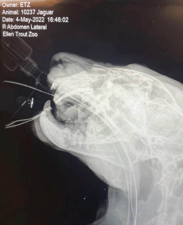
A 58-year-old Caucasian female presented with a 6-month history of right facial pain with occasional sinus drainage. Over the last 2 months, she also developed a right facial swelling. The patient had undergone a sinus lift surgery (sinus augmentation) involving the right maxilla about five years ago with placement of an implant. However, the implant failed and was removed last year. Her dental history also includes osteomyelitis of the left jaw that was treated by surgical debridement and IV antibiotics. Furthermore, the patient’s medical history is significant for two autoimmune disease conditions, Hashimoto’s thyroiditis and ankylosing spondylitis, and she also has severe osteoporosis. She was treated with IV Prolia for her severe osteoporosis, but she discontinued taking the medication 3 years ago. CBCT scan revealed an expansile lesion of the right maxilla with small focal radiopacities, and a space occupying lesion of the right maxillary sinus (Figure 1A, B). Biopsies of the right alveolus and the right sinus were subsequently performed.
AUTHORS
Ngozi Nwizu BDS, MMSc, PhD, TTS
Associate Professor Board Certified Oral and Maxillofacial Pathologist, Department of Diagnostic and Biomedical Sciences, UTHealth at Houston School of Dentistry, Houston, Texas
Steve Koo DDS
Board Certified Oral & Maxillofacial Surgeon, Piney Point Oral and Maxillofacial Surgery, Chief of Oral and Maxillofacial Surgery, Memorial Hermann at Memorial City, Houston, Texas

Figure 1A.
Cone Beam CT scan (coronal section), showing opacification of the right maxillary sinus, and an expansile mixed radiopaque-radiolucent lesion of the right maxilla.
Figure 1B.
Cone Beam CT scan (sagittal section).


Microscopic examination of the contents from the right alveolus revealed non-specific findings comprising of fragments of allograft material, interspersed with areas of hemorrhage, fragments of viable and partially viable bone, and fibrous tissue with tissue debris (Figure 2). Microscopic examination of the sinus contents showed multiple soft tissue fragments, comprising of chronically inflamed fibrous tissue displaying numerous hyperemic capillaries, areas of surgical hemorrhage, and patchy infiltrates of acute and chronic inflammatory cells, consisting of mostly neutrophils, lymphocytes, plasma cells, and histiocytes. Some of the soft tissue fragments were covered by respiratory type epithelium exhibiting inflammatory cell exocytosis. A prominent feature of the tissue specimen was the presence of multiple densely packed aggregates of microorganisms, composed of septate hyphae that branch at 45 degrees (Figure 3). These microorganisms were observed in all of the biopsy specimens obtained from the sinus.
Figure 2.
(Original magnification x 20) Biopsy revealing allograft material, interspersed with areas of hemorrhage, fragments of viable and partially viable bone trabeculae, as well as fibrous tissue and tissue debris.
Figure 3.
(Original magnification x 200) presence of a tangled mass of densely packed aggregates of fungal organisms, composed of septate hyphae that branch at 45 degrees.
What is the differential diagnosis? What is the final diagnosis? See page 516 for the answer and discussion.