
14 minute read
PRENATAL ALCOHOL EXPOSURE AND GENETIC SEX IMPACT NFIA EXPRESSION IN THE DEVELOPING CORTEX


By Aubrie Miller ‘22
INTRODUCTION
Advertisement
Prenatal alcohol exposure (PAE) can occur during pregnancy and result in fetal alcohol spectrum disorders (FASDs). PAE can cause physical abnormalities, including alterations to facial structure, growth deficits, and neurodevelopmental deficits caused by abnormal brain growth and development.1,2 PAE is the leading non-genomic cause of learning disabilities.3 Alcohol exposure during pregnancy and PAE can cause lifelong stigma for both the birth mother and the affected individual. This stigma contributes to additional hardships for individuals with FASDs including aggravating behavioral differences, mental illnesses, and substance use disorders as well as increasing barriers to achieving independence and life goals.4 Women who consume alcohol during pregnancy are often labeled as bad mothers or child abusers, and therefore deserving of harsh judgment.5 The stigma around alcohol exposure during pregnancy, combined with societal judgment, can make women reluctant to disclose alcohol use during pregnancy to their healthcare provider.6
Common binge drinking behaviors among men and women of childbearing age and lack of family planning combined with stigma against alcohol exposure during pregnancy means there is a low chance of eradicating PAE. Therefore, it is important to reduce harm both through social policies and by understanding the effects of PAE on brain development so that targets for intervention can be identified and the impact of PAE can be ameliorated.
Here, we investigated a potential mechanism of PAE’s impact on brain development. Previously,
we showed that PAE causes the premature maturation of neural stem cells (NSCs).7 In the developing brain, NSCs proliferate to make additional NSCs (self-renewal) and differentiate to produce the cellular complement of the brain. NSC differentiation occurs sequentially. First, neurogenesis produces neurons. Then, astrogliogenesis produces astrocytes that maintain the chemical environment for neurons. Finally, oligodendrogenesis produces oligodendrocytes which create myelin around axons to provide electrical insulation. For astrogliogenesis, increased expression of transcription factor Nuclear Factor-1 A (NFIA) is necessary and sufficient to induce astrocyte production.8 We hypothesized that PAE increases NFIA expression in NSCs, thereby inducing premature maturation through the premature activation of astrogliogenesis.
To determine if PAE results in premature activation of astrogliogenesis in NSCs, we examined if PAE increased the expression of Nfia mRNA during neurogenesis in the cerebral cortex using single-cell RNA sequencing (scRNAseq). scRNAseq allows analysis of transcriptional profiles within individual cells.9,10 We performed scRNAseq on two mouse models of cerebral cortical NSC development: 1) in cortical tissue on gestational day (GD) 14.5 following exposure to ethanol vapor or room air on GD 12.5; 2) in neurosphere cultures grown from GD 12.5 mouse NSCs that were not exposed (naïve) to ethanol. Experiment 1 showed the impact of ethanol exposure on NSCs and their subsequent cycles of neurogenesis. Experiment 2 allowed us to compare the effects of ethanol from experiment 1 to isolated GD 12.5 NSCs grown as neurosphere cultures. For both of these models, cells from male and female fetuses were included, allowing for the assessment of impact of genetic sex on Nfia expression. GD 12.5–14.5 is the time of cortical NSC proliferation and the beginning of neurogenesis.11 We chose to use two different models to better grasp the impact of ethanol exposure over time and examined cells at the age of exposure as well as the cells that differentiated following exposure. Furthermore, comparing these models provides insight on how the heterogeneity of the developing cortex may modulate the impact of PAE on NSCs.
posure affect Nfia expression. In GD 14.5 cortical cells, Nfia expression was higher in female stem cells, but in GD 12.5 neurosphere cultures, male NSCs have higher Nfia expression, suggesting that male NSCs may have higher levels of Nfia before female NSCs. In addition, in the GD 14.5 developing cortex, PAE increased Nfia expression in stem cells and early developing neurons.
Targeted interventions to restore normal Nfia expression may slow premature maturation of NSCs and reduce the detrimental changes to brain development following PAE. But these interventions may need to be genetic sex-specific, which impacts the timeline of Nfia expression. These interventions may be one effective way to reduce the harm of PAE and improve long-term quality of life for individuals with FASDs.
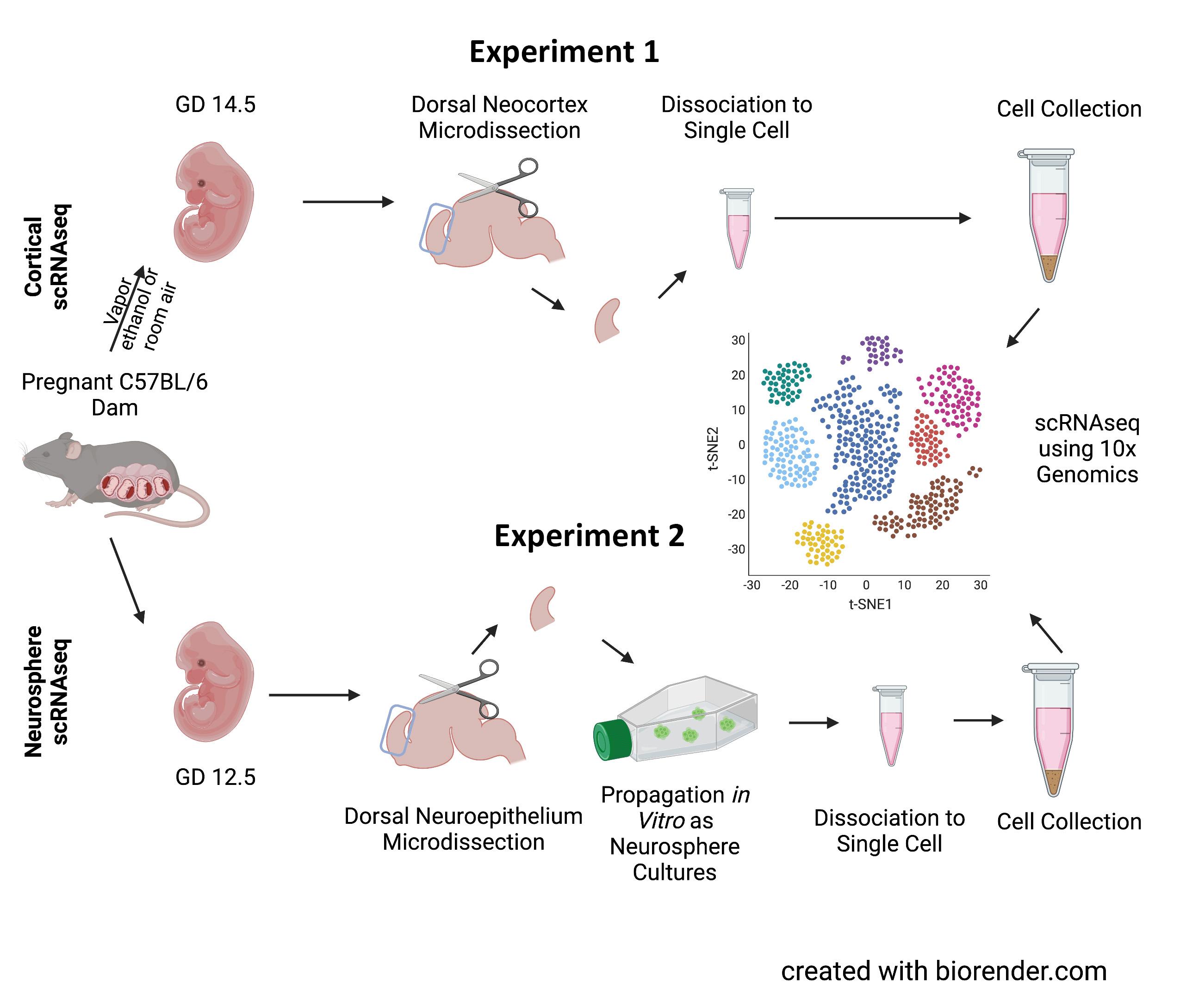
Figure 1. Paradigm for sample collection. Cortical scRNAseq (experiment 1, top) used tissues from gestational day 14.5 (GD 14.5) fetuses following the exposure to ethanol or room air vapor on GD 12.5. Following microdissection of the dorsal neocortex, tissue was dissociated into single cells which was then further analyzed using the 10x Genomics platform. Neurosphere scRNAseq (experiment 2, bottom) used cortical tissues from GD 12.5 fetuses which underwent the same dissection; however, they were then propagated as neurosphere cultures then analyzed by scRNAseq. Created with biorender.com using a premium license made available through the Texas A&M Institute for Genome Sciences and Society.
METHODS
Cortical scRNAseq
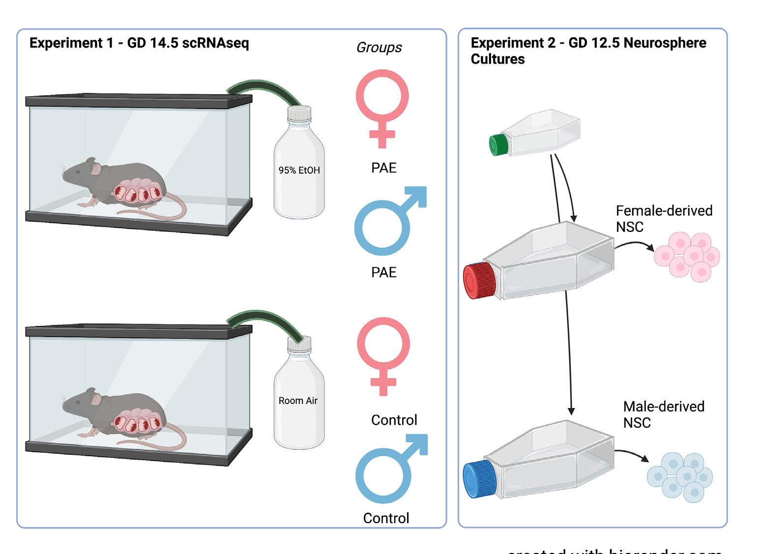
For experiment 1, pregnant C57BL/6 mouse dams were exposed to vaporized 95% ethanol or room air on GD 12.5 (Figure 1). Ethanol exposure resulted in blood ethanol concentrations ranging from 210–260 mg/ dL (equivalent to blood alcohol concentrations, or BACs, of 0.21–0.26%). Two days following exposure, on GD 14.5, the fetuses were removed and underwent microdissecFigure 2. Exposure paradigm. In experiment 1, pregnant dams were exposed to tions to obtain the dorsal either 95% ethanol or room air via a vapor chamber on GD 12.5. Subsequently, on neocortex. The collected GD 14.5, tissue from the exposed fetuses were then segregated by genetic sex. In extissues were then broken periment 2, GD 12.5 NSCs were propagated as sex-segregated neurosphere cultures down into single cell in mitogenic media. Created with biorender.com using a premium license made available through the Texas A&M Institute for Genome Sciences and Society.suspensions by manual trituration and enzymatic disaggregation. Cells were collected by centrifugation, briefly fixed in methanol, and underwent scRNAseq using the 10x Genomics pipeline (Figure 2). Initial characterization of these data has been previously published.12
Neurosphere scRNAseq
In experiment 2, similar microdissections occurred as in experiment 1. However, dorsal neuroepithelium was collected from GD 12.5 fetuses. Tissues were broken into single cell suspensions and propagated in vitro as neurosphere cultures in mitogenic media. These cells, following 5 days of growth in mitogenic media, were separated into single cell suspensions, briefly fixed with methanol, and underwent scRNAseq using the 10x Genomics pipeline. Initial characterization of these data has been published.13
Segregation of Tissues by Genetic Sex
At the time of tissue collection for both experiments, additional fetal tissue was taken to determine fetal sex. DNA was extracted from these tissues by alkaline lysis. This DNA suspension was then neutralized, diluted, and underwent quantitative real-time polymerase chain reaction (qPCR) for repetitive sequences present on the X and Y chromosomes.
RESULTS
Data obtained from scRNAseq is complex and multi-dimensional. These data can be visualized in low-dimensional space by using the t-distributed Stochastic Neighbor Embedding (t-SNE) algorithm to construct t-SNE plots. In these plots, each data point represents a single cell, and cells are clustered by similarity and separated from dissimilar cells. For both the cortical scRNAseq (Figure 3A) and the neurosphere scRNAseq (Figure 4A), a number of discrete cellular clusters were identified by the t-SNE algorithm, although 33 clusters in total were identified for
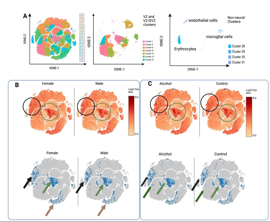
Figure 3. T-SNE plots of experiment 1, cortical scRNAseq. A.) LEFT: color coded t-SNE plot of the 33 identified cell clusters in the cortex. MIDDLE: clusters of the VZ and VZ/SVZ regions. RIGHT: non-neural clusters. B. and C.) Nfia expression in cortices stratified by genetic sex, regardless of treatment (B) or exposure, regardless of genetic sex (C). TOP: Nfia expression in all cells as indicated by color coding with log2 scale of expression in which higher expression is colored red while low rates of expression are colored yellow. BOTTOM: the bottom 10% of Nfia expression was excluded to show a clear picture of sex differences in number of cells with Nfia expression. B.) The green circle and arrow highlight cell populations with higher Nfia expression in female-derived cells while the black circle and arrow and brown arrow highlight cell populations with decreased
Nfia expression in female-derived cells. The green region contains NSCs in an
VZ cluster, black region contains neurons of lateral layer V/VI neurons, and the brown region contains neurons of rostral layer V/VI .VI. C.) The black circle and arrow highlights layer V/VI cells with greater Nfia expression in the alcohol group and the green circle and arrows highlight VZ cells with higher expression in the alcohol group. the cortical scRNAseq and 10 were identified for the neurosphere cultures. Cortical scRNAseq In previous work,14 cellular clusters were identified based on gene expression to correspond to cellular populations in the developing cortex. Here we focused the regions of the developing cortex that contain NSCs: the ventricular zone (VZ), which contains the earliest, most multipotent neural stem cells and the sub-
ventricular zone (SVZ), which contains another population of stem cells that divide more rapidly to expand the cortex (Figure 3A). In cell clusters that contained cells identified as belonging to the VZ, SVZ, or both (VZ/SVZ), we first compared Nfia expression by genetic sex (Figure 3B). We found in the central VZ cluster that female-derived cells have higher expression of Nfia than male-derived cells. We further examined the number of cells that have Nfia expression, excluding those cells in the bottom tenth percentile of expression. More female-derived cells express Nfia in this VZ cell cluster than male-derived cells. While examining the Nfia expression in the developing cortex, we noticed that sex difference in Nfia expression seen in NSCs was not similarly seen across all cell types in the GD 14.5 cortex. Contrastingly, clusters that correspond to early neurons (layers V/VI) have higher Nfia expression in male-derived cells compared to female-derived cells. We then compared Nfia in the cortical cell cluster stratified by exposure (Figure 3C). In this comparison, we found that ethanol exposure increased Nfia expression in the stem cells of the central VZ cluster and in the early layer V/VI neurons of layer V/VI. These data suggest that sex differences in Nfia expression in the developing cortex are celltype specific while ethanol may similarly regulate Nfia across cell types. For the construction of t-SNE plots for the neurosphere scRNAseq, when the data from the male-
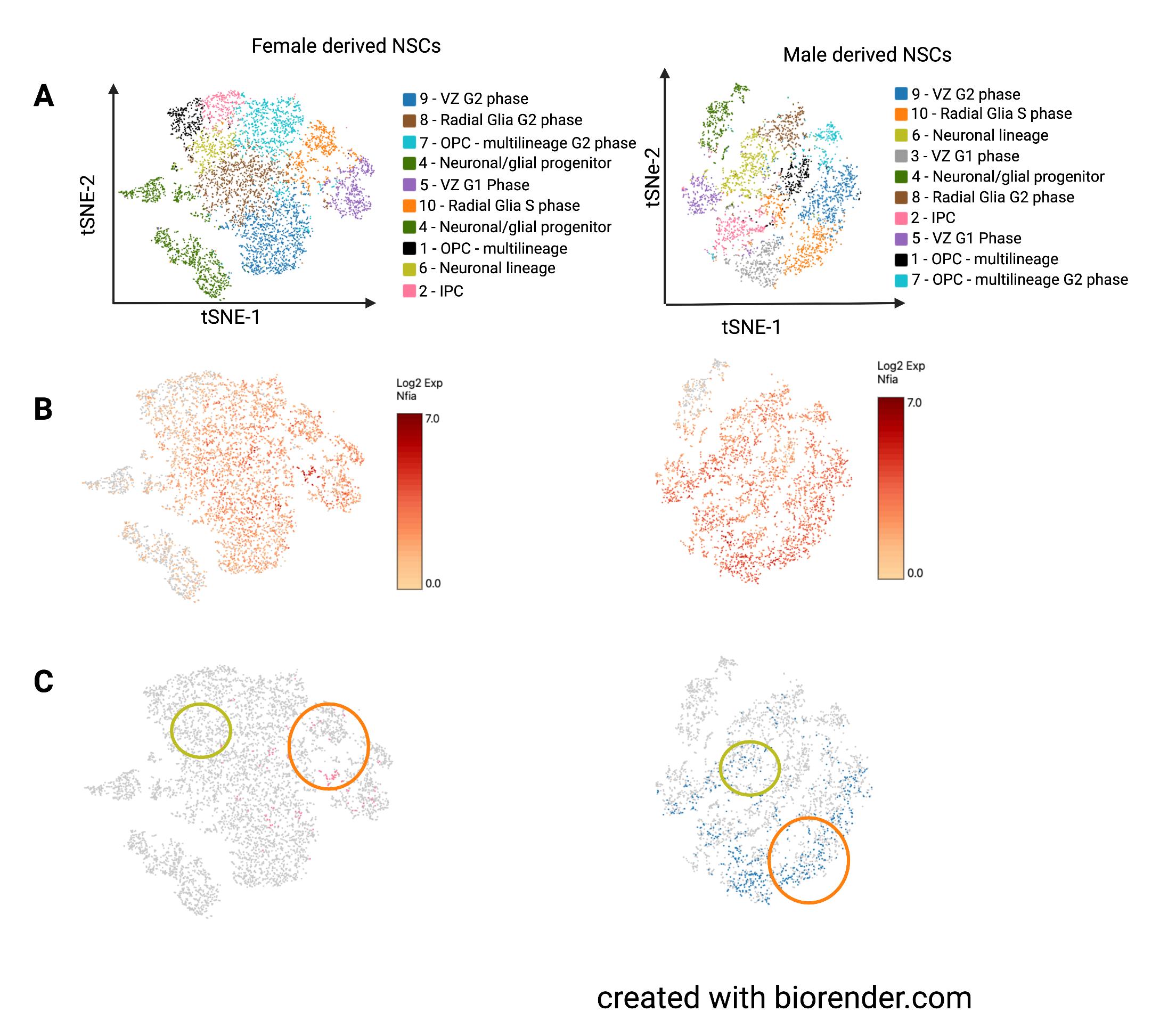
and female-derived neurospheres was combined, the cell clusters largely segregated by sex.15 Therefore, we constructed two independent t-SNE plots, one for each sex, and used gene expression data to determine closely related clusters across plots (Figure 4A).
Corresponding clusters are color-coded the same even though they may not be in the same location. For the neurospheres, almost all clusters had markers for neural stem/progenitor cells with the exception of cluster 4, which had higher expression of differentiation lineage markers. We found that Nfia is expressed similarly across NSC populations in both male and female neurospheres. However, when we examined individual cells, only a few female cells had considerable Nfia expression (>10th percentile of expression seen in both male and female neurospheres). A small group of cells expressed Nfia in female-derived neurospheres but this group had widespread expression in male-derived neurospheres. Other cell populations had almost no cells with Nfia expression in the females, while the males had widespread expression.
CONCLUSION
Here, we showed that neural stem cells in the developing cortex are not a homogenous population. Cortical scRNAseq defined 11 VZ cell clusters while the neurosphere scRNAseq had 10 clusters. We found sex-differences in the expression of Nfia in NSCs, with females showing higher expression at GD 14.5 in the central VZ NSC cluster and males showing higher expression in the GD 12.5 NSC cultures. Ethanol generally increased Nfia expression, although this effect was NSC subpopulation specific, as we did not see similar increases in expression across all the VZ/SVZ clusters. We found that Nfia expression
Figure 4. Experiment 2, neurosphere scRNAseq. A.) Both-SNE plots of cell clusters in level was widemale- and female-derived neurosphere clusters. Similar clusters were present in each plot spread in NSCs but with the exclusion of cluster 3 from the female plot. B.) Nfia expression in all cells as indicated by color coding with log2 scale of expression in which higher expression is colored red while low rates of expression are colored yellow. C.) Cells expressing Nfia, with cells also varied by NSC subpopulation. Surin the bottom 10% of Nfia expression excluded. Orange circles highlight cluster 10 which prisingly, even in contained radial glia-like neural progenitors in S phase. Yellow circles highlight cluster 6 NSC clusters with which contained the neuronal lineage marker-expressing progenitor cells.
high Nfia expression, there was not a corresponding expression of astrocytic cell markers. These data indicate that Nfia is expressed in NSCs prior to astrogliogenesis and may define NSC subpopulations. These data indicate that PAE can induce Nfia expression in NSCs and this increased expression may be targetable for intervention. However, any intervention will likely need to be tailored to each genetic sex, as we found sex differences in the expression timeline of Nfia. By decreasing Nfia expression following PAE, we may encourage NSC self-renewal, reducing harm to the developing cortex caused by PAE. Targeted interventions to NSCs have the potential to improve neurodevelopment, and therefore quality of life for individuals with FASDs. Further investigation is needed to discover the best methods of intervention of NSCs and other targets that could work alongside Nfia-targeted therapeutics to reduce harm following PAE and ameliorate more negative outcomes associated with FASDs.
ACKNOWLEDGEMENTS
Thank you to the laboratory of Dr. Rajesh Miranda for their guided support through this research and manuscript. Thank you to Dr. Nihal Salem, Dr. Alex Tseng, and Dr. Amanda Mahnke for the analysis done on the t-SNE plots. Thank you to family for their continued support throughout my journey as an undergraduate researcher.
REFERENCES
1. Lebel, C., F. Roussotte, and E. R. Sowell.
“Imaging the Impact of Prenatal Alcohol
Exposure on the Structure of the Developing
Human Brain.” Neuropsychol Rev 21, no. 2 (June 2011): 102–18. https://doi.org/10.1007/ s11065-011-9163-0. https://www.ncbi.nlm.nih. gov/pubmed/21369875.
2. Blackburn, C., and T. Whitehurst. “Foetal
Alcohol Spectrum Disorders (Fasd): Raising
Awareness in Early Years Settings.” [In English]. British Journal of Special Education 37, no. 3 (September 2010): 122–9. https://doi. org/10.1111/j.1467-8578.2010.00471.x.
3. Blackburn et al., “Foetal Alcohol,” 122–9
4. Corrigan, P. W., J. L. Lara, B. B. Shah, K. T.
Mitchell, D. Simmes, and K. L. Jones. “The
Public Stigma of Birth Mothers of Children with Fetal Alcohol Spectrum Disorders.” Alcohol Clin Exp Res 41, no. 6 (June 2017): 1166–73. https://doi.org/10.1111/acer.13381. https:// www.ncbi.nlm.nih.gov/pubmed/28370022.
5. Roozen, Sylvia, Sarah E. Stutterheim, Arjan E.
R. Bos, Gerjo Kok, and Leopold M. G. Curfs.
“Understanding the Social Stigma of Fetal
Alcohol Spectrum Disorders: From Theory to
Interventions.” Foundations of Science (2020). https://doi.org/10.1007/s10699-020-09676-y.
6. Roozen et al., “Understanding the Social,” 1166–73
7. Mahnke, Amanda H., Nihal A. Salem, Alexander M. Tseng, Annette S. Fincher, Andrew
Klopfer, and Rajesh C. Miranda. “Fetal Alcohol Spectrum Disorders: A Stem-Cellopathy?”
Stem Cells in Birth Defects Research and Developmental Toxicology (May 2018): 223–59. https://doi.org/10.1002/9781119283249.ch9.
8. Deneen, B., R. Ho, A. Lukaszewicz, C. J. Hochstim, R. M. Gronostajski, and D. J. Anderson. “The Transcription Factor Nfia
Controls the Onset of Gliogenesis in the Developing Spinal Cord.” Neuron 52, no. 6 (December 2006): 953–68. https://doi.org/10.1016/j. neuron.2006.11.019.
9. Tseng, A. M., D. D. Chung, M. R. Pinson, N.
A. Salem, S. E. Eaves, and R. C. Miranda.
“Ethanol Exposure Increases Mir-140 in Extracellular Vesicles: Implications for Fetal Neural Stem Cell Proliferation and Maturation.”
Alcohol Clin Exp Res 43, no. 7 (July 2019): 1414–26. https://doi.org/10.1111/acer.14066.
10. Salem, N. A., A. H. Mahnke, K. Konganti, A.
E. Hillhouse, and R. C. Miranda. “Cell-Type and Fetal Sex-Specific Targets of Prenatal
Alcohol Exposure in Developing Mouse
Cerebral Cortex.” [In English]. iScience 24, no. 5 (May 2021). https://doi.org/10.1016/j. isci.2021.102439.
11. Miranda, R. C. “Micrornas and Fetal Brain Development: Implications for Ethanol Teratology
During the Second Trimester Period of Neurogenesis.” Front Genet 3 (2012): 77. https://doi. org/10.3389/fgene.2012.00077.
12. Salem et al., “Cell-Type.”
13. Tseng et al., “Ethanol Exposure,” 1414–26.
14. Salem et al., “Cell-Type.”
15. Tseng et al., “Ethanol Exposure,” 1414–26.

AUBRIE MILLER ‘22
Aubrie Miller ‘22 is a biochemistry and genetics major from Fort Worth, Texas. Aubrie is passionate about women’s health and prenatal care. She hopes to go to Medical School to pursue a career in meeting women’s needs.