ents of the enteric bacterial flagellum present in E. coli, only about 20 of them are common to all bacteria.14 What is essential for flagellar function in one bacterium may not even be present in another.1
SIMILARITIES BETWEEN BACTERIAL FLAGELLA AND TYPE III PROTEIN SECRETION SYSTEMS│
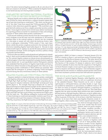
Bacterial flagella and virulence related type III protein secretion systems (T3SSs) are clearly derived from a common ancestral system sharing at least nine homologous constituents.1,12 But which was the precursor of the other? Some have argued that T3SSs preceded the BF basal construct while others have argued the opposite.3,15 We would like to suggest that both systems arose from a simpler primordial secretion system. In fact, Gram-negative bacteria have evolved many distinct mechanisms for exporting proteins across the two membranes of their cell envelopes, and some of these systems share protein constituents.16,17 T3SSs secrete substrate proteins directly from the Gram-negative bacterial cytoplasm across the inner and outer membranes of the bacterial envelope either to the external medium or directly into the cytoplasm of a host plant or animal cell with which the bacterium forms a symbiotic or pathogenic relationship.7 T3SSs consist of thin, rigid, hypodermic needle-like protein complexes anchored to the envelope by basal structures resembling flagellar basal bodies (figure 2). Because in some instances both types of systems can secrete the same proteins across the two-membrane envelope, T3SSs and flagellar secretory systems overlap structurally and functionally. T3SSs are often encoded on mobile plasmids and ‘pathogenicity islands,’ both of which can be transferred laterally between Gram-negative bacteria.5 By contrast, flagellar systems are almost always chromosomally encoded and appear to have been transmitted to progeny bacteria largely by vertical descent (ref. 12, and unpublished results). Phylogenetic analyses have established that the proteins of flagellar basal bodies and those of T3SSs form distinct branches on the phylogenetic tree when any of the homologous protein constituents of these systems are examined. This implies that the divergence of proteins comprising T3SSs and flagella occurred during the early evolutionary history of these systems.
RELATED FLAGELLAR STRUCTURAL PROTEINS│
Sequence similarity is observed between certain protein constituents of the flagellum although not all of these share the same domains. Thus, the FlgB, FlgC, FlgF and FlgG ‘transmission shaft’ rod proteins all exhibit regions of sequence similarity with the FlgE ‘universal joint’ hook protein (figure 3). For example, FlgG has 3 domains (N, C1, and C2) that are homologous to these domains in FlgE, but the central domain of FlgE (M) is absent in FlgG (figure 3). The simplest explanation is that the evolutionary precursor of these proteins resembled the smaller ones (FlgG and FlgF) and that a novel domain was inserted into FlgE to generate the larger one. The differing subflagellar locations and functions of these homologous constituents presumably resulted in part from domain insertion and in part from sequence divergence during evolution. The Flg rod proteins, the FlgE hook protein and the FlgK hook associ-
Figure 2: Comparative schematic depiction of the E. coli flagellum (left) and Gram-negative bacterial type III secretion systems (T3SS, right). Homologous structures in the basal regions of both structures are shown with the same shading. Non-homologous structures are shown as distinct structures. IM, inner (cytoplasm) membrane; PG, peptidoglycan cell wall layer; OM outer (lipopolysaccharide-containing) membrane. The multilayered MSring interacts with the IM, the P-ring anchors the structure to the PG, and the L-ring interacts with the OM. ATP hydrolysis via the FliI ATPases provides the energy required for protein export and assembly.
ated protein-1 (HAP1) all share a common C terminal domain (C2), the DUF1078 domain in figure 3, and most have similar N terminal targeting sequences, the Flg-bb-rod domain in figure 3. The latter directs the newly formed cytoplasmic proteins to the general secretory apparatus. The function of the common C2 domain is unknown, but it could be involved in assembly. It seems clear that the complexity of the flagellarrod/hook/filament complex arose in part by gene duplication events followed by sequence and domain divergence. Such mechanisms suggest a possible basis for the evolution of dissimilar protein-protein interactions since homo-multimeric proteins always have self-associative properties.
THE HOOK AND FLAGELLAR FILAMENT│
Subunits of bacterial flagellar filaments, called flagellins, vary tremendously in amino acid sequence as well as in quaternary structures of the assembled filament. These filaments can be curly or straight, right-handed or left-handed helices, and either flexible or rigid. Some are modified by methylation or glycosylation. In different strains of E. coli, there are nearly 50 sequence divergent flagellins, a surprising observation since each E. coli strain usually has just one. This observation suggests that the genes encoding these proteins have been transferred between bacteria with high frequency. Moreover, an Arctic sulfate-reducing bacterium, Desulfotalea psychrophila, has dozens of flagellins encoded within its genome. In this case, we do not know what their functions are. However, we do know that while some flagellar filaments consist of a single flagellin, others contain many. Surface exposed residues in flagellins, which are strongly antigenic, are Figure 3: Results of a search of the National Center for Biotechnology Information (NCBI) Conserved Domain Database (CDD) much more variable than the buried using the Escherichia coli FlgG flagellar basal-body rod protein as the query. The accession number for FlgG is AAN79818. Bars indicate regions of homology for different flagellar proteins: FlgF, FlgE, FlgC, FlgK and FlgB. The common N-terminal residues, which are poorly antigenic. domain is labeled Flg_bb_rod while the common C-terminal domain is labeled DUF1078. Amino acyl residue position in FliG is This surface variability provides a mechanism for immune evasion by provided at the top. The web address for the NCBI CDD is http://www.ncbi.nih.gov/structure/cdd/cdd.shtml. pathogenic bacteria and is easily explained by natural selection (see ref. 13 for a more detailed review). In spite of the tremendous sequence variation in flagellins, most if not all of them probably share a common ancestry. In fact, flagellins are also homologous to the hook-associated protein-3 (HAP3 or FlgL). Moreover, they share domains with the hook-associated protein-1 (HAP1; FlgK), which in turn shares
Saltman Quarterly│UCSD Division of Biological Sciences│Volume 4│2006-2007