Focusing on Fibre Impact on Gastrointestinal Health and Clinical Uses in Dogs and Cats

Is Regenerative Medicine The Answer to Canine Osteoarthritis?
Feed Intake Differences and Variation in the Vitamin E Status in Transition Dairy Cows
Antimicrobial Resistance A Dark Shadow on Global Public Health
Official Supporting Associations - Sponsored companies -


www.international-animalhealth.com PEER REVIEWED Volume 10 Issue 2
Your partner for contract research
Royal GD is a partner within the animal health industry worldwide, performing in vivo, in vitro and field studies. We conduct safety and efficacy studies on veterinary biologicals and pharmaceuticals in compliance with the OECD principles of Good Laboratory Practice (GLP).

Our portfolio includes, but is not limited to:
• Safety and efficacy studies of veterinary biologicals and pharmaceuticals;
• Studies to obtain vaccine or challenge-strain candidates;


• Quality control tests on final products;
• Development of models to demonstrate the efficacy or safety of veterinary biologicals and pharmaceuticals;
• Surveys on the prevalence of (emerging) infectious diseases/agents.
Get in touch and plan a meet up with our account managers and research project team: support@gdanimalhealth.com
www.gdanimalhealth.com
AHEAD IN ANIMAL HEALTH
ROYAL GD IS AHEAD IN ANIMAL HEALTH WITH EXPERT AND INDEPENDENT CONTRACT RESEARCH
MANAGING DIRECTOR
Mark A. Barker
EDITORIAL MANAGER

Beatriz Romao beatriz@senglobalcoms.com
RESEARCH AND CIRCULATION
Virginia Toteva virginia@senglobalcoms.com
DESIGNER
Jana Sukenikova www.fanahshapeless.com
BUSINESS DEVELOPMENT
Jerome D’Souza info@senglobalcoms.com
ADMINISTRATOR
Jessica Chapman jessica@senglobalcoms.com
FRONT COVER
© istockphoto
PUBLISHED BY Senglobal Ltd.
Unit 5.02, E1 Studios, 7 Whitechapel Road, E1 1DU, United Kingdom
Tel: +44 (0) 2045417569
Email: info@senglobalcoms.com www.international-animalhealth.com
International Animal Health Journal – ISSN 2752-7697 is published quarterly by Senglobal Ltd.
04 FOREWORD
WATCH PAGES
06 Low Viral Pressure Results in Better Growth
Pathogenic micro-organisms are a continuous threat for broilers as they can cause illness, immunosuppression, and poor performance. A good start of a broiler flock begins with healthy chicks in a clean house. It is important to prevent poultry from becoming infected with pathogens, especially young chicks, with cleaning and disinfection (C&D) of empty broiler houses playing an important role here. Sjaak de Wit at GD Animal explains the VIR check to assess the effectiveness of C&D in broiler houses.
08 A Pug’s Life
If you are a pug owner, you will know their characteristics already – a mixture between a four-legged clown and a toddler, which is among the many reasons why we all love them. The pug, is a brachycephalic breed, meaning flat-faced. While this does add to their cuteness, it can also lead to serious health problems. As pugs are becoming more and more popular, it is becoming more common that they are sold on the internet. Justine Marie Chambers explains what new pet owners needs to follow, whereby the end of the process you will have a healthy, genuine and lovable new puppy to add to the family, ready to have many fun times and fond memories.
REGULATORY & MARKETPLACE
10 Latest Trends in Animal Health Testing
According to P&S Intelligence, the global animal pharmaceuticals market is set to grow at a compound annual rate of 6.8 percent between 2021 and 2030, reaching a size of $72.74 billion by the end of the decade. As the market has grown and importance, due mainly to increasing consumption of meat and animal products, a booming pet industry and growing incidences of zoonotic diseases, so too have regulatory frameworks and testing techniques. Here Beccy Bell, Operations Manager at Contract Research Organization Broughton, discusses what’s on the horizon for the testing of animal health products.
12 Antimicrobial Resistance:
A Dark Shadow on Global Public Health
The opinions and views expressed by the authors in this journal are not necessarily those of the Editor or the Publisher. Please note that although care is taken in the preparation of this publication, the Editor and the Publisher are not responsible for opinions, views, and inaccuracies in the articles. Great care is taken concerning artwork supplied, but the Publisher cannot be held responsible for any loss or damage incurred. This publication is protected by copyright.
Volume 10 Issue 2 Summer 2023
Senglobal Ltd.
Antibiotics were the one of the revolutionary discoveries of the 20th century while their resistance has turned out to be one of the grievous challenges of the 21st century. The burgeoning burden of antibiotic resistance as a whole and the sharing of these resistance traits among humans, animals, and environmental reservoirs, are continuously threatening mankind. Dr. Pankaj Dhaka Guru at Angad Dev Veterinary and Animal Sciences University, Dr. Deepthi Vijay of Veterinary and Animal Sciences & Dr. Jay Prakash Yadav of the Indian Veterinary Research Institute, argue that like many emerging public health problems, the problem of antibiotic resistance reminds us that the health of humans, animals, and ecosystems are completely interconnected, and that to better understand and respond rapidly to this public health emergency at the human-animal-environment interface requires coordinated, collaborative, multidisciplinary, and cross sectoral approaches.
International Animal Health Journal 1 www.international-animalhealth.com
CONTENTS
CONTENTS
RESEARCH AND DEVELOPMENT
16 Focusing on Fibre: Impact on Gastrointestinal Health and Clinical Uses in Dogs and Cats
Dietary fibre can largely impact the composition, diversity, and richness of the microbiome, acting as a substrate for specific microbes that possess the necessary enzymes for fermentation of these complex carbohydrates. Dietary fibres can offer a varied range of physiochemical properties, reflected by the different effects the nutrient can exert on host physiology. Inclusion of dietary fibre appears to be integral for metabolic and GI health, both from a functional perspective and through its impact on microbiome composition and SCFA production. Pippa Coupe of Protexin shows how, manipulation of dietary fibre levels can offer a fundamental tool for the nutritional management of many clinical conditions in both dogs and cats.
22 Predominant Eimeria Species in Turkeys: Diagnosis and Control
Turkey coccidiosis is caused by protozoan parasites of genus Eimeria.Eimeria species are ubiquitous in intensive turkey production facilities. Seven Eimeria species have been characterised and documented in turkeys. Among the seven species, E. adenoeides, E. meleagrimitis and E. gallopavonis are considered as predominant and highly pathogenic strains of turkeys. Coccidiosis causes substantial economic losses to the turkey industry by affecting intestinal health and production performance.
Dr. Vijay Durairaj, Dr. Ryan Vander Veen & D. Steven Clark of Huvepharma, Inc. describe the pathologic manifestations of these predominant Eimeria species along with diagnosis and control measures.
28 Is Regenerative Medicine the Answer to Canine Osteoarthritis?
Veterinarians across the globe are constantly being challenged to improve their treatments for diseases such as osteoarthritis, but what criteria should they use to
make decisions between promising new treatments and tried and tested practices? In recent years, regenerative medicine has been lauded as a ‘cure’ for osteoarthritis and many other inflammatory diseases but also labelled as the ‘new snake oil’. Dr. Joanna Miller of Cell Therapy Sciences Ltd, poses the question, which is true and how do we know?
FOOD & FEED
32 Feed Intake Differences and Variation in the Vitamin E Status in Transition Dairy Cows
Vitamin E is important in the body for its antioxidant activity and a deficiency can lead to oxidative stress and immune suppression in peripartum cows. The fat-soluble vitamin is usually supplemented to dry cow diets to meet requirements and supplementation has been shown to improve reproductive performance in dairy cows. Differences in dry matter intake between cows in a herd may, however, lead to variation in the vitamin status of animals around parturition. Saskia van der Drift, et al, explain that maintaining feed intake of cows in the transition period is, thus, not only important to reduce negative energy balance, but also to prevent inadequate vitamin E uptake of cows around parturition.
TECHNOLOGY
34 Why Good Tablet Design is Important in Animal Medicine and How to Achieve it
Just as good tablet design is extremely important in the manufacture of pharmaceuticals for humans, the same applies for animal dosage forms. Tablets are used to deliver drugs in an effective and safe manner, and although less dominant in veterinary medicine, tablets or boluses are still a significant method to administer medication. Alex Bunting of IHolland explains that the importance of design should not be underestimated. Punches and dies are the most critical interface with your product, the tablet, and together everything should be measured and taken into account before tablet production.

Volume 10 Issue 2 2 International Animal Health Journal
BioSink & Autoclave Combo

This unit features a top-loading autoclave and an Astell BioSink. Any wastewater washed into the self-contained washbasin of the BioSink is sterilized using heat before it is dispatch to the drain.
Contact Astell for more information about the standard or customized BioSink & Autoclave Combo unit.


No room to swing a door?
The MNS range of autoclaves feature a vertically sliding door. This saves on the floor space that a swing door autoclave would need.
Astell Scientific Ltd. produce customizable autoclaves for every situation. Talk to us about your autoclaving needs today.

VIRUSES BIOLOGICALMA T E LAIR MICROORGANISMS PRIONS DOWN SINKS DOWN SHOWERS DOWN DRAINS DOWN TOILETS NOTHING BIOLOGICAL LEAVES STILL FUNCTIONING
Astell.com info@Astell.com +44 (0)20 8309 2031
Astell.com info@Astell.com +44 (0)20 8309 2031 NEW
Welcome to the latest edition of IAHJ. In my short commentary, I wish to highlight two important aspects which will define the Animal Health Industry in the years to come. The first is the one health concept and the send is the ever-expanding reach of AI in the field of Animal Health.
Over time the human-animal bond has been changed. For instance, the role of pets has changed from work animals (protecting houses, catching mice) to animals with a social function, giving companionship. Pets can be important for the physical and mental health of their owners but may also transmit zoonotic infections. The One Health initiative is a worldwide strategy for expanding collaborations in all aspects of health care for humans, animals, and the environment. However, in One Health communications the role of particularly dogs and cats is often underestimated. Objective: Evaluation of positive and negative One Health issues of the human-companion animal relationship with a focus on zoonotic aspects of cats and dogs in industrialized countries. Method: Literature review. Results: Pets undoubtedly have a positive effect on human health, while owners are increasing aware of pet’s health and welfare. The changing attitude of humans regarding pets and their environment can also lead to negative effects such as changes in feeding practices, extreme breeding, and behavioural problems, and anthropozoonoses. For the human, there may be a higher risk of the transmission of zoonotic infections due to trends such as sleeping with pets, allowing pets to lick the face or wounds, bite accidents, keeping exotic animals, the importation of rescue dogs, and soil contact. One Health issues need frequently re-evaluated as the close human-animal relationship with pet animals can totally differ compared to decennia ago. Because of the changed human-companion animal bond, recommendations regarding responsible pet-ownership, including normal hygienic practices, responsible breeding, feeding, housing, and mental and physical challenges conforming the biology of the animal are required. Education can be performed by vets and physicians as part of the One Health concept.
Leveraging artificial intelligence (AI) approaches in animal health makes it possible to address highly complex issues such as those encountered in quantitative and predictive epidemiology, animal/human precision-based medicine, or to study host pathogen interactions. AI may contribute to diagnosis and disease case detection, to more reliable predictions and reduced errors, to representing more realistically complex biological systems and rendering computing codes more readable to non-computer scientists,
EDITORIAL ADVISORY BOARD
Amanda Burkardt, MSc, MBA – CEO of Nutripeutics Consulting
Germán W. Graff – Principal, Graff Global Ltd
to speeding-up decisions and improving accuracy in risk analyses, and to better targeted interventions and anticipated negative effects. In turn, challenges in AH may stimulate AI research due to specificity of AH systems, data, constraints, and analytical objectives. With the development of several recent concepts promoting a global and multisectoral perspective in the field of health, AI should contribute to diffract the different disciplines in AH towards more transversal and integrative research.
In the essay by Justine Marie Chambers, titled “A Pug’s Life” it is clear how humans are intertwined with the life of our pets, and why one health education is so important now.
In the Regulatory Section, Beccy Bell, Operations Manager at Contract Research Organization Broughton, discusses what’s on the horizon for the testing of animal health products, and Dr. Pankaj Dhaka at Guru Angad Dev Veterinary and Animal Sciences University, and his colleagues, argue that like many emerging public health problems, the problem of antibiotic resistance reminds us that the health of humans, animals, and ecosystems are completely interconnected, and that to better understand and respond rapidly to this public health emergency at the human-animal-environment interface requires coordinated, collaborative, multidisciplinary, and cross sectoral approaches.
The Research Section features a report by Pippa Coupe of Protexin who explains how, manipulation of dietary fibre levels can offer a fundamental tool for the nutritional management of many clinical conditions in both dogs and cats.
I hope you all enjoy this issue of IAHJ, and I look forward to meeting you all again with the Autumn edition of IAHJ.
Kevin Woodword, Managing Director, KNW Animal Health Consulting
Fereshteh Barei – Health Economist & Strategy Advisor, Founder of BioNowin Santé Avenue Association
Carel du Marchie Sarvaas Executive Director Health For Animals
Kimberly H. Chappell – Senior Research Scientist & Companion Animal Product Development Elanco Animal Health
Dr. Sam Al-Murrani – Chief Executive Officer Babylon Bioconsulting & Managing Director at Bimini LLC
Sven Buckingham – Buckingham QA Consultancy Ltd.

Dan Peizer – Director Animal Health at Catalent Pharma Solutions

Dawn Howard – Chief Executive of the National Office of Animal Health (NOAH)
Jean Szkotnicki – President of the Canadian Animal Health Institute (CAHI)
Dr. Kevin Woodward – Managing Director KNW Animal Health Consulting
Norbert Mencke – VP Global Communications & Public Affairs Bayer Animal Health GmbH
Volume 10 Issue 2 4 International Animal Health Journal
FOREWORD
MEET NORDSON EFD
Where reliability meets advanced animal health packaging

Industry-Leading Drug Packaging & Delivery
Nordson EFD Dial-A-Dose™ and Posi-Dose® disposable dosing syringes allow users to tailor multiple, accurate doses based on treatment requirements.

• Molded from FDA-approved resins
• Wide range of nozzles for varied applications
• Oral, topical, intramammary, and rectal uses
• Self-venting feature prevents air entrapment
• Reliable, accurate, repeatable dosing
• Global customer support
 EFD dosing syringes feature a unique ‘lead-in’ aid that facilitates high-speed filling.
nordsonefd.com/ReliableAH WATCH VIDEO
Low Viral Pressure Results in Better Growth
Pathogenic micro-organisms are a continuous threat for broilers as they can cause illness, immunosuppression and poor performance. A good start of a broiler flock begins with healthy chicks in a clean house. It is important to prevent poultry from becoming infected with pathogens, especially young chicks, with cleaning and disinfection (C&D) of empty broiler houses playing an important role here.
Visual inspection doesn’t give information on the success of the disinfection. Bacterial sampling and ATP-testing provides more information about the results of the C&D regarding bacteria, but representativeness is questionable as only very small parts of the broiler house are sampled. Bacteriological testing provides also limited information about the efficacy of the C&D against viruses, especially the resistant nonenveloped viruses. PCR tests can’t distinguish between inactivated and still infectious virus particles.
With a test where the broilers act as incubator, a much better indication is generated on what was still infectious in the house at the time the day-old-chicks arrive. Viruses quite resistant to disinfectants would fit best to test for. When these resistant viruses are not prevalent anymore, other less resistant pathogens will be inactivated as well.
Based on recent results in a field study, these scores are now divided into four colours: green, yellow, orange and red. This recent field study revealed a significant correlation between the four groups within the VIR check and daily growth rates. Flocks with an yellow, orange or red score showed a reduction of the average daily growth (ADG) of 0.8, 1.6, and 2.4 grams a day over the fattening period compared to flocks with a green score. This showed that by reducing the virus pressure in the house, on average, the flocks performed significantly better. The new VIR check score with the four categories provides farmers, veterinarians, and other involved parties further tools for a more focused approach to farm hygiene and disinfection.
The test is easy to perform. Ten cloacal swabs, taken at 6 or 7 days of age are pooled on one FTA card ring, and tested on presence and quantity of these five non-enveloped viruses by qPCR at Royal GD. The results are transformed by a formula into the so called VIR-check score and colour code. Due to the way the formula is developed, vertical transmission of a virus does not influence the main score. A low VIR-check score represents a low exposure to the viruses tested, which is suggestive to a successful cleaning and disinfection procedure, including for the enveloped viruses, gram-positive and gram-negative bacteria as they are more sensitive to disinfection than the tested viruses.
In 2019 GD launched the VIR check to assess the effectiveness of C&D in broiler houses. It uses the results of a multiplex PCR on five highly prevalent non-enveloped viruses in broiler houses: rotavirus A, rotavirus D, reovirus, Avian Nephritis Virus-3 and Chicken Astrovirus to measure the success of the C&D. Several field studies including a field study in Dutch poultry showed that nearly 100 percent of the flocks tested positive on these resistant viruses at the end of the cycle, except for rotavirus D: only 50 percent of the flocks were positive. This means that an effective C&D is needed to prevent multiple early infections in the first days post arrival. Experimental infections in one-day-old chicks with four of these viruses showed a peak in shedding at day 6-7 of age So if you find them in the chicks, especially in high amounts around 6-7 days, it means that they must have become infected in the first few days of life.
The VIR-check formula transforms the quantitative results of the 5 qPCR’s into a VIR-check score between 0 and 200.

Sjaak de Wit
Prof. Sjaak de Wit, DVM, PhD, DipECPVS gained his veterinary qualification at the University of Utrecht in 1989 and completed a PhD degree, concerning diagnosis and transmission of infectious bronchitis virus, in 1997 at the University of Utrecht. His job as an immunologist and senior researcher at Royal GD has included responsibility for the quality and accreditation of serological tests for poultry pathogens, test development, applied research and on-site consultancy at farms, hatcheries and integrations. Since 2019, he is also professor Integrated Poultry Health at the Utrecht University.

Volume 10 Issue 2 6 International Animal Health Journal
WATCH PAGES
Lyophilization and Sterile Manufacturing

THE PCI WAY

Specialist expertise and experience in driving development and connecting commercialization of sterile and lyophilized life-changing therapies, delivering peace of mind for our customers.

OUR END-TO-END BIOLOGIC SOLUTIONS INCLUDE:

• Sterile Formulation & Lyophilization Cycle Development
• Lyophilization and Sterile Fill-Finish Manufacturing (including Complex Formulations)

• Analytical Support
• Clinical & Commercial Labeling & Packaging
• Refrigerated/Frozen Storage & Distribution
YOUR BRIDGE BETWEEN LIFE-CHANGING THERAPIES AND PATIENTS
www.pci.com talkfuture@pci.com
A Pug’s Life
If you are a pug owner, you will know their characteristics already – a mixture between a four-legged clown and a toddler, which is among the many reasons why we all love them.
Pugs are believed to originate from China where they were prized companions for the emperors, so it is no surprise that these little dogs strut around like kings and queens of the house. Ancient records included descriptions of these flatfaced dogs with short legs, which leads us to believe they existed in China within the royal family, and only the very important people would be able to own a pug. Pugs seemed to have stayed within the royal families as they soon moved their way over to the Netherlands, having been discovered by the Dutch when they started trading with China. There are stories of a famous heroic pug which has now become the official dog of the House of Orange, because of how he alerted Prince William of an invasion. One night when William was asleep, an assassin tried to enter his chambers, but his brave little pug, Pompey, alerted the prince to the intrusion, thereby saving the prince’s life.
These days pugs seem to be everywhere, whether it’s in the park, printed on a t-shirt or in a movie. They are classed as ‘toy breeds’ or ‘designer dogs’, which also makes them very popular, even with celebrities. Their fun-loving personalities make it easy to understand why they are so popular – which
makes it seem crazy to think that, like all dogs, these pugs are descended from the mighty wolf. Pugs are very friendly characters and are known for their comical yet sensitive personalities. The most visible trait is their stubbornness; a pug always knows what he wants and will try different ways to get it, whether it is to wait it out, or turn on the charm and give cuddles – either way, very amusing to a pug lover. Pugs can be harder to train because of their stubborn ways but, as long as you are persistent and work out what motivates them – which 99% of the time is food – then there is no reason why they cannot be trained like any other dog. Although they are big eaters, pugs are also very lively and full of beans; they love a mad five minutes, which always puts a smile on your face. Exercise is always welcomed by a pug, since they are not as lazy as they look; but at the same time, they would never turn down a Sunday lie-in. Pugs generally get on well with both humans and other dogs, being by no means shy or submissive, and loving to socialise. A pug is like a child, sulking when being told off and very sensitive to harsh tones – it will take a while to win them round again. The pug was bred to be a lap dog, therefore they are very loyal and always like to be nearby. They are true family dogs who will expect a place on the sofa or bed next to you.
The pug, like the bulldog, is a brachycephalic breed, meaning flat-faced. While this does add to their cuteness, it can also lead to serious health problems. The throat and passageways of these dogs are usually undersized, making it harder for them to breathe compared to most other dogs. If exposed to extreme heat or stress it can mean a very dangerous situation for them.

The way that pugs have been bred has led to them having an elongated soft palate, which makes it harder for them to breathe when exercising, stressed or overheating as they are unable to take long deep breaths. When this starts to happen to the pug, it can then cause more anxiety for the dog.
The signs that the dog is having problems are if the pug is engaging in noisy, open-mouthed panting or breathing, extending its neck to try to open the airways, or even cyanosis, which is turning blue due to lack of oxygen and eventually the dog collapsing. It is possible to have surgery to help a pug or other snub-nose breed which their breathing; vets can try methods to widen the nostrils and remove excess tissue around the airways to create better airflow for them.
To avoid causing breathing problems, it is recommended to avoid stressful environments and overheating. Pugs cannot be left out in the heat. Try to walk when it cools down in the evening or first thing in the morning, for short walks if necessary. Avoid using collars that can push on their airways and try to maintain the guided body weight for your pug – if you are not sure, please ask your vet. It is also advisable to carry a bottle of water and a lightweight bowl so they can stop for a drink to rehydrate and cool off if they are finding it a bit hot on their walk. If your dog is very hot, you should try to cool it down with cool water on the ears and the feet, and move them to a cool area whilst offering them cool water to drink.
Pugs are prone to a few other health problems due to their features. The pug’s eyes can have issues with ulcers and
Volume 10 Issue 2 8 International Animal Health Journal
WATCH PAGES
entropion of the eye, a condition where the eyelid rolls inwards. Another common problem is ‘dry eye’ or conjunctivitis. This is not just pugs, but other breeds too; however, it is just one of several problems they can suffer from, which is why it is important to do your research when looking to take on a pug. Having researched the breed thoroughly, your next step would be looking for a breeder. There are a lot of breeders out there from all different backgrounds, so you need to ask questions and see the parents of the puppies. Ask about the history of the bloodline; a good breeder should welcome questions and genuine interest in the puppy, which should also reassure them their puppies are going to a sensible owner.
When looking for pugs there are a few things to look out for other than puppy personalities; one being their eyes. Check that they are not bulging, watery or glazed, as this could be a sign of future problems. Next would be to assess their breathing – if you can, listen to the puppy’s breathing to make sure it is not chesty or rattling. When the puppy is calm, is it a noisy breather and having to breathe through its mouth? They should be able to breathe with their mouth closed at rest with no problem. If you are not sure, speak to your vet for advice and signs to look out for before going to view.

As pugs are becoming more and more popular, it is becoming more common that they are sold on the internet. This can bring both good and bad news for the buyer. Now that the rules and regulations of bringing pets into the UK (see www.gov.uk) have been relaxed, with a microchip, a pet passport and rabies vaccine, bringing in a puppy or two from abroad can be an easy money-maker for some people who are buying and selling these designer dogs. This is why it is so important to know their history and go to see the puppies. The Pet Travel Scheme is in place to ensure pets travelling in and out of the UK are regulated, and to ensure that healthy, disease-free animals are coming into the country, whether by air or road. As toy breeds like pugs are in high demand, it has been known that they have been brought into the country, illegally, way too young, ready to be sold again in the UK to unsuspecting new pug owners. All pets need to be at least 12 weeks of age before having a rabies vaccine, so if your new
puppy comes with a Pet Passport, please check with your vet. This is not saying that all puppies imported are illegal, but it is on the rise. If pets are imported into the UK illegally, or there are errors on the paperwork, the animals will be quarantined until the issue is resolved. This will incur quarantine charges and result in the puppy being away from their owners. If you are thinking of travelling overseas with your pug, it may be difficult, depending on the destination. Most airlines will not carry brachycephalic dogs as they do not want to carry these high-risk breeds, so you may wish to plan ahead.
If you are looking to purchase a puppy online, be aware of puppy scams, usually using free websites to advertise puppies that don’t exist. Breeds like bulldogs and pugs are offered at an attractive price, or even offered for adoption in return for covering the costs of transport. The advertiser will send you pictures and check that you are a suitable owner for their precious puppies that need homes. If the offer seems too good to be true, it usually is, and they will lure you in with cute pictures of puppies that may not even be real. You should not buy a puppy you have never seen, and if you do get as far as seeing a puppy, be sure it is a genuine breeder and not stolen, or part of a buying and selling scam. Ask for documentation like a vaccine card and receipt of purchase – even better, ask to speak to their vet. Hopefully, by the end of the process you will have a healthy, genuine and lovable new puppy to add to the family, ready to have many fun times and fond memories.
Justine Marie Chambers
Currently working in animal transport and relocation and have been pet shipping for over five years, mainly dealing with the exports of family pets around the world. Assisting people with meeting the Pet Travel Scheme and arranging the flights for their pets. As well as working with animals, also a proud pug owner.

International Animal Health Journal 9 www.international-animalhealth.com
WATCH PAGES
Latest Trends in Animal Health Testing
According to P&S Intelligence, the global animal pharmaceuticals market is set to grow at a compound annual rate of 6.8 percent between 2021 and 2030, reaching a size of $72.74 billion by the end of the decade. As the market has grown in size and importance –due mainly to increasing consumption of meat and animal products, a booming pet industry and growing incidences of zoonotic diseases – so too have regulatory frameworks and testing techniques. Here Beccy Bell, Operations Manager at Contract Research Organization Broughton, discusses what’s on the horizon for the testing of animal health products.
To perform detailed testing of animal health products, many businesses are shifting to using more advanced analytical equipment, and it is becoming common to commission independent testing, rather than complete it in-house. Often, manufacturers will turn to third-party partners for both technical and regulatory support, to streamline the process of bringing animal health products to market, as well as obtain the data needed to ensure safety.
The Shifting Analytical Environment
In process and finished product testing are how we ensure that products are safe, contain what they’re supposed to and meet the requirements of the specification. In recent years, a need for greater sensitivity, specificity, and accuracy in the analysis of animal drug products has led to the use of increasingly advanced techniques.
Thin layer chromatography (TLC) is a relatively simple and low-cost technique that can quickly identify the presence of certain compounds in a sample. Previously, TLC was a predominant method used for testing animal drug product quality, but today we are seeing a shift towards more advanced techniques, such as high-performance liquid chromatography (HPLC). This follows a similar trend in human pharmaceuticals, where HPLC is a popular analytical technique in both academic and industrial environments.
The predominant shift from TLC to HPLC can be seen in the testing of related substances. Unlike TLC, which only reports whether the concentration of related substances and impurities is below a given value, HPLC allows for the exact quantification of pharmaceutical ingredients and offers a broad range of detector options. This makes testing more accurate and flexible – important factors when ensuring drug products meet specifications.
Gas chromatography (GC) is another powerful analytical technique that has entered the veterinary testing fray in recent years. GC separates and analyses the components of a sample by vaporising it and passing it through a column filled with a stationary phase (such as a polymer or silica gel) and a carrier gas (such as nitrogen or helium). The components of the sample interact differently with the stationary phase, causing them to be separated as they move through the column. GC is frequently used to analyse volatile compounds, such as residual solvents and impurities in pharmaceutical products.
Often used in conjunction with HPLC or GC to help identify and quantify pharmaceutical compounds is mass spectrometry (MS). MS measures the mass-to-charge ratio of ions generated from the sample, allowing for the identification of specific compounds based on their unique mass spectra. As techniques go, it is highly sensitive and capable of detecting trace amounts of compounds in a sample. This technique would rarely be used in routine analysis, but may be seen in investigative analysis, for the identification of impurities, and in extractables and leachables testing.
A significant difference between animal drug products and those used for humans are the different dosage forms. Animals are unable to self medicate, and this can lead to the treatment of larger quantities of animal. For example, via the use of granules as a dosage form for adding to animal feed.
With granular dosage forms, particle size distribution (PSD) becomes an important product quality attribute, as it can impact dissolution rate and bioavailability. Typically, sieve fractions – where a sample is passed through a series of sieves with different mesh sizes, and the mass of each fraction is recorded – can be used to determine PSD. However, a demand for greater automation and higher resolution is leading to laser diffraction – which involves passing a laser beam through a sample of the drug product and measuring the scattering pattern of the laser light – being used more frequently instead.
Quality by Design
The concept of ensuring the quality of medicines by employing statistical, analytical and risk-management methodology in the design, development and manufacturing – otherwise known as Quality by Design (QbD) – has been a hot topic in the human pharmaceutical industry for some time. Increasingly, however, QbD is being applied to formulations designed for animals, highlighted by the fact that the European Medicines Agency (EMA) now “welcomes applications that include quality by design” for veterinary medicines.
By nature, QbD happens at the developmental stage, but it affects post-manufacturing testing too. Building quality into the drug product streamlines the batch release testing procedure, helping identify the most critical tests to perform and optimising the testing process. As a result, QbD is reducing lag times between manufacture and release to market for batches of medicines. These principles simply weren’t applied to the animal products of yesteryear – they are now.
Shifts in the Types of Products Tested
Aside from analytical and procedural trends, we are also seeing shifts in the types of products submitted for testing. One growing research area is the use of cannabidiol (CBD) in animal health products.
CBD, a chemical found in Cannabis sativa plants, interacts with the endocannabinoid signalling system, which influences processes like inflammation, pain, sleep, mood, and more. With human health stores stocking CBD-infused oils, topicals, edibles, drinks and gels, primarily marketed toward treating anxiety, inflammation and pain, how long before the animal health market follows suit?
Volume 10 Issue 2 10 International Animal Health Journal REGULATORY & MARKETPLACE
At the moment, the industry is proceeding with caution. Regulation for CBD products is still in its infancy, and, in the UK, there is currently no approved medicinal CBD product for animals available. The Veterinary Products Directive states, “We consider that veterinary products containing Cannabidiol (CBD) are veterinary medicines and should be regulated as such.” This makes it illegal to administer cannabinoids to pets without a prescription. There is therefore no guarantee that products available on the market will provide therapeutic benefits or are of high quality.
According to a UK Government blog “The main consideration for using these products in animals is that while the safe dosage for human use has been assessed and calculated, and the recommended dosage indicated on the product label, the safe dosage for the various species and size of animal has not been determined. You would have to “guesstimate” how much of the product to give to your pet, and hope that in doing so, trust that you are not inadvertently causing them distress or harm.”
However, a recent study known as the Hodges Review found that six percent of respondents had given CBD to their pet in the past. If this sample is an accurate representation of the population, hundreds of thousands of pet owners may have given their animals CBD products.

It seems likely that both product development and regulation will catch up with demand. Whether the products are prescribed by veterinarians or available through retail outlets, a robust testing framework will be needed to ensure the quality of CBD products for animals, where CBD levels are clearly established for labelling and dosage purposes.
Another key trend in animal health is the shift away from antibiotics. By now, most in the industry are aware of the risks posed by the use – in particular, the overuse – of antibiotics for agricultural purposes. Just to recap, two thirds of all antibiotics are used in farm animals and this is driving antibiotic resistance, which is in turn reducing our ability to combat disease. Indeed, the World Health Organisation (WHO)
identifies antibiotic resistance as “one of the biggest threats to global health, food security, and development today”.
Last year, the EU responded to this threat by banning the use of preventative antibiotics in medicated feed and, amongst other things, reserving certain antibiotics for human use only. The US Food and Drug Administration (FDA), meanwhile, recently updated its Veterinary Feed Directive (VFD) to regulate how medically important antibiotics can be legally used in feed or water for food-producing animals.
The industry is currently investing in alternative approaches, such as boosting healthy microbes. There will be significant regulatory consultancy and testing to be done on new products, to ensure they are both safe and efficacious.
Summary
The veterinary medicine market has seen several shifts in recent years, many of which bring it more closely in line with the human pharmaceuticals industry. In laboratories, scientists are turning to more advanced analytical equipment and running more thorough test procedures. The types of products tested are also likely to change, as the industry begins to warm up to CBD products for companion animals.
Beccy Bell
Beccy, Operations Manager at Contract Research Organization Broughton. has over 13 years' experience in Analytical Science with a focus on pharmaceutical and veterinary products and is experienced in stability study management from protocol generation through testing to results interpretation and trending. She is passionate and determined to improve laboratory processes to achieve maximum efficiency and deliver on client expectations.

International Animal Health Journal 11 www.international-animalhealth.com REGULATORY & MARKETPLACE
Antimicrobial Resistance: A Dark Shadow on Global Public Health
Antibiotics were the one of the revolutionary discoveries of the 20th century while their resistance has turned out to be one of the grievous challenges of the 21st century. The burgeoning burden of antibiotic resistance as a whole and the sharing of these resistance traits among humans, animals and environmental reservoirs, are continuously threatening mankind with the nightmare of succumbing to even infections like the common cold. Like many emerging public health problems, the problem of antibiotic resistance reminds us that the health of humans, animals, and ecosystems are completely interconnected, and that to better understand and respond rapidly to this public health emergency at the human-animal-environment interface requires coordinated, collaborative, multidisciplinary, and crosssectoral approaches, which in a single term can be labelled as “One Health” approaches.
Antibiotics (“anti” – against; “biotic” – relating to living organisms) are molecules that can stop the growth of microbes or can kill them outright. Most of the antibiotics introduced into clinical use to treat infectious diseases have been natural products produced by one microorganism in a particular habitat and set of environmental conditions, to affect the neighboring microbes, either to regulate their growth or to trigger their elimination.
The successful survival of bacteria for approximately 3.5 billion years on earth attests to their highly adaptive nature through genome plasticity against perilous odds of innumerable hostile circumstances, and also proves that the genomes of all bacteria can be considered as a single global gene pool, into which bacteria can dip for genes necessary for their survival against any unfavourable encounter. The discovery of antibiotics started challenging bacterial survival for some period of time, but now it seems that bacteria have completely taken over the survival race against antibiotics, with the rise of more and more 'superbugs' that could possibly announce an end to the antibiotic era. With few alternatives in the armoury, this scenario makes human existence endangered, as per a recent report from the United Kingdom – the human cost of the antibiotic-resistance crisis has been estimated to be 300 million cumulative premature deaths by 2050, with a loss of up to $100 trillion to the global economy [1]. Taking this dreadful scenario in cognizance, in 2017 the World Health Organization published its first ever list of antibioticresistant “priority pathogens” that pose the greatest threat to human health.2
Rise of Antimicrobial Resistance
The introduction of the “magic drug”, penicillin, into human therapeutics in the 1940s with a great deal of promise to counter all infectious life-threatening diseases soon went through major blows in the following decades. The pace of resistance towards these magic drugs can be estimated by observations that penicillin was given to the first patients in 1941, and resistance towards it was detected in 1942. Similarly, the drug methicillin was introduced in 1960 and resistance to the same was reported in 1961.3 The rising concern of the problem of antimicrobial resistance (AMR) is that we have
very few antibiotics in the pipeline to counter these lifethreatening ‘superbugs’, and producing new antibiotics in the 21st century has become a daunting task as the poor economic return on the investment in drug discovery has made many pharmaceutical companies halt their antibiotic research and development programmes to focus on more economically favourable arenas.
Antibiotics were developed initially to treat infectious diseases of humans, but soon their properties in veterinary science, agriculture and aquaculture were applied widely with their intense use, misuse and overuse at unprecedented rates. This broad and unrepressed use created a strong selection pressure on the surviving bacteria, which consistently has resulted in the rapid spread of resistant bacteria. Many of the resulting resistant ‘superbugs’ e.g., methicillin resistant Staphylococcus aureus (MRSA), Escherichia coli ST131, extended spectrum beta lactamase resistant Enterobacteriaceae, Klebsiella ST258, Clostridium difficile, extensively drug resistant tuberculosis bacteria, etc., have disseminated rapidly worldwide within no time. This spread is mainly facilitated by inter-species gene transmission, poor sanitation and hygiene in communities and hospitals, and the increasing frequency of global travel, trade, and disease transmission.4 The major factors responsible for antibiotic resistance are depicted in Figure 1 and discussed below in detail.
The Biological Side of Resistance
The evolution, transmission and maintenance of antibiotic resistance in a population of bacteria are driven by the complex interplay of several factors, including:
a. Rate of development of mutations
b. Level of resistance conferred by the resistance mechanism
c. Fitness cost of the antibiotic-resistant mutant bacteria as a function of drug concentration
d. Strength of selective pressures
e. Epistatic interactions and co-selection of genes that induce drug resistance
f. Development of compensatory and/or cross-resistance
g. Population bottlenecks and clonal interference
Bacteria use different responses to the selection pressure against antibiotics, disinfectants and environmental pollutants, as given below:
a) Vertical transfer of resistant mutant genes: The large numbers of bacterial cells in a population and the short generation times facilitate the development of mutants.
b) Resistance gene mobility due to horizontal gene transfer (HGT): HGT is the process by which bacteria acquire genes from other donor bacteria present in the nearby environment through processes such as conjugation, transduction or transformation. As many of the known antibiotic resistance genes are found on transposons, integrons or plasmids, which can be mobilised and transferred to other bacteria of the same or different species, there is evidence of the transfer of resistance elements between the pathogens and commensal and harmless environmental bacteria.
c) Regularity genes: e.g., mar regulon and soxRS regulon of E. coli and Salmonella enterica.
Volume 10 Issue 2 12 International Animal Health Journal REGULATORY & MARKETPLACE
d) Biofilm growth: Bacteria can encase themselves in a hydrated matrix of polysaccharides and proteins, forming a slimy layer known as a biofilm by adhering to many surfaces/tissues. Biofilm formation is important because this mode of growth is associated with the chronic nature of the subsequent infections, and their inherent resistance to antibiotic chemotherapy. Even sensitive bacteria that do not have a known genetic basis for resistance can profoundly reduce their susceptibility when they form a biofilm. There are three main prevalent hypotheses of resistance to antibiotics in bacterial biofilms.5
• The possibility of slow or incomplete penetration of the antibiotic into the biofilm.
• An altered chemical microenvironment within the biofilm, e.g., anaerobic niches in the deeper layers of the biofilm.
• The subpopulation of microbes in a biofilm forms a unique, and highly protected, phenotypic state – a cell differentiation similar to spore formation.
e) Three main intrinsic mechanisms for antibiotic resistance are: (i) inactivation of the antibiotic, (ii) efflux of the antibiotic, and (iii) modification of the susceptible molecular drug targets.
The Anthropogenic Side of Resistance
There are many human-related activities including abuse of antibiotics in medical, veterinary as well as in agriculture settings, which also spill over to the environment and continuously put selection pressure on the bacteria. The important factors for increasing resistance levels include:

In Medical Settings
• Suboptimal use of antimicrobials for prophylaxis and treatment of infections.
• Non-compliance with infection-control practices.
• Prolonged hospitalization, increased number and duration of stays in intensive care units.
• Multiple co-morbidities in hospitalised patients.
• Increased use of invasive devices and catheters.
• Ineffective infection-control practices, transfer of colonised patients from hospital to hospital.
• Grouping of colonised patients in long-term care facilities.
• Lack of stewardship.
• Lack of public awareness.
• Increasing national and international travel.
In Veterinary, Agricultural and Environmental Settings
• Many of the antibiotics that are used to treat human pathogens are also used to treat diseases in animals.
• Farm-wide administration of prophylactic antibiotics in feed and water to animals as growth promoters.
• Use of antibiotics in intensive aquaculture practices.
• Antibiotics applied in animal farming and aquaculture operations can leach into waterways and groundwater sources.
• Antibiotics sprayed on plants can drift aerially and can contaminate the nearby environment.
• Use of other disinfectant and pesticides in household chores are also posing a great selection pressure on microbes.
International Animal Health Journal 13 www.international-animalhealth.com REGULATORY & MARKETPLACE
Antibiotic Resistance Control Strategies
Antibiotic resistance is a stark reality across the globe and the challenges associated with controlling antibiotic resistance are multifaceted. The problem of antibiotic resistance is an urgent need of the hour because the antibiotic development pipeline has been drying out in recent years as a result of the innovation gap and a lack of economic interest in this market. Hence, strategies to achieve the judicious use of antibiotics and to address the challenges must be devised and communicated strategically. A strong antibiotic stewardship is needed and should be part of the “One Health” movement. As per the Food and Agriculture Organization of the United Nations (FAO), (a) improvement in awareness on antimicrobial resistance and related threats, (b) development of capacity for surveillance and monitoring of antimicrobial
resistance and antimicrobial use in food and agriculture, (c) strengthening of governance related to antimicrobial use and antimicrobial resistance in food and agriculture; and (d) promotion of good practices in food and agriculture systems and the prudent use of antimicrobials, are the four main focus areas of the FAO Action Plan on Antimicrobial Resistance.6 Some of the other antimicrobial resistance control strategies are described below:

Technological Advancements
• The prediction of antibiotic resistance can be markedly improved by including a broader analysis of bacterial fitness, cross-resistance, infection dynamics, horizontal gene transfer and other related factors.
Volume 10 Issue 2 14 International Animal Health Journal REGULATORY & MARKETPLACE
REGULATORY & MARKETPLACE
• Use of modern molecular-based rapid surveillance of the drug resistance of indicator microorganisms in humans, animals and environmental settings. For example,
• In Denmark, the Danish Program for surveillance of antimicrobial consumption and resistance in bacteria from animals, food, and humans (DANMAP).7
• In Canada, the Canadian Integrated Program for Antimicrobial Resistance Surveillance (CIPARS).8
• Implementation of an updated paradigm for assessing the evolution of resistance during the development of new antibiotics.
In Medical Settings
• Antibiotic stewardship by implementing robust surveillance system for prescriptions.
• Proper antibiotic dosage, period of administration and withdrawal period needs to be re-evaluated and rationally defined.
• Education and re-education of physicians about the long-term consequences of antibiotic over-usage.
• Hygienic measures should be implemented in hospitals to reduce the spread of infections, such as hand-washing, isolation rooms, and use of sterile gloves, gowns, etc.
• Implementation of vaccination programme for disease prevention.
• Renewed emphasis on investments into research for finding alternate, safe, cost-effective, and innovative strategies to combat AMR.
• Encouragement of alternatives of antibiotics in therapy like probiotics, phage therapy, antimicrobial peptides, herbal drugs etc.
• Education and public awareness campaigns for providers and consumers.
In Veterinary and Agriculture Settings
• Limit the use of drugs in veterinary treatment, which are commonly used in medical settings.
• Strict regulations against the use of antibiotics as feed additive and growth promoters.
• Government should encourage antibiotic-free meat production in animals.
• Limit the use of antibiotics in agriculture and encourage organic farm practices.
Conclusion
Antimicrobial resistance is a global and multi-sectoral problem encompassing the interface between humans, animals and the environment, and resistant micro-organisms and genes do not recognise geographical or ecological borders. Hence, the containment of antimicrobial resistance requires a global multi-sectoral and multidimensional “One Health” approach. The enlightened national and global leadership is needed along with sufficient technical capacity development at all different policy levels. The comprehensive national and international action plans put forth by the global agencies like FAO, WHO, OIE should be strictly implemented by adhering to the “One Health” strategy.
REFERENCES
1. Antimicrobial resistance: tackling a crisis for the health and wealth of nations. O’Neill J (2014). (https://amr-review. org/sites/default/files/AMR%20Review%20Paper%20-%20 Tackling%20a%20crisis%20for%20the%20health%20and%20 wealth%20of%20nations_1.pdf).
2. WHO publishes list of bacteria for which new antibiotics are urgently needed. News release (2017). (http://www.who. int/mediacentre/news/releases/2017/bacteria-antibiotics-
needed/en/).
3. Stryjewski, M.E. and Corey, G.R. Methicillin-resistant Staphylococcus aureus: an evolving pathogen. Clinical Infectious Diseases, 58(suppl_1), pp.S10-S19 (2014).
4. Laxminarayan, R. and Heymann, D.L. Challenges of drug resistance in the developing world. BMJ (Clinical research ed). 344, p.e1567 (2012).
5. Stewart, P.S. and Costerton, J.W. Antibiotic resistance of bacteria in biofilms. The Lancet, 358(9276), pp.135-138 (2001).
6. Food and Agriculture Organization of the United Nations (FAO). The FAO Action Plan on Antimicrobial Resistance 20162020. Rome: Food and Agriculture Organization of the United Nations, pp. 1-25 (2016).
7. DANMAP. Use of Antimicrobial Agents and Occurrence of Antimicrobial Resistance in Bacteria from Food Animals, Food and Humans in Denmark. ISSN 1600-2032 (2014). Available online at: www.danmap.org (Accessed Jan 07, 2018).
8. Government of Canada. Canadian Integrated Program for Antimicrobial Resistance Surveillance (CIPARS) 2012 Annual Report. Public Health Agency of Canada, Guelph, ON (2014). (http://publications.gc.ca/collections/collection_2014/aspcphac/HP2-4-2012-1-eng.pdf).
Dr. Pankaj Dhaka
Dr. Pankaj Dhaka serves as Assistant Professor at the School of Public Health and Zoonoses, Guru Angad Dev Veterinary and Animal Sciences University, Ludhiana, Punjab, India 141004. The author has many research papers and popular articles to his credit on zoonoses, antimicrobial resistance and food safety problems published in various international journals. The author has been awarded with various scholarships, including a Commonwealth scholarship for distance learning programme, UGC-JRF and ICAR-JRF.
Email: pankaj.dhaka2@gmail.com
Dr. Deepthi Vijay

Dr. Deepthi Vijay serves as Assistant Professor at the Department of Veterinary Public Health, College of Veterinary and Animal Sciences, Mannuthy, Kerala, India 680651. The author has published many of the research and popular articles in the field of zoonoses and public health in reputed journals. The author has been awarded with a Commonwealth scholarship for a distance learning programme.
Email: deepschinnus@gmail.com
Dr. Jay Prakash Yadav

Dr. Jay Prakash Yadav is pursuing a PhD in Veterinary Public Health at the Division of Veterinary Public Health, Indian Veterinary Research Institute, Izatnagar. The author has published many of the research and popular articles in the field of zoonoses and public health in reputed journals.
Email: dr.jayvet02@gmail.com

International Animal Health Journal 15 www.international-animalhealth.com
Focusing on Fibre: Impact on Gastrointestinal Health and Clinical Uses in Dogs and Cats
Defining Fibre
Dietary fibre is defined as edible carbohydrate polymers with three or more monomeric units including non-starch polysaccharides, oligosaccharides, and resistant starch.1,2 Unlike other macronutrients, such as fats, proteins and simple carbohydrates, fibre is resistant to the action of mammalian digestive enzymes and is fermented primarily in the large intestine by bacteria of the gastrointestinal (GI) microbiome.
Fibre can have ranging physiochemical properties meaning that its physiological effects upon the host can be widely variable, therefore fibre requires further definition to help categorise individual sources. Fibre is often categorised based on fermentability, solubility or viscosity,3 with solubility referring to the ability to dissolve in water, whereas fermentability refers to the rate of microbial fermentation. Many soluble fibres are also highly fermentable (and vice versa),4 therefore the terms are often used interchangeably.
Fibre and the Microbiome
Dietary fibre can largely impact the composition, diversity and richness of the microbiome, acting as a substrate for specific microbes that possess the necessary enzymes for fermentation of these complex carbohydrates.2 Fibres that demonstrate an ability to specifically or selectively stimulate the growth of beneficial micro-organisms to positively influence microbiome composition and host health are termed ‘prebiotics’.3 For example, fructo-oligosaccharide and acacia gum have shown to increase levels of beneficial bacteria (e.g. Bifidobacteria and Lactobacillus) and reduce potential pathogens (e.g. Clostridium perfringens) in humans, dogs and cats.5-8 Prebiotic fibres are readily fermented resulting in maximal production of beneficial metabolites including lactate and short-chain fatty acids (SCFAs).
Short-chain Fatty Acids
SCFAs are metabolites produced through the microbial fermentation of fibre, primarily butyrate, acetate, and propionate. SCFAs reduce luminal intestinal pH acting to suppress the growth of pathogens and offer a competitive advantage to beneficial bacterial species, promoting a more favourable microbiome composition.9,10 SCFAs also enhance mineral absorption and reduce degradation of peptides into toxic compounds (e.g. ammonia, amines, phenolic compounds).11 Butyrate acts as the preferred energy source for colonocytes, providing approximately 70–80% of their total energy requirement,12 and is vital for the maintenance of epithelial barrier integrity. Butyrate acts as a key regulator for normal cell colonocyte renewal, enhances intestinal mucin production, promotes epithelial tight junction formation, and is a key messenger molecule, helping to regulate local and systemic immune responses.12–17
The importance of these metabolites becomes even more apparent when their association with disease is studied. In humans, reduced levels of faecal and intestinal SCFAs, and SCFA-producing bacteria (e.g. Faecalibacterium prausnitzii and Roseburia intestinalis), have been observed in patients
with inflammatory bowel disease.18 As such, research is underway to help harness this metabolomic and microbiome data in order to develop biomarkers that can predict disease onset.19 Similarly, dogs with chronic enteropathies possess lower concentrations and abnormal patterns of faecal SCFAs, as well as reductions in important SCFA-producing bacteria (e.g. Blautia spp., Faecalibacterium spp.), decreased bacterial diversity and higher dysbiosis index.20 Further research is required to fully ascertain cause and effect, however this is a promising field for future diagnostic testing and therapeutic targets.
PHYSIOLOGIC EFFECTS OF FIBRE
Gut Transit & Faecal Consistency
Dietary fibre can help to regulate gut transit time, faecal consistency and intestinal motility, exerting different actions depending upon the fibre type. In general, soluble fibres tend to delay gastric emptying and increase small intestinal transit time.21–25 Soluble, fermentable fibres can efficiently hold water to increase stool weight and moisture, thereby acting as effective stool softeners, whilst viscous fibres can form gels to increase the viscosity of intestinal contents.24 Fermentable fibre produces SCFAs which facilitate sodium and chloride absorption within the colon to regulate fluid homeostasis and faecal moisture.22
Conversely, insoluble fibres are poorly fermented and largely retain their structure throughout the GI tract, offering little in terms of nutritional value. However, they have important physiological and functional effects, tending to promote gastric emptying, decrease intestinal transit time and help to normalise colonic motility.23–25 The proposed mechanism of action is through their ability to increase faecal bulk, leading to colonic distention and stimulation of peristalsis; or through the action of coarse fibre particles which can increase intestinal water and mucus secretions, aiding the passage of faeces through the colon.25,40 Decreased colonic transit time and increased faecal bulk help to reduce colonocyte exposure to toxins (e.g., bile acids, ammonia and ingested toxins) to support large intestinal health.40
An animal’s individual response to specific fibre sources can be variable and may deviate from these general trends, therefore clinicians should be aware that not all animals will respond uniformly and trial and error with different types and levels of fibre may be necessary.4 Potential side effects such as diarrhoea, flatulence and abdominal cramps may be seen if fibre is introduced suddenly and/or at excessive doses.40
Energy Metabolism and Appetite Regulation
Dietary fibre can impact metabolic health through its effects on nutrient metabolism (namely glucose and lipids) and appetite regulation, likely through both its physical presence in the GI tract but also through the action of SCFAs. (Figure 1) In fact, fermentable fibre intake has been inversely correlated with weight gain and obesity in humans, and can induce satiety and reduce bodyweight.37
Fibre has a relatively low energy density, meaning it contains significantly fewer calories per gram than other macronutrients such as protein, fat or carbohydrates. Research suggests that the weight of food consumed, as
Volume 10 Issue 2 16 International Animal Health Journal RESEARCH AND DEVELOPMENT
opposed to the energy content, has a greater influence on eating patterns; hence to ameliorate hunger signals during weight loss, satiety can be encouraged through substitution of more energy dense nutrients with fibre.28,29 Certain fibres possess properties (e.g. poor fermentability, high fluid-binding capacity) that increase volume of ingesta to stimulate stretch receptors in the stomach and induce early gastric signals of satiation, which appear to act via a non-cholinergic vagal pathway.26–28
Fermentable fibre likely induces satiety largely through the actions of SCFAs; local interactions with the neuroendocrine system leads to the release levels of satiety-related gut hormones, such as glucagon-like peptide-1 (GLP-1), peptide YY (PYY) and leptin.35 These hormones can act on appetite centres in the brain to reduce hunger, whilst also impacting intestinal motility to decrease gastric motility and emptying, promoting feelings of physical ‘fullness’.35–37

Viscous fibres in particular appear to have beneficial effects on metabolic parameters of glycaemia and lipidaemia via a number of proposed mechanisms. First, through delaying gastric emptying which encourages a more gradual delivery of nutrients into the small intestine, helping to improve postprandial glucose control.30–33 Secondly, gelatinous fibres can trap other nutrients (such as fats and carbohydrates) within their matrix; this limits nutrient-enzyme interactions thereby reducing the speed of digestion, whilst also physically reducing diffusion of nutrients from the lumen to the mucosal epithelium for absorption.32–34 Finally, SCFAs are absorbed into hepatic, portal and peripheral blood, where they influence cholesterol, lipid and glucose metabolism.35,36 SCFAs regulate fatty acid metabolism, increasing oxidation in multiple tissues and decreasing storage in white adipose tissue, whilst also normalising plasma glucose, increasing glucose handling and acting to reduce plasma cholesterol levels.35 Hence, SCFA’s appear to play a key role in regulation of energy homeostasis and, in turn, body composition and metabolic health.
CLINICAL USES OF FIBRE Constipation
In humans, the use of both insoluble and soluble fibres in the management of constipation has been extensively reviewed and is widely advised by numerous medical authorities.38,39 Given that the physiochemical properties of fibre form the basis of this clinical effect, results from human medicine are likely to be transferable to other species. In dogs, fibre is documented as a dietary intervention for the management
of constipation,40,43 with multiple studies demonstrating its efficacy. One study assessing the efficacy of 2% psyllium in dogs with conditions that predisposed to constipation (including perineal hernia, pelvic facture, spinal disease, prostatic enlargement), found stool consistency improved from ‘dry’ or ‘hard’ to ‘normal’ or ‘pasty’ in 62.5% of patients.41 Another study found that a high fibre fig paste significantly increased faecal quantity and reduced colonic transit time in experimentally induced constipation in healthy beagles.42 Similarly in cats, fibre is recommended for the management of constipation.40,43 One study demonstrated that a moderate fibre diet enriched with psyllium significantly improved faecal consistency in 93% of cats and resulted in a significant reduction in use of cisapride and lactulose.44 Constipation in cats is generally associated with other comorbidities that lead to dehydration, such as chronic kidney disease (CKD), diabetes mellitus and hyperthyroidism.45 Whilst increasing dietary fibre and moisture intake is recommended in the management of mild to moderate constipation cases, highly digestible diets are recommended for animals with megacolon associated with colonic dysmotility or obstipation (severe end-stage megacolon).40 In these patients colonic motility is absent, therefore the stimulatory effect of high fibre diets or supplements are no longer effective. In fact, such foods can contribute to obstipation therefore diets containing <5% DM crude fibre are recommended in the literature.40
Diarrhoea
Acute Diarrhoea
Numerous studies have been published which support the efficacy fibre-enhanced diets in the management of acute large bowel diarrhoea in both dogs and cats.46–49 Shelter dogs that presented with acute colitis had significantly improved faecal scores (P <0.04) when fed a high fibre diet compared to those fed a standard diet.46 Similarly, two abstracts by Frantz & Yamka reported that implementation of a multi-source high fibre diet in shelter puppies and kittens with acute diarrhoea resulted in faster resolution of clinical signs compared to animals receiving a control diet, with the canine study also demonstrating improved faecal scores.47,48 Finally, Rudinsky et al. found that dogs with acute diarrhoea supplemented with a highly digestible diet alone or in combination with psyllium demonstrated significantly improved time to resolution (P<0.01) compared to the same diet with metronidazole (5 days vs. 8.5 days respectively).49 Reoccurrence of colitis occurred at a lower rate in the psyllium-supplemented group compared to dogs on the highly digestible diet alone, possibly explained by the widely-accepted intestinal benefits of dietary
International Animal Health Journal 17 www.international-animalhealth.com RESEARCH AND DEVELOPMENT
Figure 1: The Interlinking Mechanisms through which Fibre can Support Metabolic Health
fibre and SCFAs. This supports the increasing rationale that antibiotic therapy is rarely indicated in the management of acute diarrhoea, and that nutritional interventions can even offer superior benefits.
Chronic Diarrhoea
The efficacy of high fibre diets in dogs and cats with chronic diarrhoea (> 3 week duration) has been assessed in several studies, primarily in animals presenting with symptoms of chronic colitis.50–52 Retrospectively, Livet et al. found that high fibre dietary interventions were most likely to result in resolution of clinical signs of chronic colitis in dogs.51 Simpson et al.50 found fibre-enhanced diets to have a reasonably similar response rate for clinical sign resolution compared to a restricted antigen diet (75% vs 85% respectively) when used in combination with steroid therapy. When looking specifically at chronic idiopathic large bowel diarrhoea, there are further studies to support the use of fibre supplemented diets.52–54 In a recent study, highly-strung working police dogs were supplemented with psyllium (a mixed, viscous fibre) for one month. Treatment response was classed as ‘good’ or ‘very good’ in 90% of patients, stool consistency was scored as ‘normal’ in 90% of dogs and there was a significant reduction in defaecation frequency.52 Another study assessed the effect of a highly digestible diet supplemented with psyllium in dogs with an average 32-week history of chronic colitis symptoms. Following the dietary intervention, 96% of dogs achieved a ‘good’ or ‘excellent’ clinical response, some dogs were able to taper down or discontinue adjunctive therapies, and many relapsed once psyllium supplementation ceased.53 Evidence
in cats is more limited, however a small study reported that diet alone, or in combination with adjunctive medication, appears highly successful in resolving clinical signs of chronic colitis, with high fibre diets or supplementation being the most common intervention used by the investigators.55 The currently available evidence presents a strong case for the selection of high-fibre diets or supplements as efficacious and feasible options for patients presenting with chronic large bowel symptoms.

Diabetes Mellitus
Increasing dietary fibre (including soluble and/or insoluble fibre types) can be a useful tool for the management of diabetes mellitus (DM) in dogs, helping to improve glycaemic control through mechanisms previously described. Several studies have reported positive results in dogs fed high fibre diets including reductions in postprandial glycaemia, urinary glucose excretion, serum fructosamine levels, and improved quality of life.56–60 Interestingly, the available research does not suggest that high fibre diets will alter insulin dose requirements on a bodyweight basis, therefore implementation of such diets in diabetic patients should not have a destabilising effect and may contribute to improved clinical symptoms.23
As the storage of excessive adipose tissue contributes to insulin resistance, abnormalities in glucose/lipid metabolism, and ongoing metabolic dysfunction, weight loss is key for the successful management of overweight, diabetic patients.61 For animals that require calorie-restricted diets, increased fibre intake can be useful due to its satiating effect and may elicit
Volume 10 Issue 2 18 International Animal Health Journal RESEARCH AND DEVELOPMENT
greater weight loss than those receiving low or moderate fibre diets.62–64 High-fibre diets may not be suitable for underweight diabetic patients or those experiencing DM-associated weight loss.65 Rather, prioritising palatability and increased energy density (often through increased fat content) with diets <10% DM crude fibre is important to promote weight gain or maintenance.66
In contrast to dogs, the nature of diabetes in cats is often non-insulin dependent with remission occurring in 50–70% of cats with appropriate dietary and pharmacological management.22 Whilst diabetic cats have shown improved glycaemic control with high fibre diets,67 the most recent ISFM consensus guidelines recommend low carbohydrate diets due to higher remission rates observed.68 However, for every surplus kilogram of body weight a 30% decline in insulin sensitivity is seen,69 therefore increased fibre intake may still be a useful tool to support weight loss and improve glycaemic control so long as other macronutrient levels are considered simultaneously.
Anal Sac Disease
Although there is no specific experimental data on the effects of fibre on the occurrence or prevention of anal sac impaction, the use of fibre supplementation is widely reported in the veterinary literature for this indication.4,70–72 Whilst further studies are required, certain deductions can be made from looking at the available data.
Anal sac impactions may occur due to an altered rectal faecal transit, inadequate faecal bulk, poor muscle tone or obesity, all resulting in retention of anal sac contents.73,74,79 Therefore, high fibre diets that are well-known to increase faecal bulk, volume and moisture content should theoretically aid natural expression. Furthermore, it is well-documented that loose stool consistency or low fibre diets are possible predisposing factors for the development of anal sacculitis.73–78 One study found that 60% (180/300) of dogs presenting with anal sacculitis were receiving all-meat/raw diets (i.e. low crude fibre), with their stools described as ‘poorly-formed’ or ‘straplike’.78 Whilst 75% of dogs had a history of diarrhoea between 7 and 21 days before the onset of clinical signs and diagnosis of anal sacculitis. These findings suggest a causal association between reduced faecal bulk and normal anal sac expression, therefore the rationale for fibre supplementation seems logical given its recognition as an effective stool bulking agent.
Weight Management and Satiety
In the UK, epidemiological studies estimate that approximately 60% of dogs and 40% of cats are overweight or obese,80,81 predisposing them to numerous conditions including osteoarthritis, diabetes and chronic inflammation. Implementation of high fibre diets for the prevention of obesity should be considered, due to the previously discussed impact of fibre on energy regulation and appetite control. Begging, scavenging and other food-seeking behaviours can make it extremely difficult for pet owners to adhere to weight loss regimes. Fibre’s satiating effects means that dietary manipulation of this nutrient can be a useful tool for mitigating such behaviours, encouraging greater owner and pet compliance. Evidence in pigs and horses demonstrates that implementation of high-fibre diets can help to reduce stereotyped behaviour, which often results from high feeding motivation (i.e. hunger).28
Several studies have evaluated the effect of dietary fibre on satiety, voluntary food intake (VFI) and feeding behaviour in dogs and cats.62,63,82–90 Bosch et al. reported that when fed high fibre diets, dogs demonstrated increased inactivity and lower levels of arousal, which can be associated with increased
satiety.82 Whilst Weber et al. found that a high protein high fibre (HPHF) diet reduced VFI in dogs to a greater extent than either macronutrient alone.83 A similar dietary intervention was employed in a different study, with the HPHF diet generating greater and faster weight loss in client-owned obese dogs than an isocaloric high-protein, medium fibre diet.84 Both Jewell & Toll62 and Jackson et al.63 documented reduced daily energy intake in dogs fed a high or moderate fibre diet; in the former study, when dogs were presented with a subsequent meal 75 minutes after the first meal, a further reduction in energy and dry matter intake was observed suggesting a prolonged satiating effect.62 Another trial in beagles found that satiety (measured through food intake) was similar between the control diet and a diet high in insoluble fibre, however, significantly fewer calories were consumed on the latter diet which supported more efficient weight loss.29 Conversely, a study assessing the impact of soya hulls on food intake and feeding behaviour found no significant effect.85 Likewise, Butterwick & Markwell reported no significant effect of soluble or insoluble fibre on food intake, however the dogs were overweight and subject to marked energy restriction to induce weight loss.86 Therefore it seems that when physiological hunger signals are high, fibre’s satiating effects can be negated.
Despite differing feeding behaviours in cats, alteration of dietary macronutrients (either by substitution of dietary protein for fibre or through fibre supplementation) can reduce spontaneous food intake through mechanisms which appear to be exclusive of diet palatability.87,88 In fact, for each 1% change from protein to fibre, VFI intake reduced by 1 g/day.89 Conversely, another study in cats assessing dry kibble diets with differing energy and fibre contents, found those fed high fibre diets employed compensatory mechanisms to increase food intake and achieve similar energy consumption overall.89 In cats, finding the ideal protein to fibre ratio for optimising food intake may be more difficult to achieve, as very high protein diets can increase VFI, whilst very high fibre diets may present palatability issues.90 Drawing on the conclusions from these studies, it appears that the properties of a fibre source in terms of water-binding capacity, effect on gastric emptying, viscosity and fermentability, may all impact the degree to which it can induce satiety due to the mechanisms previously discussed.
Conclusion
Dietary fibres can offer a varied range of physiochemical properties, reflected by the different effects the nutrient can exert on host physiology. Inclusion of dietary fibre appears to be integral for metabolic and GI health, both from a functional perspective and through its impact on microbiome composition and SCFA production. Manipulation of dietary fibre levels can offer a fundamental tool for the nutritional management of many clinical conditions in both dogs and cats.
REFERENCES
1. Valdes AM, Walter J, Segal E, Spector TD. Role of the gut microbiota in nutrition and health. BMJ 2018; 361:2179.
2. Cronin P, Joyce SA, O'Toole PW, O'Connor EM. Dietary Fibre Modulates the Gut Microbiota. Nutrients 2021; 13(5):1655.
3. Davani-Davari D, Negahdaripour M, Karimzadeh I, et al. Prebiotics: Definition, Types, Sources, Mechanisms, and Clinical Applications. Foods 2019; 8(3):92
4. Linder DE. Featuring fiber: Understanding types of fiber and clinical uses. Today’s Veterinary Practice 2017; 7(1): 69–74
5. Swanson KS, Grieshop CM, Flickinger EA et al. Fructooligosaccharides and Lactobacillus acidophilus modify gut microbial populations, total tract nutrient digestibilities and
International Animal Health Journal 19 www.international-animalhealth.com RESEARCH AND DEVELOPMENT
fecal protein catabolite concentrations in healthy adult dogs. J. Nutr 2002; 132(12):3721-3731
6. Cherbut C, Michel C., Raison V, et al. Acacia Gum is a Bifidogenic Dietary Fibre with High Digestive Tolerance in Healthy Humans. Microb. Ecol. Health Dis. 2003;15:43–50
7. Kanakupt K, Vester Boler BM, Dunsford BR, et al. Effects of shortchain fructooligosaccharides and galactooligosaccharides, individually and in combination, on nutrient digestibility, fecal fermentative metabolite concentrations, and large bowel microbial ecology of healthy adults cats. J Anim Sci 2011; 89(5):1376–84
8. Oku T, Nakamura S. Fructooligosaccharide: metabolism through gut microbiota and prebiotic effect. Food Nutr. J 2017; 2:128
9. Sun Y, O'Riordan MX. Regulation of bacterial pathogenesis by intestinal short-chain Fatty acids. Adv Appl Microbiol 2013; 85:93-118
10. Oku T, Nakamura S. Fructooligosaccharide: metabolism through gut microbiota and prebiotic effect. Food Nutr. J 2017; 2:128
11. Slavin J. Fiber and prebiotics: mechanisms and health benefits. Nutrients 2013; 5:1417–1435
12. Gasaly N, Hermoso MA, Gotteland M. Butyrate and the FineTuning of Colonic Homeostasis: Implication for Inflammatory Bowel Diseases. Int. J. Mol. Sci 2021; 22(6):3061
13. Fernández J, Redondo-Blanco S, Gutiérrez-del-Río I, et al. Colon microbiota fermentation of dietary prebiotics towards shortchain fatty acids and their roles as anti-inflammatory and antitumour agents: A review. J. Funct. Foods 2016 ;25:511-22
14. Ahmad MS, Krishnan S, Ramakrishna BS, et al. Butyrate and glucose metabolism by colonocytes in experimental colitis in mice. Gut 2000; 46:493-499
15. Burger-van Paassen N, Vincent A, Puiman PJ, et al. The regulation of intestinal mucin MUC2 expression by shortchain fatty acids: Implications for epithelial protection. Biochem. J 2009; 420:211–219
16. Salvi PS, Cowles RA. Butyrate and the Intestinal Epithelium: Modulation of Proliferation and Inflammation in Homeostasis and Disease. Cells 2021; 10(7):1775
17. Pujari R, Banerjee G. Impact of prebiotics on immune response: from the bench to the clinic. Immunol Cell Biol 2021; 99(3):255-273
18. Parada Venegas D, De la Fuente MK, Landskron G, et al. Short Chain Fatty Acids (SCFAs)-Mediated Gut Epithelial and Immune Regulation and Its Relevance for Inflammatory Bowel. Front Immunol 2019; 10:1486
19. Farup PG, Rudi K, Hestad K. Faecal short-chain fatty acidsa diagnostic biomarker for irritable bowel syndrome? BMC Gastroenterol 2016; 16:51
20. Minamoto Y, Minamoto T, Isaiah A, et al. Fecal short-chain fatty acid concentrations and dysbiosis in dogs with chronic enteropathy. J Vet Intern Med 2019; 33(4):1608-1618
21. Russell J, Bass P. Canine gastric emptying of fiber meals: influence of meal viscosity and antroduodenal motility. Am J Physiol 1985; 249(6):662-667
22. Chandler M. Dietary Fibre in Dogs and Cats- It’s Therapeutic Importance. Vet Times 2012
23. Moreno AA, Parker VJ, Winston JA, Rudinsky AJ. Dietary fiber aids in the management of canine and feline gastrointestinal disease. J Am Vet Med Assoc 2022; 260 (3):33-45
24. Mudgil, D. The interaction between insoluble and soluble fiber. In Dietary fiber for the prevention of cardiovascular disease. Academic Press 2017; 35-59
25. McRorie JW Jr, McKeown NM. Understanding the Physics of Functional Fibers in the Gastrointestinal Tract: An EvidenceBased Approach to Resolving Enduring Misconceptions about Insoluble and Soluble Fiber. J Acad Nutr Diet 2017; 117(2):251264
26. Pappas T, Melendez R, Debas H. Gastric distension is a physiologic satiety signal in the dog. Dig Dis Sci 1989; 34(10):1489-93
27. Towbin EJ. Thirst and hunger behavior in normal dogs and the effects of vagotomy and sympathectomy. Am J Physiol 1955; 182(2):377-82
28. Bosch G, Beerda B, Hendriks WH, van der Poel AF, Verstegen MW. Impact of nutrition on canine behaviour: current status and possible mechanisms. Nutr Res Rev 2007; 20(2):180-94
29. Mitsuhashi Y, Bigley K, & Bauer JE. Post-feeding satiety and weight loss effect of a vegetable-based fiber supplement in Beagles [abstract] J Vet Int Med 2008; 22(3):743
30. Müller M, Canfora E, Blaak E. Gastrointestinal Transit Time, Glucose Homeostasis and Metabolic Health: Modulation by Dietary Fibers. Nutrients 2018; 10(3):275
31. Dikeman C, Fahey G. Viscosity as related to dietary fiber: a review. Crit Rev Food Sci Nutr 2006; 46(8):649-63
32. Qi X, Al-Ghazzewi F, Tester R. Dietary Fiber, Gastric Emptying, and Carbohydrate Digestion: A Mini-Review. Stärke 2018; 70 (9-10)
33. Jenkins D, Marchie A, Augustin L, et al. Viscous dietary fibre and metabolic effects. Clin Nutr 2004; 1(2):39-49
34. Grundy MM, Edwards CH, Mackie AR, Gidley MJ, Butterworth PJ, Ellis PR. Re-evaluation of the mechanisms of dietary fibre and implications for macronutrient bioaccessibility, digestion and postprandial metabolism. Br J Nutr 2016; 116(5):816-33
35. Byrne CS, Chambers ES, Morrison DJ, Frost G. The role of short chain fatty acids in appetite regulation and energy homeostasis. Int J Obes (Lond) 2015; 39(9):1331-8
36. den Besten G, van Eunen K, Groen AK, Venema K, Reijngoud DJ, Bakker BM. The role of short-chain fatty acids in the interplay between diet, gut microbiota, and host energy metabolism. J Lipid Res. 2013; 54(9):2325-40
37. Alhabeeb H, AlFaiz A, Kutbi E, et al. Gut hormones in health and obesity: The upcoming role of short chain fatty acids. Nutrients 2021; 13(2):481
38. Van Der Schoot A, Drysdale C, Whelan K, Dimidi E. The effect of fiber supplementation on chronic constipation in adults: An updated systematic review and meta-analysis of randomized controlled trials. Am. J. Clin. Nutr 2022; 116(4):953969
39. Constipation. National Institute of Health and Care Excellence (NICE). (Accessed 05.04.2023)
40. Davenport DJ, Remillard RL, Carroll M. 2010. Constipation/ obstipation/megacolon. In MS Hand, CD Thatcher, RL Remillard, P Roudebush, & BJ Novotny (Eds.), Small Animal Clinical Nutrition (5th ed., pp. 1117–1126). Mark Morris Institute.
41. Tortola L, Brunetto MA, Zaine L, Vasconcellos RS, Oliveira MC de C, Nogueira SP, et al. Use of psyllium to control constipation in dogs. Ciência Rural 2009; 39(9)2638-2641
42. Oh HG, Lee HY, Seo MY, et al. Effects of Ficus carica paste on constipation induced by a high-protein feed and movement restriction in beagles. Lab Anim Res 2011; 27(4):275-81
43. Chandler, M. Focus on nutrition: Dietary management of gastrointestinal disease. Compendium: Continuing Education for the Veterinarian 2013; 35(6):1–3
44. Freiche V, Houston D, Weese H, et al. Uncontrolled study assessing the impact of a psyllium-enriched extruded dry diet on faecal consistency in cats with constipation. J Feline Med Surg 2011; 13:903–911
45. Benjamin SE, Dobratz KJ. Retrospective evaluation of risk factors and treatment outcome predictors in cats presenting to the emergency room for constipation. J. Feline Med. Surg 2020; 22(2):153–160
46. Lappin MR, Zug A, Hovenga C, Gagne J, Cross E. Efficacy of feeding a diet containing a high concentration of mixed fiber sources for management of acute large bowel diarrhea in dogs in shelters. J Vet Intern Med 2022; 36:488–492
47. Frantz NZ PM, Yamka RM. Novel soluble fibre food promotes stool improvements and resolution of acute diarrhoea in shelter puppies. AAVN Symposium: J Anim Physiol Anim Nutr (Berl) 2020; 104(1):406
48. Frantz NZ PM, Yamka RM. Novel food with mixed soluble fibre promotes quicker resolution of acute diarrhoea in shelter
Volume 10 Issue 2 20 International Animal Health Journal RESEARCH AND DEVELOPMENT
kittens. AAVN Symposium: J Anim Physiol Anim Nutr (Berl) 2020; 104(1):406.
49. Rudinsky AJ, Parker VJ, Winston JA, et al. Randomized controlled trial demonstrates nutritional management is superior to metronidazole for treatment of acute colitis in dogs. J Amer Vet Med Assoc 2022; 260(3):S23
50. Simpson JM, Maskell IE, Markwell PJ. Use of a restricted antigen diet in the management of idiopathic canine colitis. J Small Anim Pract 1994; 35:233–238
51. Livet V, Drut A, Floch F, et al. Contribution of dietary changes to canine chronic colitis management: a retrospective study of 25 cases. ECVIM Forum, 2013. J Vet Intern Med 2014; 28:711–748
52. Alves JC, Santos A, Jorge P, Pitães A. The use of soluble fibre for the management of chronic idiopathic large-bowel diarrhoea in police working dogs. BMC Vet Res 2021; 17(1):100
53. Leib MS. Treatment of chronic idiopathic large-bowel diarrhea in dogs with a highly digestible diet and soluble Fiber. J Vet Intern Med 2000; 14:27–32
54. Lecoindre P, Gaschen FP. Chronic idiopathic large bowel diarrhea in the dog. Vet Clin North Am - Small Anim Pract 2011; 41:447–456
55. Dennis JS, Kruger JM, Mullaney TP. Lymphocytic/plasmacytic colitis in cats: 14 cases (1985–1990). J Am Vet Med Assoc 1993; 202:313–318
56. Nelson R, et al. Effect of dietary insoluble fiber on control of glycemia in dogs with naturally acquired diabetes mellitus. J Am Vet Med Assoc 1998; 212(3):380-6
57. Graham P, Maskell I, Nash A. Canned High Fiber Diet and Postprandial Glycemia in Dogs with Naturally Occurring Diabetes Mellitus. J Nutr 1994; 124(12):2712–2715
58. Kimmel S, Michel K, Hess R, Ward C. Effects of insoluble and soluble dietary fiber on glycemic control in dogs with naturally occurring insulin-dependent diabetes mellitus. J Am Vet Med Assoc 2000; 216(7):1076-81
59. Nelson RW, Ihle SL, Lewis LD, et al. Effects of dietary fiber supplementation on glycemic control in dogs with alloxaninduced diabetes mellitus. Am J Vet Res 1991; 52:2060–2066
60. Graham PA, Maskell E, Rawlings JM, Nash AS, Markwell PJ. Influence of a high fibre diet on glycaemic control and quality of life in dogs with diabetes mellitus. J Small Anim Pract 2002; 43:67–73
61. Zoran D. Obesity in dogs and cats: a metabolic and endocrine disorder. Vet Clin North Am Small Anim Pract 2010 ;40(2):221-39
62. Jewell DE, Toll PW. Effects of fibre on food intake in dogs. Vet Clin Nutr 1996; 3:115–118.
63. Jackson JR, Laflamme DP, Owens SF. Effects of dietary fiber content on satiety in dogs. Vet Clin Nutr 1997; 4:130–134
64. Karu P, Karunakaran R, Joseph C. Effect of Dietary Protein, Fibre and Lipotropic Factor on the Management of Canine Obesity. Journal of Animal Research 2022; 12(3):351-4
65. Behrend E, Holford A, Lathan P, Rucinsky R, Schulman R. 2018 AAHA Diabetes Management Guidelines for Dogs and Cats. J Am Anim Hosp Assoc 2018; 54(1):1-21
66. Zicker SC, Nelson RW, Kirk CA, Wedekind KJ. 2010. Endocrine disorders. In MS Hand, CD Thatcher, RL Remillard, P Roudebush, & BJ Novotny (Eds.), Small animal clinical nutrition (5th ed., pp. 569). Mark Morris Institute.
67. Nelson R, et al. Effect of dietary insoluble fiber on control of glycemia in cats with naturally acquired diabetes mellitus. J Am Vet Med Assoc 2000; 216(7):1082-8
68. Sparkes AH, Cannon M, Church D, et al. ISFM consensus guidelines on the practical management of diabetes mellitus in cats. J Feline Med Surg 2015; 17(3):235-250
69. Clark M, Hoenig M. Feline comorbidities: Pathophysiology and management of the obese diabetic cat. J Feline Med Surg 2021 ;23(7):639-648
70. Van den Eijnde L. Observational study on the prevalence, predisposing factors, diagnosis, treatment, and recurrence rate of feline anal sac disease. 2020. Department of Clinical Sciences of Companion Animals Faculty of Veterinary Medicine, Utrecht University.
71. Chandler, M. Importance of fibre in diet. Sept 2016 Vet Times
72. Gallagher, A. Anal Sac Disease in Dogs and Cats. MSD Veterinary Manual. (Site accessed 05.04.2021)
73. Rutherford, L, Lee, K. Anal sac disease in dogs. In Practice 2015; 37: 435-444
74. Van Duijkeren, E. Disease conditions of canine anal sacs. J Small Anim Pract 1995; 36(1):12-16
75. Fossum, T et al. (2019) Chapter 18: Surgery of the digestive system. Small animal surgery, fifth edition (pp. 493-496) Elsevier
76. Ragni R. Anal sac disease and its surgical treatment. Companion anim 2012; 17:17-25
77. Zoran D, Washabau, R, Freda, S, Goldschmidt, M (2012). Anorectum. Canine and feline gastroenterology (pp. 778-798)
78. Halnan C. The diagnosis of anal sacculitis in the dog. J Small Anim Pract 1976; 17(8):527-35.
79. Beynen AC. Diet and anal-sac impaction in dogs. Dier-enArts 2019; 12:312–3
80. Courcier E, Thomson R, Mellor D, et al. An epidemiological study of environmental factors associated with canine obesity. J Small Anim Pract 2010; 51(7):362-7
81. Courcier E, O'Higgins R, Mellor D, et al. Prevalence and risk factors for feline obesity in a first opinion practice in Glasgow, Scotland. J Feline Med Surg 2010; 12(10):746-53
82. Bosch G, Beerda B, van de Hoek E, et al. Effect of dietary fibre type on physical activity and behaviour in kennelled dogs. Appl. Anim. Behav. Sci. 2009a; 121: 2–41
83. Weber M, Bissot T, Servet E, Sergheraert R, Biourge V, German AJ. A high-protein, high-fiber diet designed for weight loss improves satiety in dogs. J Vet Intern Med 2007; 21:1203–8
84. German AJ, Holden SL, Bissot T, Morris PJ, Biourge V. A high protein high fibre diet improves weight loss in obese dogs. Vet J 2010; 183(3):294-297
85. Sabchuk TT, Silva JR, Marx FO, Felix AP, Maiorka A, Oliveira SG. Effect of feeding frequency of a diet containing soya hulls on the food intake and behaviour of dogs. J Nutr Sci 2014; 8;(3):60
86. Butterwick RF, Markwell PJ. Effect of amount and type of dietary fiber on food intake in energy-restricted dogs. Am J Vet Res 1997; 58(3):272-276
87. Rogues J, Mehinagic E, Lethuillier D, Bouvret E, Hervera M, Lepoudere A. Reduction of cat voluntary feed intake in the short-term response to the sugar cane fibre supplementation. J. Appl. Anim. Nutr 2020; 8(3):115-125
88. Servet E, Soulard Y, Venet C, Biourge V, German AJ. Ability of diets to generate ‘satiety’ in cats [abstract] J Vet Int Med 2008; 22(3):808
89. Loureiro BA, Sakomura NK, Vasconcellos RS, et al. Insoluble fibres, satiety and food intake in cats fed kibble diets. J Anim Physiol Anim Nutr (Berl) 2017; 101(5):824-834
90. Hours MA, Sagols E, Junien-Castagna A, et al. Comparison of voluntary food intake and palatability of commercial weight loss diets in healthy dogs and cats. BMC Vet Res 2016; 12:274
Pippa Coupe
Pippa, Veterinary Product Manager, Protexin Veterinary graduated from the University of Bristol in 2015. She spent the following five years practicing as a small animal veterinary surgeon before joining the Protexin Veterinary team as Veterinary Product Manager in 2020. Pippa is passionate about nutrition and the role that diet can play both in supporting health and managing clinical disease. She particular interest in the importance of the microbiome and its pivotal role in gastrointestinal and systemic health.

International Animal Health Journal 21 www.international-animalhealth.com
RESEARCH AND DEVELOPMENT
Predominant Eimeria Species in Turkeys: Diagnosis and Control
Turkey coccidiosis is caused by protozoan parasites of genus Eimeria. Eimeria species are ubiquitous in intensive turkey production facilities. Seven Eimeria species have been characterised and documented in turkeys. Among the seven species, E. adenoeides, E. meleagrimitis and E. gallopavonis are considered as predominant and highly pathogenic strains of turkeys. Coccidiosis causes substantial economic losses to the turkey industry by affecting intestinal health and production performance. This manuscript describes the pathologic manifestations of these predominant Eimeria species along with diagnosis and control measures.

Key words: Turkey coccidiosis, Eimeria species, diagnosis, control, vaccines.

Coccidiosis is an economically important enteric disease of turkeys caused by the apicomplexan parasites of genus Eimeria. Eimeria species are obligate, intracellular parasites affecting the intestine of turkeys. They are ubiquitous in intensive turkey production systems (Figure 1) and the disease transmission occurs through the fecal oral route. Seven Eimeria species have been well recognised and documented in turkeys: E. adenoeides, E. gallopavonis, E. meleagrimitis, E. dispersa, E. innocua, E. meleagridis and E. subrotunda. Among these, three species, E. meleagrimitis, E. adenoeides and E. gallopavonis (Figure 2), are predominant in turkeys. These three Eimeria species are considered as highly pathogenic strains of turkeys.
gain. It may cause dehydration and blood-tinged diarrhoea along with mucous casts (Figure 3). In a 2022 US turkey industry survey, coccidiosis was ranked as the #9 disease affecting the turkeys.
Predominant and Highly Pathogenic Eimeria Species of Turkeys
Eimeria meleagrimitis
• Target region in the intestine: Duodenum and jejunum.
• Prepatent period: 4.3 days (103 hours).
• Gross lesions: Watery contents along with fibrin strands, mucoid exudate, and clots. Mild infection may cause petechiae on the mucosa. Severe infection results in the thickened intestinal wall along with formation of diphtheritic membrane, haemorrhage, and necrosis (Figure 4).


A.
In mild infection, pathological manifestations of turkey Eimeria species in the intestines are not very well pronounced and defined. Most of the time, it may go unnoticed as subclinical coccidiosis without any severe lesions or clinical signs, but may still adversely affect production performance. In cases of clinical coccidiosis, classical lesions in the intestinal tract along with morbidity and mortality are observed. Coccidiosis induces morbidity by impacting the feed intake, poor feed conversion rate (FCR), and reduced body weight
B.
Eimeria adenoeides
• Target region in the intestine: Ceca.
• Prepatent period: 4.3 days (103 hours).
• Gross lesions: Normal cecal contents are replaced with watery cecal contents with caseous clots/ chunks or results in the formation of fragmented cecal core or solid core. Petechiae may be seen on the mucosa (Figure 5).
Volume 10 Issue 2 22 International Animal Health Journal RESEARCH AND DEVELOPMENT
Figure 1. Raising commercial turkeys
Figure 2. Microscopic visualization of sporulated turkey Eimeria oocysts
Figure 3. Flecks of blood in feces
Figure 4. Gross lesions of E. meleagrimitis in duodenum and jejunum
Yellowish and orange mucoid exudate
Extensive necrosis and hemorrhagic mucoid exudate
Eimeria gallopavonis
• Target region in the intestine: Primarily ileum, extends up to cecal neck and rectum.
• Prepatent period: 4.4 days (105 hours).
• Gross lesions: Normal intestinal contents are replaced with caseous and/or haemorrhagic exudate. Presence of white spots on the mucosa and thickened intestinal wall (Figure 6).


with microscopic evaluation of the morphology of oocysts, helps to identify and differentiate the Eimeria species.
Microscopic evaluation. Microscopic examination of the mucosal scrapings may help to discriminate the oocysts and confirm the Eimeria species based on the morphological characteristics of the oocysts, such as size, shape, and morphology, but is often complicated as the size and shape of some Eimeria species often overlap. Presence of oocysts in the microscopic evaluation reflects the presence of infection but does not indicate clinical disease. Sometimes the oocysts of the different species might have the same size and there might be an overlap of the oocyst size in a specific location of the intestinal tract. For example, oocysts identified in the mucosal scrapings of ceca could be either E. adenoeides (L:18.9–31.3 µm, W:12.6–20.9), E. gallopavonis (L:22.7–32.7 µm, W: 15.2–19.4 µm) or E. meleagridis (L:20.3–30.8 µm. W:15.4–20.6 µm). All these three species of Eimeria are ellipsoidal in shape. In mixed infections, multiple Eimeria species may be detected with an overlap in their sizes which can complicate the diagnostic investigation. In instances where discriminating the species based on oocyst shape and size is limited, advanced molecular techniques such as PCR can help.
PCR. PCR is a valuable tool to differentiate the Eimeria species. Eimeria species-specific primers anneal to the target sequence and amplify it. PCR is an easy, quick, and highly accurate method to differentiate Eimeria species.
Differential Diagnosis
Necrotic enteritis. Diphtheritic membrane is caused by Clostridium perfringens as well as E. meleagrimitis. In coccidiosis, the Eimeria oocysts can be identified by examination of the mucosal scrapings under the microscope.
Mycotoxins. The intestinal necrosis needs to be differentiated from trichothecenes. Mucosal scrapings help to detect Eimeria oocysts. Eimeria targets only the intestine, while trichothecenes affect the GI tract including oral cavity, proventriculus, gizzard, liver, and intestine.
Cryptosporidiosis. Cryptosporidia affects both the intestines and the respiratory system, while Eimeria affects only the intestine. Cryptosporidia can be differentiated from Eimeria based on the location in the brush border of the intestine.
Histomoniasis (Blackhead disease). Based on the formation of the cecal core, histomoniasis is a top differential diagnosis. Protozoal parasites, such as Histomonas meleagridis, E. adenoeides, E. gallopavonis and E. meleagridis, can all induce the formation of cecal cores. Histomoniasis induces lesions in the ceca and liver (Figure 7), whereas E. adenoeides, E. gallopavonis and E. meleagridis, affect only the intestinal tract and do not cause any liver lesions.
Diagnosis
Diagnosis is based on the location of gross lesions in the intestine, microscopic evaluation of mucosal scrapings and PCR.
Gross lesions. Eimeria species induce site-specific lesions in the intestinal tract. The intestinal wall should be examined and evaluated externally (serosa) and internally (mucosa) for presence of any lesions, such as petechiae and haemorrhage. The thickness, fragility, and tone of the intestine should also be evaluated. The gut contents should be assessed for the consistency and presence of normal/abnormal contents. In low-grade and mild infections, lesions may recover quickly. Post-mortem investigations need to be performed during the correct timeframe to appreciate the gross lesions in the intestine. Location of the gross lesions in the intestine, along
Arizonosis. Based on the formation of the cecal core, the next differential diagnosis is Salmonella enterica subsp. arizonae (S. arizonae). S. arizonae also affects the liver, resulting in mottled and enlarged liver. Additionally, S. arizonae affects the brain, pericardium, and air sacs. Eimeria species are restricted to the intestine and can be confirmed by microscopic examination of the mucosal smear.
Control
Effective control of Eimeria in the turkey industry needs a better understanding of host-parasite interaction. The life cycle of Eimeria is complicated in such a way that the infected birds shed oocysts and sporogony occurs in the litter under optimum temperature, moisture, and oxygen (Figure 8). In addition, the oocysts are highly resistant to
International Animal Health Journal 23 www.international-animalhealth.com RESEARCH AND DEVELOPMENT
Figure 5. Gross lesions of E. adenoeides in ceca A. Watery cecal contents with caseous clots B. Presence of corrugated cecal core
Figure 6. Gross lesions of E. gallopavonis in ileum
A. Presence of hemorrhagic exudate in ileum B. Presence of caseous exudate in ileum
extreme environmental conditions. It must be noted that the unsporulated oocysts are non-infectious, while sporulated oocysts are infectious. Being an enteric parasite, Eimeria is transmitted by the faecal oral route. Raising turkeys at high density in intense turkey production facilities favours faecal oral transmission of Eimeria species. A naïve turkey ingesting a sporulated oocyst becomes infected and both merogony (asexual reproduction) and gametogony (sexual reproduction) occurs in the intestinal tract of the host. Thus, the life cycle of Eimeria occurs both inside and outside the host.

The turkey industry follows several intervention strategies to control coccidiosis (Figure 9) and inclusion of the anticoccidial drugs in the feed is a common practice. Antibiotic free (ABF), raised without antibiotics (RWA), no antibiotics ever (NAE)

and organic turkeys follow coccidiosis control methods as per their specific criteria.
Synthetic compounds/chemicals. Synthetic compounds/ chemicals are drugs produced by chemical synthesis. These compounds are administered in feed to control coccidiosis. Each synthetic product has a unique mode of action and interferes with the parasitic metabolism. The efficacy of the synthetic compounds/chemicals varies between the products. Prolonged and extensive use of synthetic compounds chemicals may result in development of multi-drug resistance strains of Eimeria in the field.
Ionophores. Ionophores are polyether antimicrobials produced by fermentation. Ionophores interfere with the ionic flow in the cell membrane and affects the permeability of Eimeria species. In the United States, ionophores are considered as antibiotics. Thus, any production system claiming with no antibiotics usage cannot use ionophores. Due to prolific use of ionophore products, drug resistant Eimeria strains have evolved.
Natural alternative products. The mode of action of natural or phytogenic alternative supplements varies between different products. Due to increasing concerns about drug resistant Eimeria strains, there is a growing interest to use natural alternative products in supplementing coccidiosis control programs.
Vaccines. Vaccination is an effective and an alternative prophylactic strategy to synthetic compounds and ionophores. Poults vaccinated with live vaccines shed oocysts in the litter and sporulation occurs in the litter. The sporulated oocysts are ingested by the growing poults and the cycling continues as long as the birds are maintained in the same barn. Repeated exposure to Eimeria species results in robust and long-lasting immunity against the particular Eimeria species included in the vaccine. Live Eimeria vaccines enhance the immunity of turkeys and reduces the severity of intestinal lesions and parasitic load and increases the body weight gain. Eimeria vaccines induce species-specific protection, i.e. protection against the particular Eimeria species included in the vaccine (Figure 10 A, B). Eimeria vaccines does not offer cross protection, i.e. vaccine does not offer protection against other Eimeria species (Figure 10 C, D). Turkeys vaccinated with particular Eimeria species will develop immunity against the same species and do not protect against other Eimeria species. (Figure 10 E, F). Ideally, the vaccines must be chosen based on the circulating field strains. Vaccines comprised of multiple Eimeria species (matching the field strains) offer more complete protection in the field (Figure 10 G). ABF, NAE, RWA, and organic turkeys may choose vaccines for their coccidiosis control. Due to the emergence of drug resistance strains, conventional turkeys are opting for vaccination. Eimeria vaccines have gained importance due to proven efficacy and can be safely used in the field without any worries about drug resistant Eimeria strains.
Coccidiosis control programs. Shuttle programs and rotation programs have been followed by the turkey industry to control drug resistance Eimeria strains. These types of programs can delay the onset of drug resistance Eimeria species, but do not completely prevent the emergence of multidrug resistant strains. Shuttle programs rotate the anticoccidial drugs within the same flock during different growth phases. In rotation programs, either the anticoccidial drugs are changed between seasons or anticoccidial drugs and vaccines are changed between the seasons.
Coccidiosis is an economically important disease in turkeys. Not all the turkey Eimeria species induce substantial losses to the turkey industry. Thus, characterisation of the

Volume 10 Issue 2 24 International Animal Health Journal RESEARCH AND DEVELOPMENT
Figure 7. Gross lesions of Histomonas meleagridis
A. Presence of cecal core B. Necrotic foci in the liver
Figure 8. Sporulation of Eimeria oocyst in litter
Figure 9. Coccidiosis intervention strategies

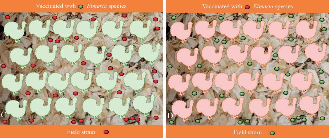

International Animal Health Journal 25 www.international-animalhealth.com RESEARCH AND DEVELOPMENT
Figure 10. A, B. Protection against particular Eimeria species included in the vaccine
Figure 10. C, D. No cross protection against other Eimeria species
Figure 10. E, F. Protection offered only against the Eimeria species included in the vaccine. No cross protection between different Eimeria species.
RESEARCH AND DEVELOPMENT
Vijay Durairaj

field strains plays an important role in understanding the field pressure. Selecting a suitable coccidiosis control method and program plays a crucial role in coccidiosis management in turkeys.
REFERENCES
1. Cervantes HM, McDougald LR, & Jenkins MC. Coccidiosis. In D.E. Swayne (Ed.), Diseases of Poultry, 14th ed., (pp. 1193-1216). 2020. Ames, (IA): Wiley-Blackwell.
2. Chapman HD. Biochemical, genetic and applied aspects of drug resistance in Eimeria parasites of the fowl. Avian Pathol. 1997. 26:221-244.
3. Chapman HD. Coccidiosis in the turkey. Avian Pathol. 2008. 37:205-223.
4. Chapman HD, Roberts B, Shirley MW, Williams RB. Guidelines for evaluating the efficacy and safety of live anticoccidial vaccines, and obtaining approval for their use in chickens and turkeys. Avian Pathol. 2005.34:279-290.
5. Clark, SR and L. Frobel. Current Health and Industry Issues Facing the US Turkey Industry. Proceedings 126th Annual Meeting of the USAHA, Virtual; Committee on Poultry and Other Avian Species. Presented Oct 3, 2022.

6. Dalloul RA, Lillehoj HS. Recent advances in immunomodulation and vaccination strategies against coccidiosis. Avian Dis. 2005.49:1-8.
7. Duff AF, Briggs WN, Bielke JC, McGovern KE, Trombetta M, Abdullah H, Bielke LR, Chasser KM. PCR identification and prevalence of Eimeria species in commercial turkey flocks of the Midwestern United States. Poult Sci. 2022. 101(9):101995.

8. Durairaj V, Vander Veen R, Clark S. Understanding highly pathogenic Eimeria species in turkeys. Durairaj V, Vander Veen R, Clark S. Understanding highly pathogenic Eimeria species in turkeys. Poultry World. 2023. Vol 39. No. 5; P 22-24.

9. Gadde UD, Rathinam T, Finklin MN, & Chapman HD. Pathology caused by three species of Eimeria that infect the turkey with a description of a scoring system for intestinal lesions. Avian Pathol. 2020. 49:80-86.
10. Lund, E.E, & Farr, M.M. Coccidiosis of the turkey. In H.E. Biester & L.H. Schwarte (Eds.), Diseases of Poultry, 5th ed., (pp. 10881093). 1965. Ames, IA: Iowa State University Press.
11. Martins RR, Silva LJG, Pereira AMPT, Esteves A, Duarte SC, Pena A. Coccidiostats and Poultry: A Comprehensive Review and Current Legislation. Foods. 2022. 7;11:2738.
12. Rose ME. Immunity to Eimeria infections. Vet Immunol Immunopathol. 1987. 17; 333-343.
Dr. Durairaj is a board-certified poultry veterinarian with more than 15 years of experience in working with poultry health, diagnostics and vaccines. He received his BVSc degree from Veterinary College and Research Institute- Namakkal, Tamil Nadu Veterinary and Animal Sciences (TANUVAS) University, India. He received his MS degree from the Center of Excellence in Poultry Science, University of Arkansas, USA. He earned his PhD degree from Poultry Diagnostic and Research Center, The University of Georgia, USA. Dr. Durairaj joined Huvepharma, Inc., USA in 2020 and currently working as a Clinical Research Manager. Previously, he worked in Boehringer Ingelheim Animal Health, USA for five years and held various positions in R&D. Dr. Durairaj has demonstrated vast experience in poultry vaccines and pharmaceutical industry by leading viral, bacterial and parasitic projects in chickens and turkeys. Dr. Durairaj has successfully conducted several research, clinical and field studies for licensing poultry vaccines. Dr. Durairaj research interests involve identifying potential vaccine candidates, develop novel vaccine strategies and therapeutics to prevent poultry diseases.
Corresponding author: vijay.durairaj@huvepharma.us
Ryan Vander Veen
Dr. Vander Veen has more than 15 years of experience in the animal health industry. He is currently the Research and Development Director at Huvepharma, Inc. Previously, he has worked for Boehringer Ingelheim Animal Health, Zoetis, and other animal health companies and has held various positions in R&D, manufacturing, and diagnostics. He received his BS degree in Microbiology from South Dakota State University and his PhD in Immunobiology from Iowa State University. Ryan has vast experience in livestock vaccine development and licensure in multiple species.
Steven Clark
Dr. Steven Clark, Veterinary Professional services Manager, Turkeys is a poultry veterinarian with over 30 years of industry experience including live production, pharmaceuticals and nutrition. Dr. Clark joined Huvepharma in 2020, specialising in turkeys, providing technical support to producers and industry, and supporting the Huvepharma portfolio of biologics and medicinal feed additives and water solubles. Dr. Clark has authored 13 peer reviewed publications and numerous technical bulletins and lay journal articles and presentations. His speciality is turkey production and anticoccidial programs and intestinal health. NC State University (DVM0 (1989), Poultry Science (19870), Internship (1990), Board Certified Diplomate (ACPV) (1997), PAACO Certified Poultry Welfare Auditor (2007), Turkey Course Master – World Veterinary Education Production Animal Health (WVEPAH, 2016)
Email: steven.clark@huvepharma.us
Volume 10 Issue 2 26 International Animal Health Journal
Figure 10. G. Protection offered by the vaccine against field strains
VISIT DRUG DISCOVERY 2023: DIVERSE THINKING – NEW PERSPECTIVES
JOIN US IN THE TECH THEATRE FOR SUSTAINABILITY PRACTICES IN DRUG DISCOVERY RESEARCH
18TH & 19TH OCTOBER 2023
ACC LIVERPOOL EXHIBITION CENTRE
To find out more and to register please scan the QR code or visit elrig.org
ADVANCEMENTS IN CELL & GENE THERAPY: NEW THERAPEUTIC HORIZONS FORUM


15TH SEPTEMBER 2023
BIOZENTRUM UNIVERSITY OF BASEL
To find out more and to register please scan the QR code or visit elrig.org.
International Animal Health Journal 27 www.international-animalhealth.com
elrig elrig_uk @ELRIG_UK elrig.org
Is Regenerative Medicine the Answer to Canine Osteoarthritis?
Veterinarians across the globe are constantly being challenged to improve their treatments for diseases such as osteoarthritis, but what criteria should they use to make decisions between promising new treatments and tried and tested practices? In recent years, regenerative medicine has been lauded as a ‘cure’ for osteoarthritis and many other inflammatory diseases but also labelled as the ‘new snake oil’.1 Which is true and how do we know?
Osteoarthritis is a progressive, inflammatory disease that affects approximately one in five of all domestic dogs.2,3 Current treatments rely primarily on symptom relief using non-steroidal anti-inflammatory drugs,4,5 weight loss programmes and nutraceuticals such as chondroitin sulphate and glucosamine. None of these prevents the progression of the disease so that ultimately surgery becomes the only option. This is expensive, traumatic for the dog and not always effective, especially in the case of elbow osteoarthritis. It would therefore be of great benefit to veterinarians and their patients if regenerative treatments were effective.
What is Regenerative Medicine?
Regenerative medicine has been defined as ‘the branch of medicine that develops methods to regrow, repair or replace damaged or diseased cells, organs or tissues’.6 Living organisms have their own inbuilt ability to regenerate tissue after damage. Natural wound healing involves cells from the immune system, mesenchymal stromal cells, growth factors and anabolic cytokines and scaffolds such as fibrin clots in a coordinated sequence of repair. Regenerative medicine uses various combinations of these factors to encourage healing in areas that are not able to heal fully. The main sources of these factors are blood (as a source of platelet rich plasma), bone marrow and adipose tissue (as a source of mesenchymal stromal cells).
Growth Factors
Platelet Rich Plasma (PRP)
PRP is a useful source of growth factors contained within the platelets and also contains fibrinogen which will clot upon injection to form a tight mesh of fibrin fibres entrapping and activating the platelets. This provides a natural scaffold which releases growth factors and paracrine signals that act as chemo-attractants to migratory cells such as mesenchymal stem cells.
PRP can be prepared from an anti-coagulated blood sample. There are many PRP preparation systems available on the market: one uses a selective filtration mechanism but most use a simple two-stage centrifugation which allows the removal of most of the red and white blood cells and leaves a platelet concentrate suspended in plasma. The relative concentrations of platelets and other blood cells vary considerably according to which system is used to prepare the PRP7,8 which could go some way to explaining the variability of clinical results reported for the use of PRP to treat osteoarthritis.9,10,11 Whilst the Cook and Fahey studies both found significant improvements in OA compared with the placebo control, the Franklin study did not find any significant improvements. There are many more less well controlled
studies and case series that have been published, but until a consistent method of PRP preparation and growth factor level estimation has been established, it will be difficult to ascertain whether PRP on its own is sufficient to treat OA effectively.
There is an increasing trend to use PRP in conjunction with mesenchymal stem cells (MSC) to treat OA. This combination has the advantage of implanting large numbers of MSCs within a growth factor rich scaffold and should therefore prime the MSCs for action.
Mesenchymal Stromal Cells (MSCs)
MSCs are a type of multipotent adult stem cell that is derived from tissues such as bone marrow, adipose tissue and other tissues of mesodermal origin. They are most abundant in adipose tissue, making up 1–5% of all the nucleated stromal cells (excluding adipocytes) extractable from canine adipose tissue.12,13,14 They have regenerative, antiinflammatory, immunomodulatory and trophic functions15 but due to their lack of telomerase activity, they do not undergo transformation even after prolonged culture expansion.16 MSCs are capable of differentiating into various mesodermal cell types such as chondrocytes, osteocytes and adipocytes, but it is their paracrine ability that is now thought to be key to their anti-inflammatory activity.17 They are able to secrete a wide range of bioactive factors that allow them to be attracted to the site of injury, reduce pain and inflammation and contribute directly to tissue repair18 and have now been shown to adapt their secretory activities according to their changing environment.19 It is no wonder that there are now more than 500 listed clinical trials involving MSCs on the US National Institutes of Health clinical studies website (http:// clinicaltrials.gov)!
MSCs can be prepared from the animal that is to be treated (autologous) or harvested from one animal and used in another of the same species (allogeneic).

Volume 10 Issue 2 28 International Animal Health Journal RESEARCH AND DEVELOPMENT
Canine MSCs in culture
Autologous Stem Cell Procedure
Autologous MSCs
Veterinary medicine most commonly uses autologous MSCs because of the perceived risk of immune rejection and, in the UK and many other countries, allogeneic cell implantation requires either an animal test certificate (ATC) or full market authorisation (MA) as the product is considered to be a drug. In the US, an investigative new animal drug application (INADA) has to be in place before culture expanded autologous or allogeneic MSCs can be administered to animals. For this reason, in the US, it has become of particular interest to regenerative medicine companies to develop patient-side preparation devices that separate the stromal vascular fraction (SVF) cells from adipose and bone marrow aspirate concentrate (BMAC) without the need for culture expansion. These preparations are not considered to be ‘more than minimally manipulated’ and therefore are not controlled by the FDA. Unfortunately, SVF contains a very small proportion of MSCs and a very large proportion of other cells, including 20% endothelial cells, 45% hematopoietic cells,20 and a large amount of cellular debris and collagen.14 BMAC has an even lower proportion of MSCs, estimated to be less than 0.01% of the nucleated cells in the preparation.21 Given that the proportion of MSCs is so low in these preparations, they should not be considered to be ‘stem cell treatments’ and should be used with caution, especially since it would not be possible to test the injectable product for microbial contamination within the short time frame.
Culture expanded autologous MSCs, on the other hand, can be prepared by authorised veterinary cell culture labs in the UK (under the Veterinary Medicines Directorate ESCC authorisation). The great advantage of culture expansion is that the MSCs can be prepared as doses of many millions of cells per joint and any of the original hematopoietic and endothelial cells will have been washed away before the final cell harvest. Further to this, the cells can be cryopreserved, a sample can be tested for microbial contamination, and a final available cell count can be made before supply to the veterinarian ready for implantation.
Autologous culture expanded MSCs are generally supplied as minimally expanded cells because they only need to be
expanded enough to treat one animal. Allogeneic culture expanded MSCs have to undergo many more population doublings before being prepared for implantation because they have been derived from only one original sample and are required to treat many thousands of animals.
Allogeneic MSCs
Allogeneic MSCs have the considerable advantage that they are ‘off the shelf treatments’. If an animal is injured, it will be possible to implant a ready-made MSC preparation without having to take a sample of adipose tissue or bone marrow and without having to wait while the cells are culture expanded. There are not yet any market-authorised canine allogeneic MSC treatments available in the UK, EU or US but there is now an equine allogeneic MSC treatment that is manufactured by Global Stem Cell Technology in Belgium.
Because MSCs are considered to be immune privileged cells, it is assumed that it is not necessary to match the recipients but there have been some studies that have found transient inflammation22 and adverse reactions following repeated intra-articular injection of allogeneic MSCs in horses.23 It is therefore not yet certain whether repeated injection of allogeneic MSCs will be possible in the treatment of osteoarthritis.
What is the Clinical Evidence of Efficacy?
The gold standard of clinical trials is a randomised, placebocontrolled study with sufficient animals in both groups to provide significant differences. One such study, treating canine OA using a single intra-articular injection of allogeneic adipose derived MSCs compared with saline as the placebo has been published by VetStem.24 The study aimed to establish both safety and efficacy of the treatment and was successful in both aims, with significant clinical improvements recorded by both validated owner questionnaires and veterinary global outcome measurements. Recently a similar study by Shah et al. in Australia came to similar conclusions18 and in addition showed that both the quality of life and osteoarthritis grade (measured by radiographic changes) were improved following mesenchymal stem cell treatment.
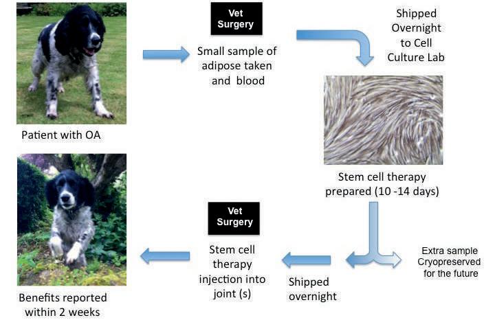
International Animal Health Journal 29 www.international-animalhealth.com RESEARCH AND DEVELOPMENT
A recent review entitled ‘Cell-Based Therapies for Animal Joint Disease’ states that ‘there are a significant number of studies that show improved functional outcomes after treatment with adipose derived MSCs for naturally occurring canine coxofemoral, cubital and scapulohumeral joint OA’.25 The studies she refers to are placebo-controlled, blinded and randomised, and include large enough animal numbers for the statistical analyses to be meaningful.26,27,12,24 Bogers also notes that the therapies have shown a ‘large effect size on lameness’ measured both objectively and subjectively with client satisfaction questionnaires. Further evidence of the cartilage regenerative effects of MSCs has recently been published by Li et al., 28 in which a beagle model of cartilage damage was treated with either MSCs in hyaluronic acid (HA), HA alone or saline. The dogs were sacrificed after 28 weeks and extensive histology, immunocytochemistry and MRI measurements showed that the MSC group had significant improvements in cartilage defects compared with the other two groups. There are also many other similar studies which do not include placebo controls but nonetheless contribute to the increasing body of evidence that canine OA can be effectively treated by intra-articular injection of MSCs.18,29
The total regenerative medicine market (human as well as veterinary) is predicted to be worth $38.7 billion by 2021 and is expanding at a rate of 23.6% per annum, it seems unlikely that this is all in pursuit of ‘snake oil’.

REFERENCES
1. Jeffery, Nicholas D., and Nicolas Granger. "GUEST EDITORIAL: Is ‘Stem Cell Therapy Becoming 21st Century Snake Oil?" Veterinary Surgery 41.2 (2012): 189-190.
2. Hielm-Björkman, Anna K., Amy S. Kapatkin, and Hannu J. Rita. "Reliability and validity of a visual analogue scale used by owners to measure chronic pain attributable to osteoarthritis in their dogs." American journal of veterinary research 72.5 (2011): 601-607.
3. Vainio, Outi. "Translational animal models using veterinary patients–An example of canine osteoarthritis (OA)." Scandinavian journal of pain 3.2 (2012): 84-89.
4. Aragon, Carlos L., Erik H. Hofmeister, and Steven C. Budsberg. "Systematic review of clinical trials of treatments for osteoarthritis in dogs." Journal of the American Veterinary Medical Association 230.4 (2007): 514-521.
One of the difficulties when comparing the efficacy of regenerative medicine treatments is that each study uses a different combination of cells, growth factors and in some cases scaffolds. A frequently used combination is MSCs injected with PRP. This combination has shown promising results26,27,30 and is increasingly becoming the optimal approach. It has been shown that platelets release factors that recruit MSCs towards the PRP clot.31,32 MSCs are able to proliferate within PRP clots and preliminary studies have shown that they are able to maintain their biological functions and three months after implantation showed evidence of osteogenesis in patients with non-union fractures.33
Is Regenerative Medicine ‘Snake Oil’?
In the past, many veterinarians have considered regenerative medicine to be an alternative and unproven treatment that has no place in modern clinical medicine. This position has now been changed to the point that the topic is included in veterinary training and the majority of veterinary practices now offer some form of regenerative medicine alongside more traditional approaches to treating osteoarthritis. The clinical evidence of efficacy of mesenchymal stem cell treatment (with or without other scaffolds and growth factors) is mounting rapidly.
5. Brown, D. C., R. C. Boston, and J. T. Farrar. "Comparison of force plate gait analysis and owner assessment of pain using the canine brief pain inventory in dogs with osteoarthritis." Journal of Veterinary Internal Medicine 27.1 (2013): 22-30.
6. https://www.nature.com/subjects/regenerative-medicine visited 10th August 2018.
7. Carr, Brittany Jean, et al. "Canine platelet-rich plasma systems: a prospective analysis." Frontiers in veterinary science 2 (2016): 73.

8. Franklin, Samuel P., Emily E. Burke, and Shannon P. Holmes. "The effect of platelet-rich plasma on osseous healing in dogs undergoing high tibial osteotomy." PloS one 12.5 (2017): e0177597.
9. Franklin, Samuel P., et al. "influence of cellular composition and exogenous activation on growth Factor and cytokine concentrations in canine Platelet-rich Plasmas." Frontiers in Veterinary Science 4 (2017): 40.
10. Cook, James L., et al. "Multiple injections of leukoreduced platelet rich plasma reduce pain and functional impairment in a canine model of ACL and meniscal deficiency." Journal of Orthopaedic Research 34.4 (2016): 607-615.
11. Fahie, Maria A., et al. "A randomized controlled trial of the efficacy of autologous platelet therapy for the treatment of osteoarthritis in dogs." Journal of the American Veterinary Medical Association 243.9 (2013): 1291-1297.
12. Black, Linda L., et al. "Effect of adipose-derived mesenchymal
Volume 10 Issue 2 30 International Animal Health Journal RESEARCH AND DEVELOPMENT
Combinations of MSCs with PRP
Canine MSCs in PRP clot
Canine MSCs in culture (CV stain)
stem and regenerative cells on lameness in dogs with chronic osteoarthritis of the coxofemoral joints: a randomized, double-blinded, multicenter controlled trial." Veterinary Therapeutics 8.4 (2007): 272.

13. Black, Linda L., et al. "Effect of intraarticular injection of autologous adipose-derived mesenchymal stem and regenerative cells on clinical signs of chronic osteoarthritis of the elbow joint in dogs." Veterinary therapeutics: research in applied veterinary medicine 9.3 (2008): 192-200.
14. Hill L, Miller J. Stem cell yields are less than 10% from Canine Stromal Vascular Fraction and further reduced after cryopreservation. In: BSAVA Congress Proceedings. Gloucester, BSAVA 2017: 557-558.
15. Spees, Jeffrey L., Ryang Hwa Lee, and Carl A. Gregory. "Mechanisms of mesenchymal stem/stromal cell function." Stem cell research & therapy 7.1 (2016): 125.
16. Zimmermann, S., et al. "Lack of telomerase activity in human mesenchymal stem cells." Leukemia 17.6 (2003): 1146.
17. Caplan, Arnold I., and Diego Correa. "The MSC: an injury drugstore." Cell stem cell 9.1 (2011): 11-15.
18. Shah, Kiran, et al. "Outcome of Allogeneic Adult Stem Cell Therapy in Dogs Suffering from Osteoarthritis and Other Joint Defects." Stem cells international 2018 (2018).
19. Lin, Paul, et al. "Serial transplantation and long-term engraftment of intra-arterially delivered clonally derived mesenchymal stem cells to injured bone marrow." Molecular Therapy 22.1 (2014): 160-168.
20. Bourin, Philippe, et al. "Stromal cells from the adipose tissuederived stromal vascular fraction and culture expanded adipose tissue-derived stromal/stem cells: a joint statement of the International Federation for Adipose Therapeutics and Science (IFATS) and the International Society for Cellular Therapy (ISCT)." Cytotherapy 15.6 (2013): 641-648.
21. Wexler, Sarah A., et al. "Adult bone marrow is a rich source of human mesenchymal ‘stem’cells but umbilical cord and mobilized adult blood are not." British journal of haematology 121.2 (2003): 368-374.
22. Ardanaz, Nekane, et al. "Inflammatory response to the administration of mesenchymal stem cells in an equine experimental model: effect of autologous, and single and repeat doses of pooled allogeneic cells in healthy joints." BMC veterinary research 12.1 (2016): 65.
23. Joswig, Amanda-Jo, et al. "Repeated intra-articular injection of allogeneic mesenchymal stem cells causes an adverse response compared to autologous cells in the equine model." Stem cell research & therapy 8.1 (2017): 42.
24. Harman, Robert, et al. "A prospective, randomized, masked, and placebo-controlled efficacy study of intraarticular allogeneic adipose stem cells for the treatment of osteoarthritis in dogs." Frontiers in veterinary science 3 (2016): 81.
25. Bogers, Sophie Helen. "Cell-based therapies for joint disease in veterinary medicine: what we have learned and what we need to know." Frontiers in veterinary science 5 (2018): 70.
26. Vilar, Jose M., et al. "Controlled, blinded force platform analysis of the effect of intraarticular injection of autologous adiposederived mesenchymal stem cells associated to PRGF-Endoret in osteoarthritic dogs." BMC veterinary research 9.1 (2013): 131
27. Cuervo, Belen, et al. "Hip osteoarthritis in dogs: a randomized study using mesenchymal stem cells from adipose tissue and plasma rich in growth factors." International journal of molecular sciences 15.8 (2014): 13437-13460.
28. Li, Lang, et al. "Mesenchymal Stem Cells in Combination with Hyaluronic Acid for Articular Cartilage Defects." Scientific reports 8.1 (2018): 9900.
29. Armitage A, Miller J, Frost L, Standen C. Clinical Efficacy of Intra-Articular Injections of Autologous Mesenchymal Stem Cells in Moderate to Severe Osteoarthritis (OA): a Case Series of 19 Dogs evaluated using both LOAD and CBPI Owner Questionnaires. In: BSAVA Congress Proceedings. Gloucester, BSAVA 2018: 474.
30. Upchurch, David A., et al. "Effects of administration of adiposederived stromal vascular fraction and platelet-rich plasma to dogs with osteoarthritis of the hip joints." American journal of veterinary research 77.9 (2016): 940-951.
31. Holmes, Hannah L., et al. "Facilitated recruitment of mesenchymal stromal cells by bone marrow concentrate and platelet rich plasma." PloS one 13.3 (2018): e0194567.
32. Roubelakis, Maria G., et al. "Platelet-rich plasma (PRP) promotes fetal mesenchymal stem/stromal cell migration and wound healing process." Stem Cell Reviews and Reports 10.3 (2014): 417-428.
33. Wittig, Olga, Dylana Diaz-Solano, and José Cardier. "Viability and functionality of mesenchymal stromal cells loaded on collagen microspheres and incorporated into plasma clots for orthopaedic application: Effect of storage conditions." Injury (2018).
Dr. Joanna Miller
Dr. Joanna Miller is the Science Director of Cell Therapy Sciences Ltd, a company specialising in providing stem cell therapy for canine and equine orthopaedic conditions. She is a cell biologist with a commercial research background and is most interested in widening the use of effective regenerative medicine in both the veterinary and human medical worlds.

International Animal Health Journal 31 www.international-animalhealth.com RESEARCH AND DEVELOPMENT
Feed Intake Differences and Variation in the Vitamin E Status in Transition Dairy Cows
Vitamin E is important in the body for its anti-oxidant activity and a deficiency can lead to oxidative stress and immune suppression in peripartum cows. The fatsoluble vitamin (which consists of a group of eight related compounds) is usually supplemented to dry cow diets to meet requirements and supplementation has been shown to improve reproductive performance in dairy cows (Moghimi-Kandelousi et al., 2020). Differences in dry matter intake between cows in a herd may, however, lead to variation in the vitamin status of animals around parturition.
Aim
The aim of our study was to assess the association between dietary intake and plasma concentrations of vitamin E in transition dairy cows. For this research, Royal GD and Schothorst Feed Research B.V. worked together to investigate how feed intake differences around parturition can influence the vitamin status of cows in the transition period.
Materials & Methods
The study was performed at the dairy farm of Schothorst Feed Research B.V. (Lelystad, the Netherlands). Sixty cows of all parities were enrolled in the study at approximately four weeks before the expected calving date. Animals were housed in a loose housing system equipped with individual feeding places and individual daily feed intake was registered from four weeks before the expected calving date until four weeks after calving. Animals had ad libitum access to a basal diet (~5% residuals) and water. The dry cow diet consisted of grass silage, corn silage, straw, a protein supplement, and 1 kilogram of concentrates per cow per day. The basal diet for lactating cows consisted of grass silage, corn silage, pressed beet pulp, and a protein supplement. Concentrates were fed to lactating cows according to a fixed scheme based on parity and days in milk.
Roughages were sampled once a week and pooled per batch for analysis. Concentrates were sampled at delivery. Vitamin E concentrations in feedstuffs were analysed using high-performance liquid chromatography. Total vitamin E concentrations were 108 IU/kg DM in the dry cow diet and 87 IU/kg DM in the lactation diet. Plasma samples were collected at approximately one week before the expected calving date and around four weeks after parturition. Vitamin E concentrations in plasma were analysed using highperformance liquid chromatography. Feed intake data were available for 57 out of 60 cows. Prepartum and postpartum plasma vitamin E concentrations were available for 56 and 54 cows, respectively. Descriptive statistics were performed and the association between the average dietary intake of vitamin E in the three weeks preceding the week of sampling and the vitamin E concentration in plasma of cows was assessed by linear regression using separate models for prepartum and postpartum data.
Results
Descriptive data of intake and plasma parameters are shown in Table 1. There was considerable variation in the dry matter intake of transition dairy cows, even under conditions with
unlimited dietary access via individual feeding places, as was the case in our study. As a consequence of these feed intake differences, there was also quite some variation in the dietary vitamin E intake of the animals (Figure 1). Plasma vitamin E concentrations were larger at four weeks after parturition than at one week before calving (P<0.001). Dietary intake of vitamin E was significantly associated with plasma vitamin E concentrations (P<0.01) in both prepartum and postpartum cows (Figure 2).

Conclusions
Dietary intake of vitamin E was associated with plasma vitamin E concentrations in transition dairy cows. This shows that differences in dry matter intake of cows in the transition period can lead to variation in the vitamin E status in animals fed on the same diet. Maintaining feed intake of cows in the transition period is, thus, not only important to reduce negative energy balance, but also to prevent inadequate vitamine E uptake of cows around parturition.
Volume 10 Issue 2 32 International Animal Health Journal FOOD & FEED
Parameter N Average SD Median Min Max DM intake (kg/day) Prepartum (week -4 to -2) 57 14.6 2.2 14.4 10.6 19.9 Postpartum (week 1 to 3) 57 19.8 3.4 19.6 11.1 28.3 Vit. E intake (IU/day) Prepartum (week -4 to -2) 57 1545 318 1529 947 2376 Postpartum (week 1 to 3) 57 1785 452 1759 713 2906 Plasma Vit. E (µmol/L) Prepartum (week -1) 56 12.5 4.1 12.1 4.1 23.1 Postpartum (week 4) 54 14.4 3.9 14.4 6.4 21.3
Table 1: Descriptive statistics of dry matter intake (DM), vitamin E intake, and plasma vitamin E concentrations in transition dairy cows.
Figure 1: Weekly average dietary vitamin E intake in transition cows (n=57) from 4 weeks before until 4 weeks after calving.
REFERENCES
1. Moghimi-Kandelousi, M.H., Alamouti, A.A., Imani, M., Zebeli, Q. 2020. A meta-analysis and meta-regression of the effects of vitamin E supplementation on serum enrichment, udder health, milk yield, and reproductive performance of transition cows. J. Dairy Sci. 103: 6157-6166.


Saskia van der Drift
Saskia graduated as a veterinarian from Utrecht University and completed a PhD thesis on ketosis in dairy cows. She also obtained a MSc degree in Veterinary Epidemiology and Economics. From 2012, she works as senior researcher at Royal GD with focus on biomarkers and dairy cow nutrition and metabolism.
Ant Koopmans
Ant graduated as a veterinarian from Utrecht University. During the study described in this article, she worked at Schothorst Feed Research B.V. as researcher ruminant nutrition and health. From 2022, she works at Royal GD as senior veterinarian with focus on dairy cow nutrition and health.

Sanne Carp – van Dijken
Sanne graduated as a veterinarian from Utrecht University. During the study described in this article, she worked at Royal GD as senior veterinarian with focus on dairy cow nutrition and herd health.

Rianne Grotentraast

Rianne studied Animal and Livestock Farming at Aeres University of Applied Sciences in Dronten and has been working as R&D laboratory technician at Royal GD since 2012. Developing new tests for livestock farming is her passion.
Manon Holstege
Manon studied Health Sciences at VU University in Amsterdam, with a specialization in infectious diseases & public health. Since 2014, she works as an epidemiologist at Royal GD in Deventer, focussing on methodological aspects of various studies.
International Animal Health Journal 33 www.international-animalhealth.com
FOOD & FEED
Figure 2: Association between average dietary vitamin E intake in the 3 weeks prior to sampling and plasma concentrations of cows prepartum (week -1) and postpartum (week 4).
Why Good Tablet Design is Important in Animal Medicine and How to Achieve it
I Holland
Just as good tablet design is extremely important in the manufacture of pharmaceuticals for humans, the same applies for animal dosage forms. Tablets are used to deliver drugs in an effective and safe manner, and although less dominant in veterinary medicine, tablets or boluses are still a significant method to administer medication.
Getting tablet design correct has an impact upon anticounterfeiting, tooling strength, tablet coating, durability and functionality. It also helps to avoid downstream manufacturing problems such as tablet sticking, picking, lamination, capping and premature tooling failures. It is essential to consider these at the beginning of the design process, ensuring a problem-free, high-quality end product.
Several elements need to be considered when designing tablets for animals; here we will consider the most important.
Tablet Shape and Profile
The first thing to consider is the tablet shape and optimum tablet profile. There are two basic tablet shapes, round and non-round; however, the complexity of non-round shapes can be very varied and require specialised tool manufacturing capability. Boluses, which are commonly used for large animals are cylindrical shaped to prevent choking. Due to their size (typically 3 to 16 g) it is important to get the shape correct. ‘Bolus formulation poses challenges because of the high drug to-excipient ratio. Less room is left for diluent, binders, and other adjuvant needed to overcome objectionable features of the drug or to facilitate bolus manufacture.’
These higher numbers of active ingredients in animal formulations bring challenges related to particle size, flow, compressibility, moisture sensitivity, ingredient interaction, content uniformity and quality control (QC) testing. For example, some active ingredients may be available in granular form, while some may be available only in fine powder form. Because of this, the ingredient blend may have many different particle sizes and ingredients with a variety of characteristics. This variation demands the shape and profile to be correct during manufacture.
Once the base shape has been decided, tablet size must be determined. Consideration should be given to the type of press available for tablet manufacture as this can limit the size of the tablet.
Next follows selection of the tablet. The type of profile required is influenced by several factors: the granule, embossing requirements, coating process, packaging and the company’s branding.
Thought should also be given to the volume of the tablet and if it will be coated. Successful coating is dependent on tablet profile. Coated tablets, whether film- or sugar-coated,
present challenges for the tablet designer. The complexity of the coating process is vast. Many of the variables are within the manufacturer’s control but expert tablet design can help eliminate some potential problems.
Coated tablets are a popular choice in animal medicine. Administering tablets to animals is not a guaranteed process due to the uncertainty of the tablet being swallowed. Many animals also chew the tablet, exposing the disagreeable taste of the drug. For this reason, flavours or sweeteners are combined or a coating is used. ‘Tablets can be coated to differentiate the product by colour, to help mitigate offensive tasting compounds, or to prevent dusting in the bottle.’
Typically, the centre of a tablet is lower in hardness, so during the coating process core erosion may take place. This is when the tablet comes into contact with the coating pan and other tablets, causing wear. This vulnerability, caused by mechanical stress during coating, can be reduced by avoiding very deep concaves and ensuring a robust design. For filmcoated tablets, double radius profiles are the preferred choice. For shallow tablets with hard, sharp edges, the coating process will damage the exposed edge of the tablet, resulting in chipped edges and sometimes cracks. Therefore, flat and shallow tablet profiles should be avoided. Double radius designs ensure a strong tablet edge and a balanced profile, which will roll in the coating pan. Another benefit of the double radius design is that it can accommodate most marking and branding requirements, because it increases the usable surface area available for this.
Poor marking and branding design on the tablet can lead to bridging (where the coating collects in the detail on the face of the tablet because the coating does not fully follow the contours of the marking on the tablet core, but bridges over leaving a void under the coating) and infilling (when too much coating material has filled the detail, making it indistinct) during film-coating. Possible causes for this can include:
Inadequate adhesion of the film coating – the coating supplier should be consulted to improve the adhesion characteristics of the coating.
Inappropriate marking design where the angle may be too acute or too deep (bridging) – The marking should be redesigned in consultation with the tooling supplier. It can also be due to the stroke or section of the embossing being too wide or too shallow (infilling).
Inappropriate coating procedure, e.g. spray rate, drying time, etc. – the coating supplier should be consulted.
Another problem is twinning – tablets sticking together during coating. This is normally caused by the flat surfaces of the tablets coming into contact and adhering to each other. To avoid this, a slightly curved surface can be applied, which reduces the contact area and eliminates the potential for twinning.
Tablet Breakability
Good tablet design will enable the tablet to be broken easily and accurately, ensuring that when the tablet is broken,
Volume 10 Issue 2 34 International Animal Health Journal TECHNOLOGY
the required tolerance for dosage is achieved. Uneven breaking of a tablet may result in significant fluctuations in the administered dose. The degree of inaccuracy may be associated with breakline design, tablet hardness, and / or formulation.
The following factors should be considered when selecting a breakline:
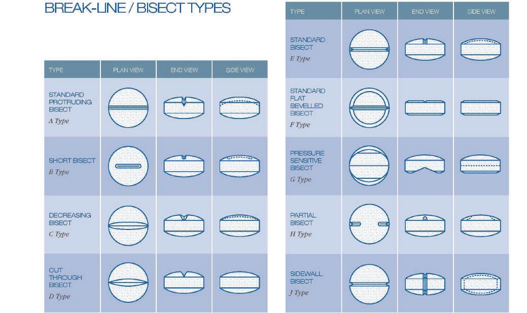
• Accuracy of breakage, which is important for equal dosage.
• Holding of the tablet and ease of breakage. This relates to tablet size and hardness.
• Inclusion of other detail, such as a logo, and its influence on the breakline. Product identification must be maintained to ensure brand integrity when the tablet is divided.
• Robustness of the tablet during compression, coating and packing. Because the tablet’s physical qualities are changed by adding a breakline, it may become weaker.
• The breakline should penetrate into the tablet whilst maintaining an optimised radius and angle. A larger radius usually makes the breakline less effective.
Where a breakline is functional and present on both sides of the tablet, alignment of the breakline on the upper and lower punch tips is critical and requires the turret to have a lower key facility. Also, pay attention to breaklines that stand above the punch tip edge. Upper and lower punches must be set correctly for effective tablet ejection and take-off. If not set correctly, damage can occur to the breakline on the punch tip, resulting in catastrophic failure and damage to the tooling and the tablet press. It can also cause severe chipping of the tablet on ejection.
Tooling Performance

Tablets are becoming more complex in both shape and profile, increasing the demand for tooling strength, durability and overall performance. This has to be a major consideration when designing tablets for veterinary medicine.
One of the most important features of any tablet design is the blended land. Often, tablet manufacturers elect not to apply a land as it may not be visually acceptable on the finished tablet. Lands that are applied incorrectly, either unevenly or made too large, can present a range of issues, including: flashing or lamination during compression; chipping of the land during take-off, or build-up of coating on the edge of the tablet which eventually will chip.
International Animal Health Journal 35 www.international-animalhealth.com TECHNOLOGY
Figure 1 – Tablet Breaklines
Figure 2
Figure 2 shows a stress and fatigue analysis of a punch tip using finite element analysis, or FEA. FEA is a softwarebased numerical technique for calculating the strength and behaviour of engineering structures. It is used to calculate deflection, stress and strain to determine fatigue limits of both material and design.
The answer is to always include a blended land as, when applied correctly, it will optimise tablet and tooling strength and performance.
Figure 3 shows the application of a blended land to a punch tip. The correct method of applying the land is to ensure that the flat area on the tip edge is maintained, whilst blending the intersection between the profile and the flat. This is achieved by applying a radius to the finished punch tip.

A correctly selected and applied blended land provides benefits to handling, loading, setting, tooling strength, the visual appearance of the tablet and ultimately, your brand.
Tablet Branding
When considering the visual appearance of the tablet, it is important to think about the type of font and logos used for branding. Typefaces and designs must take into account practicality of tablet manufacture.
Caution is required during the design process when applying branding to your tablet. Failure to consult with an expert tablet design team could result in a product that looks good on paper but is not practical to produce.
For tablets with a logo, the design and placement are very important. The tablet designer should always seek to maximise the face area to avoid picking and lack of distinction.
Figure 4 shows a good example of the importance of spacing on logos. The top example clearly shows the embossing within the safe zone for this particular tablet shape. The bottom example shows embossing that goes beyond the safe zone and, on the side view, you can see how the embossing protrudes.

Occasionally, when there is a need to exceed the safe zone, the best practice is to ensure the detail is spaced far enough away from the edge of the tip. As a general guide, the embossing should sit below the landed edge of the punch tip. If this guide is not followed then the embossing will be unprotected and prone to damage, causing further downstream problems during the tabletting process.
The correct font style is also very important to avoid tabletting problems such as ‘picking’. Picking is compressed granule that has adhered to the detail on the punch face, resulting in ‘picking out’ of parts from the tablet face.
To reduce picking, the best practice should be to design font styles that have large open counters and no sharp corners, which could act as a trap for granule. Selection of the right font style can also help to avoid coating problems, tooling failures and lack of distinction.
When the font has been chosen, it is important to ensure clarity of definition. The profile of the embossing is equally important to reduce picking, and ensure good tablet coating and tooling strength.
Volume 10 Issue 2 36 International Animal Health Journal TECHNOLOGY
Figure 3 – Blended land
Figure 4
The best practice is for the stroke angle to be between 70 and 80 degrees. The stroke depth should be 50% of the width and the stroke break radius blends 30% of the stroke depth (Figure 5).
Another technique used to minimise picking is a reduced counter. The counters, which are sometimes referred to as islands, are very vulnerable to picking and granule can easily get trapped in these areas on the punch tip face. The counter is modified to increase the surface area by reducing stroke depth, which will minimise the tendency for the product to pick.
‘Sticking’ of course is another major issue in the design and manufacture of tablets. Sticking differs from picking in that it is granule adherence to the punch tip face, rather than in and around the embossing. This is not normally associated with design, however when picking occurs on a tablet this in turn can result in sticking on the punch tip face by providing a key to which further granule will adhere.
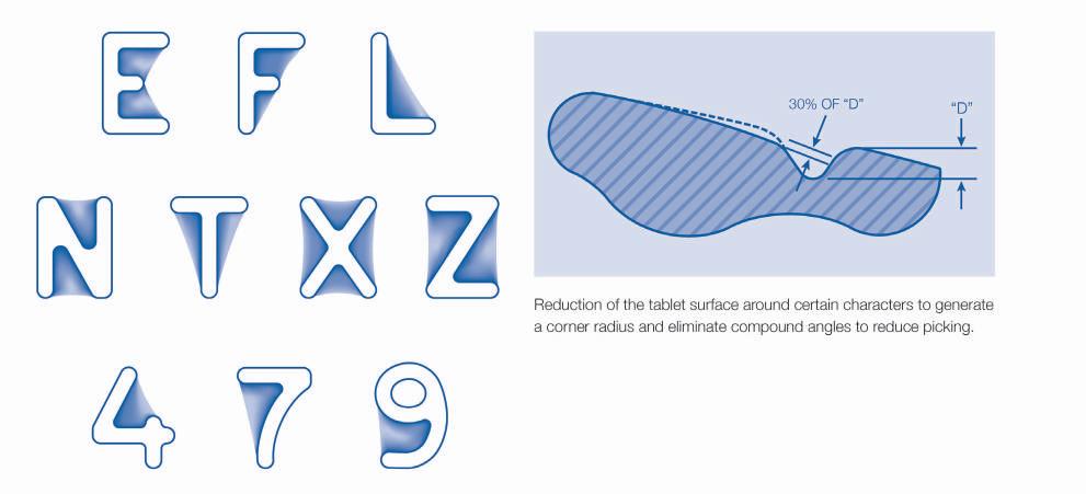
Anti-counterfeiting Techniques
As counterfeiters become more technologically advanced, basic tablet designs are more easily reproduced.
There are many anti-counterfeiting technologies available to manufacturers. Within tablet manufacture, this is usually done by applying anti-counterfeiting features directly to the tablet.
An expert tablet designer can employ techniques to make this more difficult. These are not always visible to the naked eye but ensure that a branded tablet can be identified as an original.
Several anti-counterfeiting techniques can be used on each product, to help reduce the risk. For example, altering the thickness of the embossing in places, changing the angle of the lettering, or simply having the logo on different inclines. Although hard to see with the naked eye, expert tablet designers can see the difference between the original and the counterfeit.

International Animal Health Journal 37 www.international-animalhealth.com TECHNOLOGY
Figure 5
Figure 6
Conclusion
Good tablet design is imperative, whether it is for human or animal medicine, and it is something that should be strongly considered. It is important to consult with an expert tablet designer as early on in the process as possible, who can ensure that designs of tablet or boluses are not only unique and visually appealing, but are also robust and producible in a rigorous tablet manufacturing environment. By making just a few simple changes to a design it can stop future problems, from picking and sticking to counterfeit issues.
The importance of design should not be underestimated. Punches and dies are the most critical interface with your end product, the tablet, and together everything should be measured and taken into account before tablet production.
REFERENCES
1. Veterinary Pharmaceutical Dosage Forms: A Technical Note - Ramteke KH*, Joshi SA, Dighe PA and Kharat AR
2. Pharmaguideline.com

3. J Marriott & R Nation, Department of Pharmacy Practice, Victorian College of Pharmacy, Monash University, Melbourne.
4. Protecting your Brand with Anti-Counterfeiting Solution –John Mack

5. Eurostandard Educational Collection, 2010 – I Holland Ltd
Alex Bunting
Alex manages the marketing team at I Holland, is a graduate of English and member of the Institute of Digital Marketing. He joined I Holland in April 2008 having spent the previous years working in Environmental Science. Alex was instrumental in the design of the 2010 edition of the widely adopted Eurostandard, educational animations and hosts I Holland’s extensive webinar program.
Volume 10 Issue 2 38 International Animal Health Journal
TECHNOLOGY
December
12th & 13th, 2023
MARSEILLE

December 15th, 2023
ONLINE MEETINGS
Inside the event:
• One-on-one meetings
• Conferences
• Pitch sessions
• Exhibition
• Project - CEO matchmaking
1,000+ delegates
12 TH EDITION
Register now and benefit from 10% off the full pass with the code: BF23_IAHJ_2706
Applied on the regular fee, from July 21st
The
35+ countries represented
www.biofit-event.com


@BIOFIT_EVENT
9TH EDITION
October 17th & 18th, 2023 Rennes, France
Register now and benefit from 10% off the full pass with the code: NE23_IAHJ_2706

Applied on the regular fee
delegates
650 25+ countries represented
INSIDE THE EVENT
One-on-one meetings
Conferences
Pitch sessions
Exhibition
International Animal Health Journal 39 www.international-animalhealth.com
The leading European partnering event for early-stage deals and investment rounds in the Life Sciences field
| @NutrEvent
European meeting place for innovation in Food, Human and Animal Nutrition
www.nutrevent.com
I hope this journal guides you progressively, through the maze of activities and changes taking place in the animal health industry.

IAHJ is also now active on social media. Follow us on:
www.twitter.com/AHMJournal www.facebook.com/Animal-Health-Media www.plus.google.com/+Animalhealthmediajournal www.animalhealthmedia.tumblr.com/
Volume 10 Issue 2 40 International Animal Health Journal AD INDEX Page 3 Astell Bio Page 39 BIOFIT
27 ELRIG
Page 5
Page 39
Subscribe today at www.international-animalhealth.com
IBC CPHI Page
BC Moredun Scientific
Nordson
NutrEvent Page 7 PCI Pharma Services IFC Royal GD
or email info@senglobalcoms.com
24-26 October 2023

Exclusive Entry Into The Global Pharma Market
Barcelona, Spain Book

41 International Animal Health Journal At the heart of Pharma
Stand Now
Your
Pharma’s largest event is making an exciting return to Barcelona on 24-26
SCAN ME
October 2023.
Animal health contract research
Expert delivery of contract studies supporting product development and commercialisation.

Efficacy studies
• Broad portfolio of infectious disease models
• New model and protocol development
• VICH-GCP compliant studies
• Studies in all species of livestock
• Aquaculture studies
Target animal safety testing
• GLP compliant studies
• All species of livestock
• Aquaculture studies

• All types of biological & pharmaceutical products
Facilities
• GLP accredited animal accommodation
• Conventional farm accommodation
• Category 3 containment
• Specific Pathogen Free
• Gnotobiotic units
• Aquaculture Testing Facilities (Seawater and Freshwater)
• GLP accredited laboratory facilities
www.moredun-scientific.com
Volume 10 Issue 2 42 International Animal Health Journal
Moredun Scientific, Pentlands
Park, Bush Loan, Penicuik EH26 0PZ, Scotland, UK Tel: +44 (0)131 445 6206 Email:
us
For further information please visit www.moredun-scientific.com or contact:
Science
info@moredunscientific.com Follow
