Phoma Leaf Spot Phoma leaf spot is caused by the seedborne pathogen Phoma betae and commonly affects sugar beet (Fig. 12), table beet (Fig. 13), fodder beet, and chard, in addition to oat and Cheno podium album (lambsquarters). The same fungus also induces seedling disease problems in sugar beet and table beet. It also can persist in the crowns, and it may cause root rot and sugar beet storage rot in harvest piles later in the season and a type of crown rot of mature roots. Although occurring almost everywhere these different beet types are grown, Phoma leaf spot is of little economic importance, except as a source of inoculum for infections of seed stalks and seed clusters.
Symptoms
Individual leaf spots are usually light brown and round to oval (1–2 cm in diameter) and have dark, concentric rings near the perimeter (Fig. 14) as opposed to the smaller lesions with solid centers associated with Cercospora leaf spot (see Foliar Diseases Caused by Fungi and Oomycetes; Fig. 4). Small, spherical, black pycnidia develop in the dark rings, and conidia are produced within the pycnidia. On seed stalks, brown to
black necrotic streaks form and develop grayish, pycnidiabearing centers.
Causal Organism
P. betae is an imperfect fungus and is the most commonly found form in nature. P. betae produces pycnidiospores (conidia) in black, ostiolate, lenticular to globose pycnidia that are immersed in host tissue. The conidia (2.6–4.9 × 3.8–9.3 µm; sizes vary greatly because of environmental influences) are hyaline, one celled, and oblong. The perfect stage, Pleospora bjoerlingii (Fig. 15), develops in the autumn under lesion surfaces. Perithecia (160–205 × 230–340 µm) are black and somewhat hemispherical. Asco spores (8.5–10 × 19.5–25 µm) are pale yellow-green and usually muriform. Further descriptions of the pathogen are found in other sections (see Seedling Diseases, Phoma Root Rot, and Postharvest Deterioration of Sugar Beet).
Disease Cycle and Epidemiology
The fungus is seedborne and can survive as conidia in pyc nidia and as mycelium in soil and crop residue for up to 26 months after a beet crop has been harvested. During moist weather, the pycnidia exude a cirrhus (gelatinous mass of spores) from which spores are then dispersed mainly by splashing rain (Fig. 16). When the fungus is present in its perfect stage, ascospores produced in the perithecia are primarily carried to beet foliage by wind. The disease is most severe during periods of high humidity and temperatures of 15–32°C (Fig. 12).
Fig. 12. Severe Phoma leaf spot infection in a sugar beet field. (Courtesy R. M. Harveson)
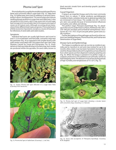
Fig. 14. Phoma leaf spot of sugar beet showing a circular lesion with concentric rings. (Courtesy L. E. Hanson)
Fig. 13. Phoma leaf spot of table beet. (Courtesy L. J. du Toit)
Fig. 15. Ascus and ascospores of Pleospora bjoerlingii. (Courtesy W. M. Bugbee)
11