
3 minute read
Sabri Ülker Advanced Imaging Lab


SOLVING STRUCTURAL AND ARCHITECTURAL QUESTIONS
(at the molecular scale)
Advertisement
The establishment of the Sabri Ülker Advanced Imaging Lab has given us power to visualize how metabolically critical cells and their organelles, such as endoplasmic reticulum and mitochondria, adapt to different nutritional, hormonal, and stress states.
Imaging is a vital component of life sciences research that allows scientists to observe and quantify the intracellular and intercellular events at various space and time scales. The Sabri Ülker Advanced Imaging Lab (SAIL) possesses instrumentation that was acquired with the generous support of the Ülker family. Their financial support has enabled us to image live and fixed biological samples, such as cell lines and metabolic tissues, under different nutritional conditions—all in high resolution. We are able to capture information such as cell morphology, lipid content, organelle abundance, distribution, localization, motility, and interaction. More specifically, we have imaged hepatocytes, adipocytes, and pancreatic cells and were able to determine the effect of nutritional stress in endoplasmic reticulum (ER) and mitochondria dynamics and interaction in high spatio-temporal resolution. Additionally, we were also able to measure intracellular calcium dynamics in the different cellular compartments such as cytosol, mitochondria, and ER. These measurements enabled us to discover a dysfunction in calcium signaling in cells such as hepatocytes that is a key problem that occurs in obesity.
The SAIL enables collaborations with other research programs that would not otherwise be possible. For example, Dr. William Mair’s lab studies the effect of aging in organelle dynamics in cellular models and also in Caenorhabditis elegans (C. elegans), a free-living transparent nematode about 1.5 mm in length. The microscopic capacity offered by our imaging lab created new research opportunities for his team. Additionally, our imaging technology capacity will allow us to collaborate with new faculty members, such as Dr. Nora Kory, who is now affiliated with the Sabri Ülker Center. She will be utilizing high-resolution microscopy to ask interesting questions that explore mitochondria form and function in aging and metabolic diseases. Previous work in the Hotamışlıgil Lab has shown that within liver cells, changes to the structure and function of the ER are key events in adapting to metabolic stress. We are now investing in even higher capacity and resolution instrumentation, which will give us tremendous power to visualize how the organelles in liver cells adapt to different nutritional and hormonal states, such as fasting and feeding, by reorganizing its structure. Drs. Ana Paula Arruda, Güneş Parlakgül and Hatoon Baazim are now embarking on similar analysis on one of the most challenging of cells, the adipocytes, and the adipose tissue to explore their subcellular architecture and its regulation under metabolic and nutritional challenges. We are also working and planning to further expand our imaging, computational, artificial intelligence, machine learning, and visualization capacities in the near future.
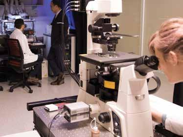
(Top) The imaging platforms at the Sabri Ülker Advanced Imaging Lab allow scientists to study the molecular events in cells and tissues over time. Live cell imaging enables visualizing the localization, interaction, and abundance of molecules such as proteins and lipids during metabolic processes. (Bottom) This microscope is equipped with a fluidic injection/ pump system for delivering various chemicals and agents during imaging. It allows us to determine calcium dynamics in the cells.
Cos-7 cell showing endoplasmic reticulum (in red), mitochondria (in green), nucleus (in blue), and peroxisomes (in purple). This cell line has been widely used to study organelle communication and other cell biology questions regarding subcellular organization.


