
Can we improve head and neck radiotherapy? REMOVE THE MASK COMPARISON STUDY Mince pies – a doubleblind randomised control trial BIG DEBATE Our panel discuss clinical risk management SURVEY Treatment of brain metastases with stereotactic radiosurgery WORKFORCE Racial healthcare inequalities and the NHS Volume 33 | Issue 4 | WINTER 2022 ipem.ac.uk


A time of reflection
Usman Lula outlines the content in the latest issue of Scope, from an evaluation of the medical physicist role to a mince pie study.


Welcome to the final issue of Scope for 2022. The end of the year is often a time for reflection, so we thought it would be the perfect opportunity to critically evaluate the role of medical physicists and ask what changes may be needed for a sustainable future. After a very useful and interesting exchange with James Clark Ross, he has supplied a brilliant piece on just this – “Scaling back and modernising the medical physicist”. He asks some fundamental questions around the work we do – and I totally agree – how much of our work pertains to science (rather than just practice)? How much value would we add to our role if we were to add more science to our work, realised through a top-down management approach? Interested? Turn to p44.
Lately, I’ve been sitting down with our Scientist Training Programme (STP) students and discussing the new changes to the STP syllabus. Rather than perform a series of practical activities to achieve competency, they are now required to provide reflective pieces based on their experiences in various clinical areas, pathways, meetings and dialogue with staff members. As I was talking through the process, we took the “radiotherapy mould room experience” as an example for reflective writing. And in thinking about the deeper levels of reflective writing, the cover piece on “Remove the Mask” (kindly supplied by
Paul Doolan) really helped the students appreciate how we could focus our requirements to include the patient needs and what we could do to improve the patient experience. After all, the patient is at the heart of everything we do in the NHS. My next topic to consider with them will be the “patient-specific verification” piece in this issue (thanks to Dan Johnson) – not just the fact about why, what and how we do this, but to consider the wider questions around such activities.
With the seasonal celebrations coming up we may ask ourselves how the iconic pudding from the Middle Ages has come to play such a central role. Or you may be looking forward to biting into your favourite mince pie and wondering how you can be sure the mince pie you plan on eating is the tastiest. Well, to round off a year of Scope issues, Azzam Taktak kindly supplied the perfect feature in this issue on comparing mince pie brands to statistically establish which has the best taste.
There are lots of other fantastic features to whet your appetite during the festive period. So, take your pick, sit back, relax and enjoy. Happy 2023.
Strategic aims for Scope magazine
I’d like to thank the readers, the IPEM Office, the Redactive publishing team and the Scope Commissioning Board for feature contributions and continued support in
developing Scope magazine. We now have two strategic aims for Scope:

1. Improving the quality, variety and balancing of feature content – which is
by and large being worked on and developed with each issue. We are working on collating a list of conferences for the different IPEM areas so that
we can contact speakers to include cutting-edge and innovative features.
2. Improving reader engagement – we are currently thinking about
ways in which we could improve this.
We are hoping to conduct a Scope magazine reader survey in the new year, so please watch this space.
U F UPFRONT COMMENT IPEM.AC.UK 3 WINTER 2022 IPEM.AC.U K
Usman Lula Chair of the IPEM Scope Magazine
CONTENT
CHAIR OF IPEM SCOPE EDITORIAL ADVISORY BOARD
How much value would we add to our role if we were to add more science to our work , realised through a topdown management approach?
FEEDBACK
Discuss, debate, share. mycommunity.ipem.ac.uk/login
WEBSITE

ARCHIVES

IPEM SCOPE 4 WINTER 2022
News, events, support. ipem.ac.uk
CO Scope is the quarterly magazine of the Institute of Physics and Engineering in Medicine
Fairmount House, 230 Tadcaster Road,
1ES
|
|
|
of IPEM Scope Editorial Advisory Board: Usman I. Lula Principal Clinical Scientist, 1st Floor, Radiotherapy, Building, Medical Physics – University, Hospitals Birmingham NHS Foundation Trust, Queen Elizabeth Hospital, Queen Elizabeth Medical Centre, Birmingham, UK B15 2TH 0121 371 5056 | usman.lula@uhb.nhs.uk Commissioning Editor Ejay Nsugbe Commissioning Editor Clara Ferreira Commissioning Editor Natasa Solomou Commissioning Editor: Usman I. Lula 0121 371 5056 | usman.lula@uhb.nhs.uk Commissioning Editor: Dr Paul Doolan Medical Physicist, German Oncology Center,
Avenue, 4108 Agios Athanasios, Limassol, Cyprus
8025 | paul.doolan@goc.com.cy
is published on
of the
in
(IPEM) by
2728
Advertising
| +44 (0)20 7880 7556 Scope is published quarterly by the Institute of Physics and Engineering in Medicine but the views expressed are not necessarily the official views of the Institute. Authors instructions and copyright agreement can be found on the IPEM website. Articles should be sent to the appropriate member of the editorial team. By submitting to Scope, you agree to transfer copyright to IPEM. We reserve the right to edit your article. The integrity of advertising material cannot be guaranteed. Copyright: Reproduction in whole or part by any means without written permission of IPEM is strictly forbidden. © IPEM 2022. ISSN 0964-9565 Beyond training more of our colleagues in clinical risk management, it’s important to start applying the standards to our day-to-day work in order to build up experience.
page 16 UPFRONT 03 / CHAIR’S COMMENT 07 / NEWS 10 / IPEM NEWS 12 / IPEM'S SCIENCE, TECHNOLOGY AND ENGINEERING FORUM 14 / POLICY UPDATE 16 THE BIG DEBATE 16 / CLINICAL
Our panel look at DCB0129 and DCB0160 – mandatory risk management standards for England – and look at the implications, from the level of experience needed to be a clinical safety officer, to the possibilities and impacts of outsourcing. 29
Back issues of Scope online. ipem.ac.uk/scope
IPEM
York, YO24
T: 01904 610821
F: 01904 612279 office@ipem.ac.uk
ipem.ac.uk
Chair
1 Nikis
00357 2520
Scope
behalf
Institute of Physics and Engineering
Medicine
Redactive Publishing Ltd redactive.co.uk Publisher: Tiffany van der Sande tiffany.vandersande@redactive.co.uk | +44 (0)20 7324
Editor: Rob Dabrowski Design: Glen Wilkins, Joe McAllister Picture researcher: Claire Echavarry Production: Aysha Miah-Edwards aysha.miah@redactive.co.uk | +44 (0)20 7880 6241
sales: scope@redactive.co.uk
– Claire Tarbert, Principal Clinical Scientist, Medical Devices Unit
RISK MANAGEMENT
NTENTS
GENERAL
20
/ REMOVE THE MASK
We hear from a patientinitiated project at the Image X Institute in Australia that aims to make head and neck radiotherapy a less harrowing experience for patients.
24 / A HISTORY OF RADIOTHERAPY TRIALS QUALITY ASSURANCE
Edwin GA Aird looks back over the beginnings of interdepartmental audit.
29 / TREATMENT OF BRAIN METASTASES WITH STEREOTACTIC RADIOSURGERY
Principal Stereotactic Radiosurgery Radiotherapy Physicist Anna Bangiri looks at the processes and procedures across the country.
34
/ PATIENT-SPECIFIC VERIFICATIONS
How many passing results do we need before we have the confidence in beam models to calculate dose and linacs to deliver it accurately?
48 / RACIAL HEALTHCARE INEQUALITIES AND
Elisa Ly and Sara Majid look at inequality in the health service and ask what action is needed.
ENDNOTES
54 / BOOK PITCH: HANDBOOK OF RADIOTHERAPY PHYSICS –THEORY & PRACTICE –SECOND EDITION


38
/ BATTERY PERFORMANCE IN HIGH-RISK MEDICAL DEVICES

An exploratory study evaluating the performance of two batteries currently in use in the NHS.
Philip Mayles, Alan Nahum and Jean-Claude Rosenwald outline the ideas behind their new second edition.
44

/ SCALING BACK AND MODERNISING THE MEDICAL PHYSICIST
James Clark Ross critically evaluates the role of the medical physicist and asks what changes are needed for a sustainable future.
F.
We hear from four participants who have undergone an NHS Digital training course.
IPEM SCOPE 5 WINTER 2022
Volume 32 | Issue 4 | Winter 2022
THE NHS
/ COMPARISON
OF GUSTATORY RECEPTORS’ RESPONSE TO SEASONAL NUTRIMENTS:
DOUBLEBLIND RANDOMISED CONTROL TRIAL
51
STUDY
A
Azzam
G. Taktak compares mince pie brands. 52 / CLINICAL RISK MANAGEMENT
38


IPEM SCOPE 6 WINTER 2022 Please visit us at www.pycko.co.uk Email: bill@pycko.co.uk or Call 01476 401992
FEEDBACK
Discuss, debate, share my.community.ipem.ac.uk
WEBSITE News, events, support ipem.ac.uk
UPFRONT
Reduce racial disparities in blood measurements
Ateam from the US has published key findings that illustrate how a new device measures haemoglobin more accurately in individuals with darker skin pigmentations.
The clinical study at the University of Texas at Arlington (UTA) featured 16 healthy volunteers and measured their haemoglobin and oxygen content using the newly developed technology.
The team compared the results to those obtained using a commercially available pulse-oximeter for accuracy and variability.
Racial disparities in haemoglobin and blood oxygen measurements are an urgent public health issue, as current devices are inaccurate in people with dark skin.
The findings from the UTA team’s research show that the new technology has the potential to address this clinical unmet need.
The researchers said their intent is to develop a wearable device, such as a watch or a monitor, that would read the blood through the skin.
Most currently available methods for monitoring haemoglobin require blood samples and expensive equipment. The available noninvasive spectroscopic methods have a high degree of variability and are often inaccurate in people of colour due to differences in skin melanin. There is a significant need
for a reliable, noninvasive device to estimate haemoglobin, irrespective of skin colour, say the authors.
Currently available pulse-oximeters use red-infrared light and are based on technology first designed more than 50 years ago.
The UTA team’s device relies on the spectroscopic properties of haemoglobin in the blue-green light spectra.
Dr Vinoop Daggubati, a study lead author, said: “We have used the green-blue light and have successfully tested the device in preclinical and clinical studies.
“Our group has addressed the issues around shorter wavelength, scattering of light and the impact of skin melanin. The scientific community should open its mind to the concept of green light for these measurements. The device has huge potential to eliminate this racial disparity.”
The device consists of light emitting
diodes with wavelengths ranging 520–580 nm, and a photosensor component. The probe is gently placed on the back of the subject’s wrist and reflected light is measured as an electrical signal, with digital recordings.
Using a specific algorithm accounting for melanin (as determined from Von Luschan’s chromatic scale) and employing software, the results can be displayed on screen as Hb values and ratio of tissue oxygen saturation.
The results of the investigational non-invasive device were comparable with the invasive, point-of-care method. bit.ly/3U0cJTf
520
VOLUNTEERS
The number of people who were recruited for the study.
–580 NM
The range of wavelength of the light emitting diodes on the device.
NEWS / TECHNOLOGY / POLICY / DEBATES
NON-INVASIVE DEVICE
FAST FACTS +50 YEARS AGO When the pulse-oximeters technology was first designed. 16
IPEM.AC.UK 7 WINTER 2022
NEUROIMAGING
Nanoprinting electrodes for customised treatments of disease
Researchers have created a new type of microelectrode array for brain–computer interface platforms.
The technology – CMU Array – holds the potential to transform how doctors are able to treat neurological disorders, the team claims.
The ultra-high-density microelectrode array (MEA) is 3D printed at the nanoscale and fully customisable. This means that one day, patients suffering from epilepsy or limb function loss due to stroke could have personalised medical treatment optimised for their individual needs.
The team, from Carnegie Mellon University in the US, applied the newest microfabrication technique, Aerosol Jet 3D printing, to produce arrays that solved the major design barriers of other brain computer interface (BCI) arrays.
ALDEHYDES RADICAL SYSTEM FOR CANCER THERAPY

One approach to treating cancer is photodynamic therapy using photouncaging systems, in which light is used to activate a cancer-fighting agent in situ at the tumour.
A team from the Institute of Industrial Science at the University of Tokyo, has developed a new platform that uses, for the first time, organorhodium(III) phthalocyanine to do so.
Rahul Panat, one of the study authors, said: “Aerosol Jet 3D printing offered three major advantages – users are able to customise their MEAs to fit particular needs; the MEAs can work in three dimensions in the brain; and the density of the MEA is increased and therefore more robust.”
MEA-based BCIs connect neurons in the brain with external electronics to monitor or stimulate brain activity. They are often used in applications like neuroprosthetic devices, artificial limbs, and visual implants to transport information from the brain to extremities that have lost functionality. BCIs also have potential applications in treating neurological diseases, such as epilepsy, depression, and obsessivecompulsive disorder. However, existing devices have limitations.
bit.ly/3SICs1F
Conventional photodynamic techniques depend on the formation of reactive oxygen species to destroy tumour cells, but many tumours contain environments that lack oxygen. Photo-uncaging systems, where the agent is administered in an inactive form and then activated, or “uncaged”, in the location of the tumours, address this issue.
They uncage alkyl radicals, which are known to be capable of inducing cell death both with and without the presence of oxygen. Alkyl radicals are converted into terminal aldehydes in the presence of oxygen, and these terminal aldehydes can also induce cell death.
rsc.li/3Szz6y5
Soundwave sensor mask
A research team led by the City University of Hong Kong recently invented a smart mask, integrating an ultrathin nanocomposite sponge structure-based soundwave sensor, which is capable of detecting respiratory sounds of breathing, coughing and speaking. Using machine-learning algorithms and a high sensitivity soundwave sensor operable across a wide bandwidth, the smart mask has opened new avenues for its application in the identification of respiratory diseases, as well as a voice interaction tool.
bit.ly/3FMBq0W
Long COVID and 2D chest X-rays
In a new study, researchers at the University of Iowa have developed what is called a contrastive learning model. This model “learns” from composite 2D images constructed from 3D CT images to detect compromised lung function in long-COVID patients. Another technique, called “transfer learning”, then conveys lung diagnostic information from a CT scan to a chest X-ray, thus allowing chest X-ray equipment to detect abnormalities – the same as if those patients had used a CT scan.
bit.ly/3FEgjy4
AI-enabled screening
New research finds that clinicians who were high adopters of an AI-enabled clinical decision support tool were twice as likely to diagnose low left ventricular ejection fraction as low adopters of the tool. The study found wide variation in the rate of adoption of AI recommendations. Clinicians who were high adopters tended to be less experienced in dealing with patients with complex health issues, but age, gender, years of experience and number of patients cared for were not significant factors.
bit.ly/3sXM8tS
NEWS IN BRIEF U F UPFRONT NEWS IPEM SCOPE 8 WINTER 2022
METASTASIS
Lymphatic drug delivery and total-body irradiation
Scientists have developed a lymphatic drug delivery system (LDDS), where anti-cancer drugs are injected directly into the metastatic lymph node (LN).
When combined with total-body irradiation (TBI), the new LDDS has a superior anti-tumour effect compared to conventional chemotherapy on early stage LN metastasis.
TBI provides a uniform dose of radiation to the entire body, and has shown positive results
in activating immune responses and altering the tumour micro-environment. Meanwhile, LDDS is mainly used for treating metastatic LNs locally.
The team from Tohoku University in Japan wanted to investigate the dual therapy of LDDS and TBI for LN and distant metastases on metastasis model mice. They used irradiation gamma rays (a one-time dose of 1.0 Gy) and anti-cancer drug CDDP
adjusted with a solvent to have an osmotic pressure of 1987 kPa and a viscosity of 11.3 mPas.
An in vivo bioluminescence imaging system, a high frequency ultrasound system, and histology showed the new therapy was more effective than employing LDDS or TBI alone. bit.ly/3REuYvh
RNA-BASED TOOL ILLUMINATING BRAIN CIRCUITS

Duke University researchers have developed an RNAbased editing tool that targets individual cells, rather than genes.
It is capable of precisely targeting any type of cell and selectively adding any protein of interest.

Researchers said the tool could enable modifying very specific cells and cell functions to manage disease.
DIGITAL TWIN
WHAT ARE DIGITAL TWINS?
Digital twins are virtual representations of devices and processes that capture the physical properties of the environment and operational algorithms/techniques in the context of medical devices.
WHAT COULD YOU USE THEM FOR?
They may allow healthcare organisations to determine methods of improving medical processes and enhancing patient experience.
WHY ARE WE LOOKING AT THIS NOW?
A new paper looks at “integrating digital twins and deep learning for medical image analysis in the era of COVID-19”.
WHAT IS THE CONTEXT?
During the COVID-19 pandemic,
medical devices, such as CT scanners and X-ray machines, are constantly collecting and analysing medical images. When collecting and processing an extensive volume of image data, machines and processes can suffer from system failures, creating critical issues.
WHAT HAPPENED IN THE STUDY?
A digital-twin-based smart healthcare system was integrated with medical devices to collect information regarding the health condition, configuration, and maintenance history of the system.
WHAT DID THEY FIND?
The experimental outcomes reveal the efficiency of the detection architecture, which yields a mean average precision rate of 0.94. To read more, visit bit.ly/3SYFTkc
Using an RNAbased probe, the team demonstrated they can introduce into cells fluorescent tags to label specific types of brain tissue – a light-sensitive on/off switch to silence or activate neurons of their choosing; and even a self-destruct enzyme to precisely expunge some cells, but not others.
Neurobiologist Josh Huang said. “We could actually modify specific types of cell function to manage diseases, regardless of their initial genetic predisposition. That’s not possible with current therapies or medicine.”
go.nature.com/3fvTTEr
IPEM.AC.UK 9 WINTER 2022
IMAGES: SHUTTERSTOCK / ISTOCK / DUKE UNIVERSITY
UP CLOSE
IPEM
FIRST FEMALE PRESIDENT ELECT
The first woman to become President Elect of IPEM has been appointed to the role. Dr Anna Barnes, an IPEM Fellow, will become the Institute’s first female President next year, after her appointment as President Elect was confirmed at the IPEM Annual General Meeting.
Dr Barnes said: “My aim is not to be the first and only female President this decade and I feel really privileged to take on this role at this exciting time for IPEM.
“I’m really looking forward to my presidency and to pushing forward on important matters like equity of opportunity, diversity of thinking and inclusion across academia, industry and public healthcare.”
A Clinical Scientist in the School of Biomedical Engineering and Imaging Services at King’s College London, and a Director of the King’s Technology Evaluation Centre at KCL, Dr Barnes has been involved with IPEM throughout her career.
Dr Robert Farley, IPEM’s President, said: “I’m very excited and privileged to be able to work with Anna. She has many exciting ideas for taking IPEM, medical physics and clinical engineering forward and I wholeheartedly support her aim to build on the diversity and inclusion work done by IPEM and to make it sustainable for the future.”
Closing Part II Training Scheme applications
the current requirements and application process will remain largely the same. This will introduce:
● A new induction day and opportunities to network throughout
● Skills workshops supporting CPD and covering topics such as scientific report writing and critical reflection
● Case presenter sessions
● Annual reviews with external advisors
● Additional guidance and support in preparation for assessment by an HCPCapproved body.
I
PEM has been reviewing how it can further support trainees seeking registration as Clinical Scientists.


Earlier this year IPEM’s Professional and Standards Council approved redeveloping the current Part II Training Scheme into the Clinical Scientist Guided Training Scheme.
The Clinical Scientist Guided Training Scheme will build on the current Part II Training Scheme, continuing to make use of an IPEM-appointed External Advisor, whilst
Dr Jemimah Eve, IPEM’s Head of Workforce Intelligence and Training, said: “We will continue to support those already enrolled on the Part II Training Scheme, there will be no requirement for any of our trainees already on Part II to move over to the Clinical Scientist Guided Training Scheme, however, this opportunity will be available to them. We will also be closing applications to the Part II Training Scheme on 31 December 2022.”
To be the first to hear when the training scheme is ready to accept applications, please contact training@ipem.ac.uk
CLEAN SWEEP BY IPEM MEMBERS AT AWARDS
It was a clean sweep by IPEM members at the Chief Scientific Officer’s Excellence in Healthcare Science Awards.
The awards celebrate the contributions and achievements of the healthcare science workforce and the impact they have on patient outcomes, by championing inspiring case studies of quality improvement, innovative partnerships, and pioneering
service delivery.
Five individual IPEM members picked up awards:
● Former IPEM President Professor David Brettle, Chief Scientific Officer at Leeds Teaching Hospitals NHS Trust, received a Lifetime Achievement award
● IPEM Fellow Professor Wendy Tindale OBE, Consultant Clinical Scientist and Scientific Director at Sheffield Teaching Hospitals NHS Foundation Trust, received a Lifetime Achievement award
● IPEM Fellow Claire Greaves, Head of Medical Physics and Clinical Engineering at Nottingham University
IPEM SCOPE 10 WINTER 2022 U F UPFRONT TECH NEWS CLINICAL
SCIENTISTS
CSO AWARDS 2022
IMAGES: ISTOCK SHUTTERSTOCK
AWARD WINS
Institute of Physics awards
An IPEM Fellow and an IPEM member have won prestigious Institute of Physics awards for their work.
Professor Gail ter Haar, Team Leader in Therapeutic Ultrasound in the Division of Radiotherapy and Imaging at the Institute of Cancer Research (ICR), won the Peter Mansfield Medal and Prize for her work in therapeutic ultrasound and the development of methods for the treatment of cancer in the clinic.

Professor ter Haar said: “I feel very honoured to receive this award. I see it as recognition not only of me, but also for the exciting field of therapy ultrasound and all my collaborators over the years.”


Dr Sharon Ann Holgate was
awarded the 2022 Institute of Physics William Thomson, Lord Kelvin Medal and Prize for her work in communicating science to a wide variety of audiences and for positive representations of scientists from non-traditional backgrounds.
Dr Holgate said: “This award means a great deal to me, not least as due to my physical disability I have faced many obstacles to progressing my career. I am hoping my award can encourage other people, no matter what their background is or what challenges they face, to pursue a career in their chosen sector of science.”

HONOURING MEMBERS
President’s Gold Medals for ServiceExceptional 2022
Three members of IPEM have been honoured for their long and exceptional service to the Institute.
The recipients of the President’s Gold Medal for Exceptional Service in 2022 are:
● Dr Elizabeth Parvin – an Honorary Associate in the School of Physical Sciences for the Open University. She has been heavily involved in outreach work and has been Secretary of the Course Accreditation Committee since 2017 and organises the processes around accreditation. She is a MLAF Assessor and is Chair and Secretary of the MLAF Assessors’ Group. Dr Parvin was also a Trustee from 2008–09.
● Robin McDade – an IPEM Fellow and Advanced Specialist Clinical Technologist in the Nuclear Cardiology Department at Glasgow Royal Infirmary.
● Professor Richard Lerski – an IPEM Fellow and retired Chief Scientific Officer of the Medical Physics Department at Ninewells Hospital and Medical School in Dundee.
Hospitals NHS Trust, was also honoured with a Lifetime Achievement award
● Professor Chris Hopkins, Consultant Clinical Scientist and Head of Innovation & the TriTech Institute at Hywel Dda University Health Board, was the recipient of the Research and Innovation award
● David Stell, a Medical Equipment Engineer at St George’s University Hospitals NHS Foundation Trust in London, was the recipient of a Rising Star award. David is the current recipient of the IPEM PhD in Work Bursary.
IPEM.AC.UK 11 WINTER 2022
Science, Technology and Engineering Forum

With bookings now open, we look at some of the speakers and themes for the first IPEM Science, Technology and Engineering Forum.

The inaugural IPEM Science, Technology and Engineering Forum (STEF) will bring together IPEM members and guests to consider the latest developments across the medical physics, clinical engineering and technology landscape, and the major healthcare science challenges that will impact the professions in the very near future.
It will champion the importance of professional knowledge and innovation, identifying and raising awareness of the key challenges that lie ahead for physics and engineering in medicine, and will present an opportunity to come together to collaborate, innovate and accelerate knowledge and understanding as a community.


STEF is taking place at the University of Strathclyde in Glasgow on 28 February and 1 March 2023 and will showcase some of the most significant developments in different areas of medical physics and clinical engineering. Sessions will be built around radiotherapy, imaging and engineering, but will intentionally cut across traditional specialism boundaries, and explore the need for alignment and collaboration between healthcare, academia and industry.
Speakers and lectures
One of the highlights of STEF is likely to be the Woolmer lecture, which is going to be given by Professor Sir Jonathan Van-Tam MBE 1 on the opening day.


The former Deputy Chief Medical Officer, Professor Van-Tam played a key role in the UK’s response to the COVID-19 pandemic as part of the UK Scientific Advisory Group for Emergencies (SAGE).

He regularly took part in the daily televised news briefings, where he became known for explaining scientific concepts in layperson’s terms and often used football analogies to illustrate his points.
Professor Van-Tam trained as a physician in Nottingham and his career has taken him to many different fields, including Public Health England, the pharmaceutical and vaccine industries, the World Health Organization and roles in academia. The title of Professor Van-Tam’s Woolmer lecture is “Communicating Science”.

Professor Bas Raaymakers 2, of the University of Utrecht in the Netherlands, will give the John Mallard lecture. Professor Raaymakers is working to improve cancer therapy by investigating, as well as developing, new high precision radiotherapy. He has helped to design and build a hybrid MRI accelerator to facilitate high precision, soft-tissue based image guidance for radiotherapy.
Science Leadership Strategy


Several of the grand challenges identified in


IPEM’s Science Leadership Strategy, which was launched at IPEM’s Annual General Meeting in September, such as climate change, workforce, safety and security, will be discussed, particularly in the context of how tackling these challenges will require the harnessing of emerging trends, technologies and enabling platforms expected to make a significant impact on the health and care landscape. Trends identified are:
● Alignment and collaboration
● Smart digitisation
● Personalised (or person-centred) health.
Biennial Radiotherapy Physics Meeting
IPEM’s highly regarded Biennial Radiotherapy Physics Meeting will also be a key element of STEF and is incorporated into the conference by including a dedicated radiotherapy stream and ensuring topics and issues in radiotherapy are discussed and debated with colleagues
IPEM SCOPE 12 WINTER 2022 U F UPFRONT TECH NEWS
1
IMAGES: ISTOCK
from other professions across the main programme. Radiotherapy professionals are encouraged to engage in debates on contentious topics so that the community’s views can be written up for publication.
Topics will include:
● Online adaptive radiotherapy
● How to train and commission AI-based systems, how to ensure these continue to operate safely over time, and who is responsible when things go wrong


● Implementation of the IPEM 2020 high-energy MV code of practice: challenges and opportunities
● Update on particle therapy in the UK
● 3D printing
● In-house software development


● The role of the Clinical Safety Officer in managing the risk of radiotherapy information systems.
Prizes and awards

The conference will also feature the presentation of a range of IPEM prizes and awards, from Gold Medals to journal prize winners.
Among those attending is Dr Ben Oldfrey, a Research Fellow at University College London, who won the Jack Perkins Prize for the best paper in IPEM’s Medical
Engineering & Physics journal.
He will present his award-winning paper “Additive manufacturing techniques for smart prosthetic liners” at the conference. Belinda Gorell was the first recipient of the IPEM PhD in Work Bursary. A Clinical Scientist in the Radiation Protection Service at Velindre University NHS Trust, Cardiff, she will give a presentation on her research on developing an evidence-based approach to neonatal and paediatric radiation dose optimisation methods.
Book your place
Bookings for STEF are open and you can register for your place on the IPEM website at ipem.ac.uk/what-s-on/ipem-sciencetechnology-and-engineering-forum. The website also has information on the venue and accommodation.
STEF – don’t just come to listen, come to participate!
2 IPEM.AC.UK 13 WINTER 2022
EXTERNAL RELATIONS MANAGER
At crisis point
Sean Edmunds , the Institute’s External Relations Manager, outlines the latest policy news and Institute updates.
Y


ou might be forgiven for feeling a sense of déjà vu as this year ends as the previous one had, with concerns over workforce shortages still making the headlines.
A “crisis point” was reached by radiotherapy departments treating cancer patients due to a lack of investment, according to a letter from 34 heads of radiotherapy physics departments, sent to the then Health and Social Care Secretary Sajid Javid about cancer care.
On its knees
A special report by BBC’s Newsnight programme and the Health Service Journal looked at the number of cancer patients waiting three months or longer for treatment following GP referral.
The report also talked about the letter from the heads of radiotherapy physics departments about a system in crisis, with out-of-date equipment and a workforce “on its knees”.
The letter pointed out radiotherapy is one of the most important weapons in the fight against cancer, as well as being the most cost-effective treatment, and is recognised as the most COVID-safe treatment.
It added radiotherapy had been “systematically overlooked, marginalised and monumentally underfunded in the UK [and] is at a crisis point. The consequences of successive governments failing to harness and fund radiotherapy is devastating for patients”.
When Thérèse Coffey MP became the third Health and Social Care Secretary this year, she repeated her own personal
mantra of “ABCD” - ambulances, backlogs, care, doctors and dentistry – with no mention made of workforce shortages across the NHS in general, or within healthcare science in particular.
Speaking on BBC Radio 4’s Today programme to outline “Our Plan for Patients”, Ms Coffey said her priorities included access to primary care, with an expectation that patients would be seen by a GP within two weeks for a routine appointment.
Brutal workforce crisis
However, writing in the Daily Telegraph, former Health Secretary Jeremy Hunt, who was the Chair of the House of Commons Health and Social Care Select Committee, said the NHS was facing “a brutal workforce crisis”, and had the “biggest waiting lists in history”.
IPEM has consistently reported over a period of several years concerns about workforce shortages in healthcare science and lack of investment in equipment, including:


● Staffing in radiotherapy centres being barely “adequate”
● Little room for training new staff or implementing the latest treatment
technologies to improve care

● A struggle to recruit clinical technologists and difficulties in finding maternity and sick cover, leaving services strained, which has become even more critical due to staff absences caused by COVID
● The diagnostic radiology and radiation protection workforce being at less than half the level recommended by established staffing models.
Urgent investment
Matthew Dunn, IPEM’s Vice President Medical Physics, said: “There is no point in seeing a GP quickly if you then cannot get the treatment you need. The government must urgently address the workforce shortages across the NHS, and those in medical physics and clinical engineering in particular.”
And Dr Anna Barnes, IPEM’s President Elect, added: “We need the government to tackle the lack of investment and inadequate numbers of training places in healthcare science as a matter of urgency.
“They will not make inroads into the backlogs, particularly in cancer diagnosis and treatment, without urgent investment in people and equipment.”
IMAGE: GETTY IMAGES
We need the government to tackle the lack of investment
P U POLICY UPDATE IPEM SCOPE 14 WINTER 2022
CHALLENGES AND DRIVERS OF CHANGE
IPEM’s Science Leadership Strategy was officially unveiled at the Annual General Meeting.
It provides a framework to identify the key challenges and drivers of change and anticipates how these will impact the operating environment within which IPEM members work, focussing IPEM’s activity and scientific outputs, and growing the Institute’s reputation and credibility.
IPEM supported a statement from the Imaging and Oncology Forum about the service pressure being faced by diagnostic imaging. The Forum,
of which IPEM is a member, has called on UK governments, healthcare policy bodies and hospital management teams to recognise the risks to their services and to patients and to support imaging services as they seek to build capacity to meet rising demands.
IPEM has representation on the Molecular Radiotherapy Consortium, an independent alliance of clinicians and patient advocates to bring about improvement to MRT service provision in the UK.
The Consortium’s aims include:


● Creating a cohesive,
multidisciplinary forum representing all relevant experts
● Provide a space for knowledge exchange, shared learning and collaboration
● Ensure long-term, sustainable infrastructure for MRT research and service delivery
● Develop coordinated guidance for MRT delivery

● Measure and track gaps in MRT service provision and identify

key areas for improvement. Finally, Nicky Whilde, Chair of the Radiotherapy Professional Standards Panel, attended a Westminster Health Forum on the “Next steps for cancer prevention, diagnosis, treatment and care in England” as part of the government’s 10-year Cancer Plan.
To read the Science Leadership Strategy, visit ipem.ac.uk/about/ science-leadership-strategy

IPEM.AC.UK 15 WINTER 2022
THE STRATEGY PROVIDES A FRAMEWORK TO IDENTIFY THE KEY CHALLENGES AND DRIVERS Please visit our new website phoenix-dosimetry.co.uk or call 01252 871990 Dosimetry Solutions from Phoenix Dosimetry Ltd The UK Distributor for Thermo and specialists in Dosimetry Equipment and ‘Harshaw TLD Systems’. RadEye B20 measures Alpha, Beta, Gamma surface contamination with optional H*10 fi lter and B-20 then becomes a dose/doserate meter Ionisation Chambers 0.6cc NE2571/ NE2581 and NPL-2611 Secondary Standard Patch Panels for bunkers improve cable management, simple to install, custom made Dosimetry Cables: Any length, any connector, now with 2 year warranty Bart’s Solid Water and Phantoms Fast Repair Service: typically 1-2 weeks for Mini and Legacy NE/Thermo Equipment. Free inspection service with no obligation quote NEW Polimaster EPD Real Time Personal Dosemeter Small (badge size), 63x50x18mm, Bluetooth enabled, Wireless charging, compatible with Radsight software. 0.1 Micro Sv/h to 1 Sv/h, 15KeV-1.5Mev
THE BIG DEBATE
Clinical risk management
QIn what way does your role enable you to contribute to the standards?
CLAIRE TARBERT
I work in the Medical Devices Unit at NHS GGC where we design and develop medical devices (including software) under an ISO 13485-certified quality management system. We currently perform risk management in line with ISO 14971, which the DCB standards were originally based on. Application of the DCB standards is not currently mandatory in Scotland, however some boards are beginning to implement them on a voluntary basis. NHS GGC is one of them.
JAMIE FAIRFOUL
Working in radiotherapy physics, I and other clinical scientists will be able to use the standards in commissioning and implementing complex radiotherapy systems, which cross the boundaries of clinical information and medical devices. We should be working with our suppliers to raise their awareness of the standards, and it should become our expectation that our suppliers engage with the standards and be open and transparent in sharing how they comply.
GEOFF TAYLOR
My role in the manufacturing and supply sector is allowing me to help shape the standards for the future. There is still much confusion over the application of the standards amongst industry and things are very much at a formative stage. That’s where being an IPEM member is a great benefit by enabling that voice to be heard to try and bring all parties together. We are all committed to the same end no matter what side of the fence we are on but we need to align on what those requirements actually are.
BOB WHELLER
A major aspect of my role involves collaboration and knowledge sharing internally (often with IT colleagues) and with other organisations. I’ve found a highly effective approach to engagement is to invite stakeholders to see healthcare technology first hand. My close relationship with our radiotherapy department gives access to an ideal environment for visitors to see the connected medical devices and visualise our approach to risk management in action.
JUSTINE NORTH
I have been a Registered Nurse for 30 years, working in the acute sector, specialising in respiratory, neurology, renal and emergency care. I have been involved in Health IT (HIT) implementations as a Clinical Informatics Specialist, leading a team of clinicians and admin staff who were responsible for process redesign for staff, whilst also forming a close relationship with the organisations Clinical Safety Officer (CSO) to ensure that any process change/IT solution didn’t have a negative impact on patient safety. My role for the last 12 months at NHS Digital is within the central Clinical Safety Team as a CSO is to assure the Clinical Safety Case Reports (CSCR) of HIT systems prior to implementation, ensuring they are compliant with the standards. As part of the central team we are responsible for ensuring the standards are reviewed and updated.
CLAIRE TARBERT
I’m not sure! The competencies required are quite broad; enough technical expertise to understand and identify inherent design flaws, and enough clinical experience to understand the potential impact. It’s unlikely that a

IMAGE: ISTOCK IPEM SCOPE 16 WINTER 2022 U F UPFRONT DEBATE
Q What is the appropriate level of experience and background to be a CSO?
Our panel look at DCB0129 and DCB0160 – mandatory risk management standards for England – and look at the implications of their publication.
CLAIRE TARBERT


Principal Clinical Scientist
Medical Devices
Unit, Department of Clinical Physics and Bioengineering, NHS Greater Glasgow and Clyde (NHS GGC)
 JAMIE FAIRFOUL


Head of Radiotherapy Physics
North West Anglia
NHS Foundation Trust
newly qualified clinical engineer, technologist, physicist or doctor would meet those criteria. Training in clinical risk management should be essential, plus training beyond the basic level for qualification in the relevant discipline. Previous experience as part of a clinical risk team may also be appropriate.
JAMIE FAIRFOUL
These standards will apply to a range of projects in radiotherapy, from installation and commissioning of whole record and verify (R&V) systems or treatment planning systems, to smaller software applications used for QA analysis. The CSO role would be best suited to a senior clinical scientist, at medical physics expert level. This would ensure they had an appreciation of the clinical risks in their area of expertise, and the ability to transfer this knowledge to the risk management of the project. Clinical scientists working at this level should also have the experience to challenge those completing the project work to build a comprehensive safety case.
GEOFF TAYLOR

Systems are now complex and go beyond a medical device. Engineers with a proven background and experience in clinical engineering that can be demonstrated have a valid role to play as CSOs. I believe that there should be a register of those people that are able to take on this role, and that ultimate sign-off of safety cases should be multi disciplinary. Clinicians and engineers both have vital experience and knowledge in their fields and if working alone, something is missed. Engineers are known for having an inquisitive mind and really do like to ask the “what if” question.
BOB WHELLER

The clinical safety standards, which have been in place for

GEOFF TAYLOR
IT Solutions Specialist
Mindray

BOB WHELLER
Head of Radiotherapy Technology Services


Clinical and Scientific Computing Lead, Department of Medical Physics & Engineering, Leeds Teaching Hospitals
NHS Trust
JUSTINE NORTH
Registered Nurse and Clinical Safety Officer
NHS Digital
MEET
IPEM.AC.UK 17 WINTER 2022
THE PANEL
well over 10 years, apply to HIT Systems. They are as applicable to a small organisation as they are to a large organisation. The standards require the nomination of CSOs who are suitably qualified and experienced clinicians; who hold current registration with an appropriate professional body and who are knowledgeable in risk management and its application to clinical domains.
JUSTINE NORTH
A CSO needs to be a clinician with a current professional registration; doctor, physiotherapist, pharmacist, radiographer, nurse, etc. They should have experience with risk management and ideally, they should attend NHS Digital’s Clinical Risk Management Foundation Course. A CSO needs to be at the heart of all HIT deployments and have patient safety front and foremost to achieve successful implementations. The CSO should ensure rigorous assurance of HIT products before procurement and throughout the programme/project lifecycle, such as hazard assessment workshops and ensure the Clinical Safety Case Report (CSCR) is maintained.
Q In what way do you think the standard impacts on medical devices?
CLAIRE TARBERT
Software that meets the definition of a medical device should be developed under an ISO 14971 risk management framework. However, this is not always the case, particularly for in-house devices. Making DCB0129 mandatory should ensure a more rigorous approach is taken regardless of in-house development. DCB0160, which seems to have similarities to an equipment management specification, may have a bigger impact. The NHS has well-established device risk management programmes, however, they’re typically focused on physical devices. DCB0160 would be a valuable tool in extending the good work already done to software.
JAMIE FAIRFOUL
There is a huge overlap in radiotherapy between what we would call “clinical information systems” and medical devices. Radiotherapy R&V systems are the perfect example – they can hold demographics, diagnosis data, ongoing clinical assessment data, but are also responsible for the accurate storage, transfer, and recording of complex data files, which drive radiotherapy plan delivery on the treatment machines. So the standards should impact directly on the risk management of these systems. As clinical scientists become more aware of and experienced in utilising these regulations, there will also
be some transfer of practice between the clinical risk management strategies outlined in the standards, and our routine software and device management procedures.
GEOFF TAYLOR
From speaking with colleagues and clinical safety officers in hospital, I know this is a source of debate. Of course medical devices have plenty of regulatory standards around them and now need more adding. However, on flip side, when that device is connected to and becomes part of a system which is covered by the DCB0129/0160 standard, where does the line stop? If that device is now forming part of the user interface to the IT system it could very well be at the device where an error is introduced that could form a hazard and, ultimately, if left unchecked could go on to cause harm. Errors are best corrected at source. Yes the onwards IT system could have controls in it to trap that error but surely it could be best detected and controlled within the device itself. So yes, I believe there is an impact to medical devices, and this I think is one of the formative areas where there is work to be done with the standard.
BOB WHELLER
The term “connected medical device” is increasing being used to describe computerised medical devices that are connected to a computer network. The range of such devices is broad, but being connected normally implies that they operate as part of a system. The standards explicitly define HIT systems to include those that are also controlled by medical device regulations.
JUSTINE NORTH
The standard includes consideration of medical devices. Work needs to be done to align more with the new medical device regulations as they come online to ensure efficiency and reduce any duplication.
CLAIRE TARBERT
There is an overhead (documentation) associated with these standards. Outsourcing to a centre of excellence may minimise the impact of that on already stretched clinical teams. However, it is likely
IPEM SCOPE 18 WINTER 2022 U F UPFRONT DEBATE
Q What are the possibilities and impacts of outsourcing and how would this be done?
to slow down development further. It’s important to build up in-house expertise so that they can be applied locally and the standards don’t become a barrier to innovation.
JAMIE FAIRFOUL
I could see a possible role for outsourcing clinical risk management of large projects to an independent professional – potentially, a trust could hold a list of staff with CSO experience that could work across specialisms to provide a clinical risk management resource. In some ways this would work well – an independent eye can bring clarity to the risk analysis process, and it would allow organisations to get the most use out of the professionals they have with CSO experience. In practice I think this could have severe limitations. A CSO qualified to oversee implementation of a drug prescribing system, for example, may not have a full appreciation of the risks attached to a radiotherapy treatment planning system replacement, which could have implications for the quality of the risk management achieved.
GEOFF TAYLOR
Outsourcing the role of a CSO is viable, but does bring about challenges. They are not experts in a particular product or system. They are experts in conducting risk assessments, but still require the team of specialists from the manufacturer to be part of that initial brainstorm and will rely on open and honest answers when assessing what could go wrong with our product.
BOB WHELLER
There are many organisations that will provide ongoing contracted CSO services. In my experience many of the consultants are, or have been, senior clinicians and I can see value in these services. There is always value in a fresh pair of eyes on our HIT systems but some HIT Systems are extremely complex. I feel it’s unlikely that external consultants will always possess the detailed knowledge required unless they work in hospitals using similar complex systems.
JUSTINE NORTH
Outsourcing a CSO would enable targeted and dedicated
time during a programme lifecycle to perform the activities required for producing a safety case. However, in my experience, this doesn’t grow a culture of clinical safety within an organisation, often leaving the organisation without the skill to either move on with new programmes or upgrades. Outsourcing can be costly.
Q What should happen next?
CLAIRE TARBERT
Beyond training more of our colleagues in clinical risk management, it’s important to start applying the standards to our day-to-day work in order to build up experience. It would be useful to have a forum to share resources and learning with other engineers and physicists, but also with our eHealth colleagues. This would certainly be an area where we could add value to their work and vice versa, and a good opportunity to work more closely together.
JAMIE FAIRFOUL
Firstly, I think it is important to raise awareness of these standards in the medical physics community –steps towards this are obviously underway, which is excellent. IPEM continuing to invest in training the medical physics workforce is also important – many of us working in radiotherapy physics will have been assuming a role not unlike a CSO in much of the work we do, but having the base skills and appreciation of the standards is key to ensuring we do this successfully.
GEOFF TAYLOR
I would like to see a UK working group or steering group setup where there is focus on clinical safety standards for the future. How the standard is applied and what the boundaries are needs to be looked at, especially with regards to medical devices as an essential part of the system. As I said before, knowledge and take-up of the standards by manufacturers is variable, although I have no firm evidence for that and it is based on my own personal findings. Some practical groups where some clarity is provided to manufacturers would be very beneficial, more often than not many people view these standards as just another administrative burden or just a tick the box exercise.
BOB WHELLER
I’m trying to influence national, regional and organisational policy to ensure the appropriate governance is in place. I see this as the platform to justify resourcing the work required to address this new generation of clinical risks.

IPEM.AC.UK 19 WINTER 2022
IT’S IMPORTANT TO START APPLYING THE STANDARDS TO OUR DAY-TO-DAY WORK TO BUILD UP EXPERIENCE To view the standards in full, visit bit.ly/ SC_DCB0129 and bit.ly/SC_DCB0160 For accounts of IPEM-commissioned training from NHS Digital around the standards, turn to page 52.
REMOVE THE MASK
IPEM SCOPE 20 WINTER 2022 G F GENERAL FEATURES
Can we improve head and neck radiotherapy?
Remove the Mask is a patient-initiated project at the Image X Institute in Australia that aims to make head and neck radiotherapy a less harrowing experience for patients.
“
Painful”, “anxiety inducing”, “causing panic attacks”, and “making them sob uncontrollably”; these are but a few of the ways in which head and neck (H&N) cancer survivors describe their experience with thermoplastic masks. Thermoplastic masks, or immobilisation masks, are skin-tight personalised masks that immobilise patients during radiation delivery 2. Mask use is standard of care for H&N radiotherapy treatments as motion can cause the radiation beam to miss the tumour and hit nearby healthy organs. As such, patient immobilisation is currently the preferred way in H&N radiotherapy to ensure that the entire tumour receives the correct dose of radiation while sparing vital nearby organs such as the brainstem, spinal cord, salivary gland, and eyes. However, the use of immobilisation masks comes at a great
cost to patients. Immobilisation masks can lead to an increased skin toxicity and up to 50% of H&N cancer patients treated with immobilisation masks experience significant distress and claustrophobia, which can lead to post-traumatic stress disorder. Mask anxiety can also lead to treatment refusal which is associated with a 10% reduction in three-year survival.
The current standard of care for H&N radiotherapy is to use immobilisation masks to force the patient into being still. The Remove the Mask project aims to shift this paradigm into one where the treatment is adapted to the natural motion of a human body, thereby enabling mask-free H&N radiotherapy treatments that are more accurate, safer, and more comfortable than what is available for patients today. To do so, our team is working on integrating three technologies that are already being used in the clinic to improve radiotherapy treatments: surface imaging, real-time tumour tracking and beam adaptation.
IPEM.AC.UK 21 WINTER 2022
Surface imaging system
Surface imaging is a technology that uses advanced software to extract 3D data from images captured by optical cameras. The most commonly used surface imaging technologies are structured light cameras which project a structured pattern onto a surface and use the distortion in that pattern to measure distances, time of flight cameras which measure the length of time it takes light to travel back and forth between the camera and the imaged surface, and stereo depth cameras which use two cameras that triangulate the position of every pixel and estimate distances similarly to how humans perceive depth.
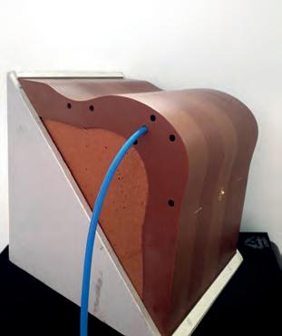
In radiation oncology departments, surface imaging systems are commonly used for patient positioning and to ensure that patient motion during treatment is within acceptable margins. In H&N cancer radiotherapy, surface imaging has been used in conjunction with open-face masks, immobilisation masks that leave a portion of the face free, to reduce the burden that immobilisation masks pose on patients while preserving treatment accuracy. However, current implementations of surface imaging in the clinic can’t completely replace immobilisation masks as current surface imaging technologies struggle to accurately track non-rigid surfaces. This is not a major issue when tracking an area like the pelvis, however non-rigid motion such as opening the mouth, smiling, yawning, etc. can significantly decrease the accuracy of surface imaging technologies and makes completely replacing masks with current surface imaging technology challenging. To address this issue, we have developed and built the Remove the Mask Surface Imaging System (RtMSIS).
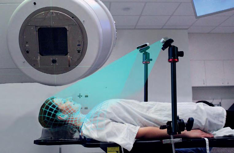
The RtMSIS is a fully functional surfaceguided radiotherapy system that uses low-cost and widely available stereo depth cameras (Intel RealSense D415) in conjunction with an open-source surface imaging code. RtMSIS uses two cameras, one on each side of the patient, positioned about 30 cm above the patient’s chest 1 The use of a dual-camera system allows for images to be combined to obtain a complete 3D image of the patient’s head and neck while avoiding field-of-view obstruction
by the treatment unit. In phantom and preliminary human studies, the RtMSIS has been shown to have an accuracy of <2 mm for translations and <1 degree in all rotational directions.
To solve the issue of non-rigid surface motion, we have developed a solution that relies on the knowledge that we are imaging a human face. To track a moving human face, the RtMSIS uses facial recognition technology to segment facial landmarks and identify the different sections of a human face. The facial landmark information is fed to a Bayesian model in conjunction with the motion estimate from a rigid registration algorithm to obtain the true head motion. The accuracy that can be achieved by this technique is currently being investigated in a human trial held at the University of Sydney’s Image X Institute.
Real-time tumour tracking
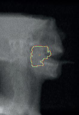
Surface imaging captures information on where and how the outside of the patient moves. However, tumours are located inside of patients and can move without creating any external changes. To ensure that all tumour motion is captured, we have improved an x-ray imaging technology that
we have pioneered and which uses the onboard imager of a standard medical linear accelerator to track tumour motion in realtime during radiotherapy. This technology has been tested in multiple clinical trials for various tumour sites outside of H&N cancer. However, the current technology cannot be directly applied to H&N cancer as it requires the surgical implantation of radio-opaque markers, a process that is risky for H&N tumours.
Our approach to translate this technology to H&N treatments is to use a machine learning algorithm trained to directly segment the tumour in kV images, avoiding the need for markers 3. This type of marker-less tracking technology is being developed for other tumour sites but applying it to H&N treatments poses an additional challenge; machine learning algorithms require a lot of data to train and there is a paucity of data for H&N cancer. To augment our data, we have developed a novel CT deformation algorithm that is based on a bio-mechanical model of the head and neck that allows us to simulate highly realistic head motion. Thanks to this algorithm, we can use prior knowledge of how the average patient moves in a mask-less radiotherapy setting to generate
IPEM SCOPE 22 WINTER 2022 G F GENERAL FEATURES
THE RTMSIS USES FACIAL RECOGNITION TECH TO SEGMENT FACIAL LANDMARKS
1
1
2
3
realistic patient-specific dataset that spans the entire possible range of motion. Those CT images are then used to properly train our machine learning algorithm. The accuracy of our marker-less H&N tumour tracking system is currently being tested using clinically acquired patient data. Preliminary results suggest a precision of <2 mm.
Advantage of imaging with multiple modalities
There are two types of motion that can affect tumour position: head motion, which is motion originating from the spine such as head nodding or shaking and during which the entire skull moves, and intracranial motion which can originate from jaw movements or internal biological processes such as swallowing and breathing. It is important to differentiate between the two types of motion because they each present individual challenges.
Head motion occurs due to a patient’s involuntary movement and is usually not periodic (patients who try to stay still usually do not move their head back and forth). It usually presents as a slow drift in one direction or as a sudden quick change in position. Immobilisation masks are used principally to prevent this type of motion and they also somewhat restrict jaw movement.
In a mask-less treatment scenario head motion affects not only the position of the tumour, but also the position of all surrounding organs. As such, it is necessary to gate the treatment and reposition the
patient if motion exceeds the acceptable threshold. A rigid registration algorithm based on surface images is ideal to track this type of motion since there is usually no non-rigid facial motion involved.

Intracranial motion in a H&N radiotherapy treatment setting is usually associated with biological processes and involuntary facial expression. While this type of motion can be minimised with the use of additional immobilisation equipment, it is often, at least partially, treated as negligible. However, studies have shown that H&N tumours and laryngeal tumours in particular can experience motion of up to 7.8 mm during treatment even when the patient is wearing a mask. Since intracranial motion can occur independently to head motion, neither surface imaging nor an immobilisation mask alone can compensate for this type of motion. We plan to use our real-time kilovoltage tumour motion tracking system to inform our motion compensation strategy for intra-cranial motion.
Once we know the entire patient H&N anatomy and can track how it changes during treatment, the final step is to adjust the radiation beam to ensure that it is always targeting the tumour, thereby preserving the efficacy of the treatment and minimising the risks of side effects. For this step, we will adapt a beam adaptation technology that we developed for prostate and lung cancer patients. This technology adjusts the beam shape in real-time by adjusting the aperture of the multi-leaf collimator using real-time information
about the position of the patient as well as a real-time optimisation algorithm that ensures the patient is receiving the most accurate treatment.
Conclusion
The Remove the Mask project started when a H&N cancer survivor and patient advocate shared her mask anxiety story with the University of Sydney’s Image X Institute. This story led to developing an alternative to immobilisation masks and to make future H&N radiotherapy treatment more comfortable for patients. The technology to do so already exists. Surface tracking, especially head tracking, is a very active field of study and not only for its use in radiotherapy.
Clinical trials testing novel tumour tracking methods such as marker-less tracking are currently ongoing and show very promising results. Treatment adaptation has already started to move into the clinic. While there is still work to be done with regards to combining these technologies, it is only a matter of time before we can finally remove the mask.
IPEM.AC.UK 23 WINTER 2022
Youssef Ben Bouchta and Mark Gardner are Postdoctoral Research Associates at the Image X Institute and Paul Keall is the Director of the Institute. They would like to acknowledge Cancer Australia, which is funding the Remove the Mask project, and the entire dedicated Remove the Mask team.
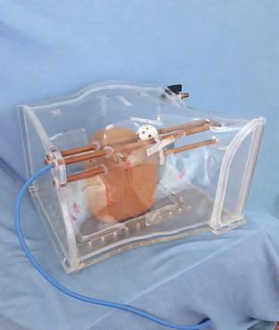
Illustration of RtMSIS replacing an immobilisation mask and tracking H&N motion.
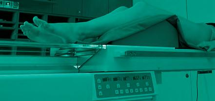
Different immobilisation masks utilised in head and neck cancer radiotherapy.
2
Illustration of the kV tumour tracking technology (right) and actual data from the kV imaging system showing the true tumour location in red and predicted tumour location in green (far right).
3 IMAGES: JULIA JOHSON & MARK GARDNER


IPEM SCOPE 24 WINTER 2022 G F GENERAL FEATURES
A HISTORY OF RADIOTHERAPY TRIALS QUALITY ASSURANCE


The beginnings of interdepartmental audit
IPEM Fellow and former Mount Vernon Hospital Head of Physics Edwin GA Aird on the early history of Radiotherapy Trials Quality Assurance (RTTQA).
In the mid-1980s David Thwaites and Stan Klevenhagen formed a group that decided it would be a good idea to check dosimetry for UK radiotherapy centres.
In Europe and US this had been done with thermoluminescent dosimeter (TLD), but the new group thought that using an ion chamber would be more accurate and give more information. It was also decided to use a semi-anatomical phantom; so that, as well as measuring dose under standard conditions, dose could also be measured under treatment conditions. A phantom was built from water-equivalent plastic (WEP), which had holes for a Farmer chamber for measurements at five points within a
tumour volume. A section of lung type WEP could be inserted so that lung corrections (which were still quite simple at that time) could be assessed. There was also an oblique surface for one of the three beams, to treat a typical central lung tumour.


The first audits
A nationwide survey was designed on a “round-robin” basis (there was no grant money with the project), so the phantom and dosimeter were ferried around the country and passed from centre to centre within eight regional groups. Each region would then move the equipment to the next region and the nominated auditor for
each region would then make the survey together with the in-house team.
Between 1987 and 1991, 64 centres were visited; 161 machines (including 61 Cobalts). Only two centres had a discrepancy of greater than 5%; but one of those centres had an extraordinary discrepancy of 25% (for more information, see Thwaites 1988 enquiry, which also led to the mandatory requirement for a quality assurance (QA) management system in every radiotherapy department in the UK).
Grant money was found to extend the audits to include electrons. This National Group has now become a standard feature within the community covering all aspects
IMAGE: ALAMY IPEM.AC.UK 25 WINTER 2022
of dosimetry within radiotherapy.
It’s important to understand the status of the technology used in radiotherapy at this time. There were still several cobalt 60 sets in use, most centres had at least one 5-6MV linac and many centres were installing high-energy linacs (10-20MV). The imaging on simulators was reasonable, but nothing like it is today. The use of computerised tomography (CT) scanners was in its infancy; field shaping was achieved with lead blocks. Treatment planning using computers was in its early stages with the outlines and tumour entered by hand.
We will see that as equipment changed and the techniques for radiotherapy delivery changed the audit and QA needed to keep abreast with these changes: CT planning; multileaf collimator (MLC) (stationary and moving), intensity modulated radiotherapy (IMRT) in all its forms and volumetric modulated arc therapy (VMAT).
The “Interdepartmental audit” became a regular feature of radiotherapy physicists. But then it became obvious that more extensive QA of the whole process of delivering a particular type of radiotherapy was needed, particularly when a new clinical trial was planned.
The sequence of new trials that I was involved with, mapped below, tells this story well:
1. CHART (1989) conventional 2D radiotherapy, mainly simple computer planning
2. START (1998) tangential field issues (oblique surfaces and lung correction), beginnings of a 3D understanding of dose distribution.
3. RT01 (1998) the beginnings of conformal therapy (3D planning, with shaped blocks or MLC).
4. Then, with radiotherapy centres beginning to perform IMRT, came the PARSPORT (2003 H&N) trial, which combined all the new techniques of planning and delivery; followed by the hypofractionated and concomitant boost breast trials, including: IMPORT High (2014, which also introduced the issue
of QA for image-guided radiation therapy (IGRT)).
Continuous hyperfractionated accelerated radiotherapy trial
Together with the Gray Laboratory, Prof Stan Dische (Director of Cancer Services at Mount Vernon Hospital (MVH)) and Michel Saunders had put together a proposal for a clinical trial of continuous hyperfractionated accelerated radiotherapy trial (CHART) in both H&N and Bronchus. Since this was a radical change of treatment for these cancers Prof Dische wanted to be sure that the actual treatment aspects were very safe. So he was very keen to have QA associated with the trial.
My interest in QA was already extensive and, because of my involvement with a French QA study for a European Trial in 29 centres (QA organised by IGR (Paris) 1987-1988 reported 1994), I could offer my services to Prof Dische. At Mount Vernon we formed a sub-group of the CHART team: senior radiographer, engineer and physicist (C Williams; G Mott; E Aird).
We designed an elaborate set of tests that initially tested many aspects of the linac and simulator performance. Treatment planning was tested by making use of two WEP phantoms: a bronchus phantom and a head and neck phantom. The plans could be compared with a gold standard; but the actual doses within the phantoms were also measured using an ion chamber. This allowed for an immediate assessment of the complete performance of each of 14 treatment centres. The total equipment at that time was extensive and transport was achieved using a Volvo Estate car.
Standardisation of breast radiotherapy trial
The standardisation of breast radiotherapy trial (START) was set up by John Yarnold (the Institute of Cancer Research and Royal Marsden Hospital (RMH)), to broadly investigate a comparison of two breast fractionation schemes : 50Gy in 25 fractions vs 40Gy in 15 fractions. (A “southern” fractionation scheme vs a “northern” one). MVH was asked to form a QA group to
40Gy/15Fr CONTROL TEST ARM 1 TEST ARM 2 40Gy/15Fr 40Gy/15Fr 56Gy/23Fr Sequential dose escalation 48Gy/15Fr Sequential dose escalation 53Gy/15Fr Sequential dose escalation 36Gy/15Fr 36Gy/15Fr
TOTAL EQUIPMENT AT
EXTENSIVE
TRANSPORT
ACHIEVED USING A VOLVO ESTATE CAR The QA for IMPORT High: set a new standard of QA for breast cancer planning and delivery IPEM SCOPE 26 WINTER 2022 G F GENERAL FEATURES
THE
THAT TIME WAS
AND
WAS
ensure that standards of care were identical in all centres participating in the trial. This group (Karen Venables; Elizabeth Winfield, Amanda Deighton) started this work by considering some of the difficult questions arising from different standards used throughout the country. From the outcome of these studies they wrote a process for ensuring that almost identical delivery of dose to the newly defined “treatment point” could be achieved. It seems strange now that it was necessary to do this. They also had to consider how to deliver the postclavicular fields safely and consistently; this was around the time that the Radiotherapy Action Group Exposure (RAGE) study was reporting where, for example, suspected damage to brachial plexus nerve may have been caused by radiotherapy).
Outcomes of START QA
1. Various phantoms built to assess dose.
2. Plan checking was introduced.
3. A 3D phantom was built and 36 sets of data were obtained.
4. A “help desk” was established to help with interpretation of the protocol
5. The organisation of participants meetings to discuss issues within the trial (attended by all professionals involved). These became a regular feature to several of the trials using RTTQA facilities.
The START QA studies set an excellent standard for breast radiotherapy in the UK. John Yarnold continued to develop clinical trials in breast radiotherapy for the next 15–20 years. QA has been necessary for each trial, and the development of QA in clinical trials is nicely mapped by the development of breast radiotherapy trials: in particular the IMPORT trials, which have made use of IMRT and VMAT etc. Hypofractionation in the FAST and FAST FORWARD trials has allowed centres throughout the world to confidently use shorter fractionation schemes during the COVID epidemic.
RT01 Trial
To continue with the development of QA in trials chronologically, the next trial, which involved the RMH physics department rather than MVH, was RT01. This was a Medical Research Council (MRC)-sponsored trial (as it turns out the “01” was a very
hopeful , but false: there were no more entirely sponsored MRC trials), led by Prof David Dearnaley (the Institute of Cancer Research and RMH).
Within the UK RT01 trial the MRC also funded a QA programme. Philip Mayles led the group that took on the QA for planning and delivery of radiotherapy to the prostate (and surrounding glands when needed).
This included a planning and dosimetry audit at participating centres using a purpose-built phantom. Of special interest was the prostate phantom with its “silver prostate” to allow the phantom to be very accurately aligned before ion chamber measurements were taken.
Geometrical setup was visually assessed via field shaping around the phantom GTV (to within the order of 1 mm). Within the phantom, ion chamber positional uncertainties were estimated as 0.6 mm (95% CL, k=2). Setup errors are not eliminated, but minimised and estimated.
Many papers were produced and this work has contributed to high standards of care in the UK radiotherapy community.
LIST OF QA MODULES FOR RT01:
● Outlining exercises and questionnaire to be completed.
● A process document giving details of the centre’s treatment procedure to be submitted demonstrating that a written
quality system is in use.
● A treatment planning exercise should be carried out based on a standard set of data supplied to each centre.
A BRIEF DESCRIPTION OF THE ADDITIONAL BENEFITS OF QA.
● This was an excellent opportunity for centres to learn from both the QA centre and from each other.
● Several groups were created to bring together centres with the same equipment to create forums (including clinical oncologists) for specific discussion points. At the time of implementation of this trial 3D target volume definition was not routinely performed in all centres, and this trial required the staff to work to a steep learning curve.
● This allowed for the speeding up of implementation of complex radiotherapy with greater confidence and safety.
The development of RTTQA
It became evident to the NHS that the work from this group of trials had provided in the UK with a very high standard of care within the radiotherapy community; and, with the support of medics, it was proposed that a group should be formed to support
IPEM.AC.UK 27 WINTER 2022
Left: A WEP phantom with a lung insert. 5 measuring points in target; 1 in contralateral lung. Right: Formed from a plastic sheet on a plaster of Paris mould of the breast of an average size volunteer. The lung slices were cut from CT slice images of a patient of similar build. Courtesy of K Venables
clinical trials that had NHS sponsorship. The National RTTQA Group was founded in 2002 under the auspices of NCRI UK to carry out QA for all NIHR Clinical Research Network portfolio trials that include a radiotherapy component. This group became known as RTTQA and was led by Elizabeth Miles and nationally funded.
The central group works through four NHS sites, Mount Vernon Cancer Centre (MVCC), RMH, Velindre Cancer Centre (VCC), and Clatterbridge Cancer Centre (CCC) with central coordination from the MVCC site.
Each of these national lead QA centres contributes specialist expertise in sitespecific areas collaborating with others when further expert knowledge in niche areas is required.
RTTQA vs local RTQA
In the clinical trial setting there is a distinct difference in the purpose and outcome of central RTQA, as performed by the RTTQA group, and local RTQA completed at a departmental/centre level. It is, however, important to emphasise that neither functions in isolation and there is always close interaction between the two, particularly when new techniques are being evaluated.
The QA for IMPORT High (see p.26, top) set a new standard of QA for breast cancer planning and delivery, with the following components:
1. Dosimetry: small field dosimetry checked, for IMRT and Rot IMRT.

2. Initial information: A questionnaire and process document (concepts first used with RT01); pre-trial visits to discuss new techniques and any questions within the documents, clarification.
3. Phantoms: IMRT and Rotational IMRT credentialing phantoms.
4. Volumes: benchmark cases; volumes and planning techniques (exchange with RTTQA group for critical comments).
5. Individual plan review
6. Clips in tumour bed and CBCT (kV or MV); so introducing QA for IGRT for the first time.

The elements of QA became standardised
EACH OF THESE NATIONAL LEAD QA CENTRES CONTRIBUTES SPECIALIST EXPERTISE
after these first complex trials as:
● Verification of electronic data transfer.
● A process document; using a template provided by the chief investigator (or QA centre) describing the procedure to be followed for planning and delivery according to the trial protocol.
● A facility questionnaire: to be completed by centres entering patients into the trial that demonstrates that the centre has appropriate resources and has developed a procedure to deliver the radiotherapy prescription required by the trial protocol.
● Outlining benchmark case: one or more test cases are sent to participating centres for outlining to check that the clinical oncologists understand the trial protocol and meet the standard of volume outlining defined within it before any patients are entered into the trial.
● Planning benchmark cases: which are pre-outlined test cases to be planned by the local team to check full understanding of the protocol.
● Audit: this may consist of various types – inspection, dosimetry, measurements using film or detectors in various phantoms including the special IMRT/ VMAT phantoms.
As the number of trials grew – they increased from 20 in 2010 to 50 in 2020 –they included the various forms of complex planning and delivery of IMRT, more and more visits were needed, particularly in those centres taking on several clinical trials at once. This made for a challenging workload both for the RTTQA team and the local team (now able to officially form under a new funding arrangement). To alleviate these pressures the RTTQA group
implemented a system of streamlining of the IMRT QA processes whereby a centre already credentialed for IMRT use in a particular trial would not be required to undergo the full credentialing for a trial using similar techniques.The group also developed a flexible postal audit system to cover some of these audits, so far performed at eight centres since its introduction in 2014 reducing workload for both the central group and local centres.
Summary

This has been an account of the formation of RTTQA and some of the trials I have been involved with. Since 2005, the number of trials cases increased markedly. The last one I was closely involved with, led by Patty Diez, together with NPL, was the INTERLACE trial (Ca cervix) . The main aim of our contribution was to measure the prescription dose at 2cm from an HDR line source; a considerable challenge. We managed to visit 47 centres in the UK.
The RTTQA group is one of four main audit groups in the UK alongside the IPEM RTSIG Interdepartmental Dosimetry Working Group, National Physical Laboratory (NPL) and the UK SABR consortium. These groups have worked together to provide integrated national audit for the more advanced techniques.
Recent national collaborative audits include intensity modulated RT audit, rotational IMRT audit (VMAT/ tomotherapy), UK SABR lung dosimetry audit and the HDR brachytherapy audit, which have helped to support the implementation of advanced techniques, identify potential dosimetry issues and set standards. For more information on clinical trials the RTTQA group is currently working on, visit rttrialsqa.org.uk
IMAGE: GETTY IMAGES IPEM SCOPE 28 WINTER 2022 G F GENERAL FEATURES
The current landscape in England
TREATMENT OF BRAIN METASTASES WITH STEREOTACTIC RADIOSURGERY
Anna Bangiri, Principal Stereotactic Radiosurgery
Radiotherapy Physicist at Nottingham University
Hospitals NHS Trust, looks at the processes and procedures across the country.

In 2013, NHS England published the Clinical Commissioning Policy for treating patients with cerebral metastases. A formal commissioning process followed and a total of 16 centres were commissioned to provide stereotactic radiosurgery (SRS) services across England. A mixture of platforms were approved, including Leksell Gamma Knife (GK), Cyberknife (CK), and linear accelerators (linacs), both Varian and Elekta. Patients could be treated either with a surgical frame attached to their
head or with a frameless technique. Six years after the initial commissioning process, we carried out a short survey to establish what is the current landscape for treating patients with brain metastases in England.
The background GK is the gold standard in treating brain metastases since its inception in 1967 by Lars Leksell. It uses a stereotactic frame surgically fitted on to the patient’s skull on the day of treatment. With the
IPEM.AC.UK 29 WINTER 2022
G F GENERAL FEATURES
introduction of volumetric arc therapies (VMAT), linacs gained the capability of delivering high doses in a very focused way. This has further improved with the introduction of flattening filter free (FFF) beams as well as HyperArc. The former allows for high doses of radiation to be delivered up to four times faster and the latter allows complex deliveries where the gantry, the collimator, the MLC and the couch all move synchronously. In combination, all these innovations have allowed linacs to produce treatment plans that are comparable to those produced with a GK.
The GK, even though it is the gold standard, is not easy to implement in a hospital setting due to the various restrictions that come along with it. Considerations for the use of sealed sources need to be made, as well as the fact that only a limited range of patients can be treated on it. For example, it is only in the last few years, with the introduction of GK Icon, that fractionated treatments can be performed.
Treatment pathways
The treatment pathways between GK and linac are substantially different and this needs to be considered. With GK, the stereotactic frame is fitted on the patient, an MRI scan is performed, the
done on the same day, or an older scan can be used. The patient will then leave and return once the treatment plan has been created and the patient-specific quality assurance (PSQA) carried out.
extensive PSQA. The planning time, along with the treatment delivery time, will also depend on whether it is single or multiple isocentre delivery. This can introduce time delays in the pathway to treatment.
lesions are outlined and a treatment is planned and delivered within the same day. With a linac-based approach, including CK, however, the pathway is dissimilar and longer. The patient will have an immobilisation device fitted and a CT scan carried out on the same day. If there is an MRI scanner in the department, a planning MRI can also be
These processes can be time consuming and are very dependent on the systems being used, as well as on the type of treatment delivery. For example, a 3D conformal plan can be time consuming to create, especially for multiple lesions, but requires minimal PSQA. On the contrary, dynamic conformal arc (DCAT) or VMAT plans might be quicker to plan but require
At the time of commissioning, there were no recommendations, either national or international, and each centre had to establish its own treatment pathway. Recommendations for the implementation of an SRS treatment pathway came out in 2019 by Hartgerink D. et al. In this paper, it was recommended that the interval between MRI scan and treatment should be kept at a maximum of two weeks, ideally less than a week. This is to avoid a potential geographical miss due to tumour progression. Seymour et al. showed that local control at six months can drop from 95% to 56% if the interval between MRI and treatment is longer than two weeks. At one year, the local control rates drop from 75% to 34%. Furthermore, Plunkett et al. reported that certain tumours could grow by up to 200% depending on initial tumour volume and primary

IPEM SCOPE 30 WINTER 2022 G F GENERAL FEATURES
THE GK, EVEN THOUGH IT IS THE GOLD STANDARD, IS NOT EASY TO IMPLEMENT IN A HOSPITAL SETTING DUE TO THE VARIOUS RESTRICTIONS THAT COME ALONG WITH IT
histology. Garcia et al. reported a volume increase of 1.35 times the original, after 14 days, and Bronnimann et al. showed that a 1 mm margin can be outgrown in six days and 14 days for melanoma and non-small cell lung cancer, respectively, if a linear growth model is used. When an exponential model is used the interval drops to three and eight days respectively. The literature clearly shows that it is paramount to try and minimise the interval between MRI and treatment for patients with brain metastases. Considering the different platforms and treatment pathways, the above mostly affects linac-based treatment approaches.
The methods
The survey included questions regarding the multi-disciplinary team meetings (MDT) and the corresponding referral process, average and maximum intervals between MRI and treatment, as well as whether a specialised planning MRI was used for outlining the tumours. A total of 16 centres were commissioned
with one centre covering two areas. The questionnaire was sent to all commissioned centres and replies to the survey were received by all centres bar one, resulting in a response rate of 94%.
Two hospitals applied jointly during the commissioning process but each have their own MDT and treat patients on different platforms therefore, those answers were considered separately.
The results
The spread of treatment platforms is four GK (23%), three CK (18%), three Elekta (18%) and seven Varian centres (41%)
One centre uses two different platforms to treat their patients hence the total is 17, as shown in 1. For 11 out of 16 centres (69%), the cases are discussed at a NeuroOncology MDT, whereas the remainder five centres discuss patients in a dedicated SRS MDT 2. The majority of the centres accept referrals through MDT-specific forms (94%), with some using a MDT-specific email address as well, and only one centre using email referrals without a specific
form. The time cut-off for referrals at the MDT is one to two days before, with two hospitals being more flexible. These accept referrals in the same morning, if the radiologist is happy to review the images prior to the MDT discussion.
All the centres barring one, use a MRI scan that has been done specifically for planning purposes, and this is in addition to the diagnostic MRI scan. Only two hospitals have MRI scanners available within the radiotherapy department and can therefore scan the patient at the same time as the planning CT. The remainder (88%) use the diagnostic MRI scanner of the treating hospital for the planning MRI scan.
Only one hospital routinely accepts MRI scans that have been carried out in a hospital other than the treating one. The MRI scan can be up to two weeks old before a new one is requested for planning purposes. Another two hospitals would accept MRI scans done in another hospital but only if the scan was done within one week of the planning CT.
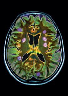
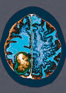
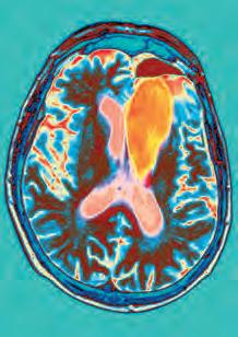
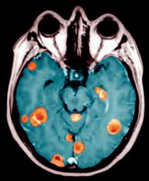
IMAGES: GETTY IMAGES, SCIENCE PHOTOLIBRARY IPEM.AC.UK 31 WINTER 2022
■
MDT ■
MDT Discussion 69% 31%
Top left: Coloured axial magnetic resonance imaging scan through the brain of a patient with metastatic brain cancer. Top right : Coloured CT scan of a patient with brain cancer. Bottom left and right: Coloured axial magnetic resonance imaging scans of patient with brain cancer. 7 3
2 Brain metastases patients are mainly discussed at Neuro-Oncology MDTs with fewer centres preferring a SRS Dedicated MDT. 4
Neuro-oncology 3
SRS Dedicated MDT
■ Varian ■ Cyberknife ■ GK ■ Elekta
1 Treatment platforms used across the 16 centres treating brain metastases in England. The above total is 17 as one centre uses two different platforms to treat their patients.
Treatment platforms
The average and maximum MRI-totreatment times vary greatly between hospitals. All the GK centres have a maximum MRI to treatment time of one day, and the average is in general a few hours. On the contrary, the linacbased centres report maximum MRI to treatment times of 28 days, with a median at 14 days (range 10 days-28days).
The average MRI to treatment time is 11.4 days with the median at 11 days and a range of three to 21 days 3.A total of nine centres (56%) believe that an MRI scanner is required within the radiotherapy department to treat SRS patients 4
The remainder of the centres thought that is was not necessary as long as there was timely access to the MRI scanners of the radiology department in the hospital. The majority of the hospitals treat between three and five patients with brain metastases every week.
The discussion

The responses above clearly show that there are some differences in the treatment pathways for SRS patients within England. The referral process is more or less similar across all hospitals, with most cases discussed at a neurooncology MDT. The Varian platform is favoured for treatment by nearly half of the centres. Both GK and CK, despite being commercially available for SRS treatments years before the introduction of modern linac-based SRS, were not a widely preferred option. This was mostly due to the restrictions mentioned earlier, as well as the fact that they can only accommodate a limited number of patients per day and cannot treat all sites and tumours. Varian, in combination with Brainlab, produced the first linac that had integrated imaging possible at all couch angles, with a six degrees of freedom (6DoF)
couch to allow for submillimetre positional accuracy, and a treatment delivery that did not require the use of cones.
Out of the three hospitals that might accept MRI scans done elsewhere, it is only the one that routinely does so that has the longest MRI to treatment interval of 28 days. This can likely be attributed to the fact that it often takes up to a week for a MRI scan to be reported, and if this is added to the time lapse until the MDT discussion and the CT scan taking place, one can easily see why the interval reaches four weeks. The other two centres that commented they might accept MRI scans from other departments had set a time cut-off of one week and this did not seem to affect the maximum MRI to treatment times. There was one hospital that, despite using a diagnostic MRI scanner within the hospital for the planning MRI scans, still reported maximum MRI to treatment times of three
IPEM SCOPE 32 WINTER 2022 G F GENERAL FEATURES
to four weeks. A few hospitals commented that a good relationship with the radiology department of the treating hospital was paramount to ensure the swift treatment for these patients. Some hospitals have slots reserved on their diagnostic MRI scanners every week, whereas others liaise with the department depending on the needs of the service on a weekly basis.
Looking at the literature, it is evident that these patients need to be treated as quickly as possible, with the main focus being on minimising the time between the MRI and CT scans and the treatment. Tumours grow, some faster than others, and this needs to be considered especially when delays are introduced in the treatment pathway due to reporting, planning and PSQA. In the commissioning policy and the service specification it was distinctly mentioned that the patients need to be seen within one week from the MDT discussion and as they are considered palliative patients, the 14-day target applies. If a new planning MRI scan is not ordered after MDT discussion, then a minimum of three weeks MRI to treatment interval exists. This highlights the issue of the lack of MRI scanners in radiotherapy departments as well as the more general
issue of access to a diagnostic MRI scanner for all patients. In Scotland, the devolved government has made plans to introduce MRI scanners in all radiotherapy departments by September 2023. With the recommendations by Hartgerink et al., a similar case is made for all centres that are providing SRS treatments in England. Careful considerations should be made when delays occur and what the potential impact might be on patient outcomes.
The conclusion
A survey was carried out to assess current practise across England for treating patients with brain metastases using SRS. A clear difference can be seen in the treatment pathways between GK centres and linac-based approaches, including CK. The average MRI to treatment times
for all centres are within the 14-day target specified by NHS England. Two centres have maximum MRI to treatment times of over two weeks. Overall, the majority of the centres thought that a MRI scanner is required within the radiotherapy department for the provision of service, and considerations should be made for future implementation, as is currently done in Scotland. All centres should aim to follow published guidance with respect to timings from MRI to treatment and what the impact of delays is on patient outcomes.
Anna Bangiri is Principal Stereotactic Radiosurgery Radiotherapy Physicist at Nottingham University Hospitals NHS Trust. To see the questionnaire email anna.bangiri@nuh.nhs.uk
IMAGE: GETTY IMAGES IPEM.AC.UK 33 WINTER 2022
10 8 6 4 2 0 1 day 2-7 days 8-14 days 14-21 days Average MRI to treatment time No of centres Days 3 The average MRI to treatment times for all centres that took part in the survey. A GOOD RELATIONSHIP WITH THE RADIOLOGY DEPARTMENT OF THE TREATING HOSPITAL WAS PARAMOUNT TO ENSURE SWIFT TREATMENT ■ Yes ■ No Is MRI required in the RT Department 56% 44% 4 More than half of the centres believe that RT departments that treat patients with SRS should have a MRI scanner.
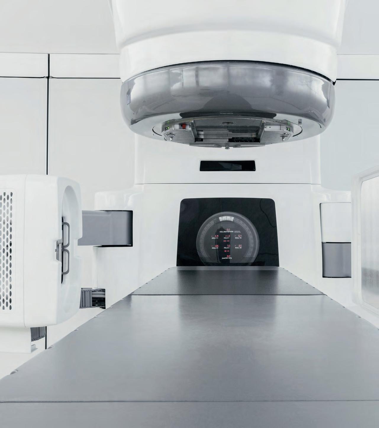
PREPARATION AND DELIVERY OF PATIENT-SPECIFIC VERIFICATION IS TIME CONSUMING AND OFTEN DONE OUT OF HOURS, SO WHY DO WE DO IT? IPEM SCOPE 34 WINTER 2022 G F GENERAL FEATURES
PATIENT-SPECIFIC VERIFICATIONS
Have we done enough?
Patient-specific verifications use a linear accelerator to test an individual patient’s radiotherapy treatment plan.
Every radiotherapy centre will have its own processes, but they will all follow the same basic path. An Intensity Modulated Radiotherapy (IMRT) plan is created using a treatment planning system (TPS), the final dose is calculated on the patient’s CT study set.
The plan is signed off by a clinician, exported to the record and verify system (RV) ready to be delivered by the radiographers to the patient. The doctor is happy, the dosimetrist is happy, the radiographers are happy – the physicists aren’t. Things need to be tested. So, the TPS is used to recalculate the plan on a phantom, the linac is then used to deliver the plan to the phantom. The dose measured in the phantom is compared to the dose calculated by the TPS, provided the agreement between the two is within an established tolerance, the plan passes. Now the physicists are happy.
Phantoms, broadly speaking, sit in one of two camps: ones that contain multidetector arrays and ones that house a single ionisation chamber; the former can normally assess the whole treatment volume, the latter is only capable of measuring a single point at a time. Detector array readings are normally subject to gamma analysis or, in more advanced systems, DVH analysis. Gamma analysis is computationally light and intuitive, but the result is difficult to interpret in a clinical context and is not sensitive to some delivery errors. DVH analysis is computationally quite heavy, but the results neatly illustrate changes to the planned and delivered dose to the patient. Measurements taken with a chamber can be done in solid water or some sort of anatomical phantom.

Why do we do it?
Preparation and delivery of patientspecific verification is time consuming and often done out of hours, so why do we do it? 3D conformal plans used the multi leaf collimator (MLC) to create
different shaped apertures that, broadly speaking, conformed to the shape of the target volume. These shapes and weights of the beam were tweaked by a dosimetrist until an optimal dose distribution was reached. IMRT plans are a different beast altogether. An optimisation algorithm determines the weights and shapes of beam segments so that the dose distribution achieves pre-defined objectives. Inverse planning can create highly-conformal dose distributions allowing higher target doses and lower doses to organs at risk, this is achieved by splitting the beam into a number of segments, each segment has a small fraction of the total delivery going through, the shapes vary in size and complexity. The smaller, more complicated the shape, the more uncertainty there is associated with dose calculation and the bigger impact inaccuracies in leaf position will have on the delivered dose. This fundamentally different approach to the planning and delivery of radiotherapy tests the dose calculation algorithms harder and stresses the machine much more than 3D conformal techniques. With the sheer variety of variable solutions it was and still is difficult to put together a QA programme that checks all the attributes that the optimiser might ask of the machine. Given the sensitivity of the plans to machine parameters – even a 1mm shift in the position of the leaf bank can cause a clinically significant different to the dose distribution –
IPEM.AC.UK 35 WINTER 2022
IMAGES: SHUTTERSTOCK
Patient-specific verifications represent a considerable workload. How many passing results do we need before we have the confidence in beam models to calculate dose and linacs to deliver it accurately? Lead Clinical Scientist Dr Dan Johnson looks at the issues.
patient-specific testing of each plan wasn’t just prudent, it was the only way to give us the assurances we needed that the dose was being delivered as it was intended.
So, what are we testing when we do a patient-specific verification? Well, maybe a good place to start is to consider what we’re not testing. Crucially, what we’re not testing is the patient dose calculation. Patient-specific verifications require the dose to be recalculated on a phantom scan set; the ability of the dose algorithm to model the interaction of radiation with the patient’s tissues is not tested. Assurances about the dose algorithm’s ability to do this are made during the commissioning of the TPS and the secondary MU check confirms on a per-patient basis that no gross errors have occurred in this process. So, what are we checking with a patient-specific verification? I believe there are three things (see box, below-right).
Taking the first point: It is reasonably easy to calculate the dose delivered by an open field. With smaller fields, the leaf tip transmission, leaf leakage and leaf transmission make a bigger contribution to the total calculated dose. These features are more difficult to model and the uncertainty associated with the calculated dose increases. IMRT optimisers tend to build up the target dose using a number of small segments. The beam model’s ability to calculate a plan made up of these segments will be tested a lot more than in the case of a 3D conformal plan made up of, broadly speaking, larger fields. By recalculating the patient dose on a phantom, then measuring it on a linac, the ability of the dose algorithm to accurately calculate dose for these, typically quite-tough, scenarios is tested.
Taking the second point: IMRT can be delivered through step and shoot or Dynamic MLC static beam techniques, but the most prevalent approach is VMAT on account of being clinically equivalent to the other techniques but much easier and quicker to deliver. With a VMAT delivery the radiation is delivered a variable rate while the gantry and MLCs move. This is a huge change from 3D conformal deliveries and relies on an accurate, synchronous interplay between a number of complex components. Furthermore, small (<1mm)
inaccuracies in leaf position can have a significant clinical impact on the dose distribution that simply wouldn’t be seen if the equivalent error was present for the delivery of the 3D conformal plan. Patient-specific testing demonstrates that the linacs are capable of performing this complex operation with sufficient precision to deliver the dose accurately.
Taking the third point: data transfer is now handled with reasonably simple, often in-house, checking software.
To summarise: the move from 3D conformal plans to IMRT significantly increased the demands on both the hardware and software that we are using to deliver radiotherapy. Because of the bespoke nature of radiotherapy planning, it was impossible to devise a machine QA system that tested every scenario of what could be asked of the machines, consequently it was necessary to perform patient-specific QA to ensure that this new technique was delivering dose accurately. Locally, we have performed literally thousands of patient-specific verifications. There have been issues with less than five. These issues were not serious and would have had negligible clinical impact. Anecdotally, I believe this is the case in many radiotherapy centres in the UK. With these huge banks of data demonstrating
that the linacs and TPS are capable of delivering IMRT with the necessary accuracy, can we stop doing patientspecific verifications?
Routine basis
Well, in short, no. There will always be times when it is necessary to perform patient-specific QA, however, it is my opinion that we can - and should - move away from performing them on a routine basis. And, like us, I believe a lot of centres have started to move away from patientspecific QA for all IMRT treatments. But can we go further? Can we stop doing them on a routine basis full stop? To address the first
WHAT TO CHECK WITH PATIENT-SPECIFIC VERIFICATION?
1. The ability of the TPS to calculate dose in phantom material for specific, complex plans.
2. The ability of the machine to deliver the plan accurately.
3. That the data transfer between TPS, R&V and linac control system has happened correctly.
IPEM SCOPE 36 WINTER 2022 G F GENERAL FEATURES
point of testing – I believe that the sheer volume of passing results demonstrates that the TPS’ dose algorithm is capable of accurately calculating the dose for the segments that the optimiser produces. And, for the most part, the optimiser and calculation algorithm are something of a constant. Upgrades and patches will change aspects of the TPS, maybe add a new feature, but the beating heart of the TPS – the optimiser and dose algorithm –remain unchanged. So, provided these two aspects of the TPS remain unchanged, we have established that they are fit for purpose and do not need to be tested anymore. So, in centres with an established IMRT service, the purpose of patientspecific QA is to ensure that the linac is capable of delivering the treatment plan accurately. With this in mind, the question becomes – why wouldn’t a machine be able to deliver a plan accurately? I think there are two main reasons:
1. Linacs are complex machines with moving parts and perishable components. Performance is dynamic. Delivering IMRT plans accurately demands a level of precision in the machine’s performance that is often considerably tighter than the interlock tolerance. Regular testing of IMRT plans on a per-machine basis is therefore necessary to ensure that each linac remains capable of delivering these plans accurately.
2. A plan is produced that asks more of the machine than previous plans and the machine simply cannot work with sufficient precision to accurately deliver the planned dose.
Can we develop a system that offsets the risk of these scenarios occurring that does not include patient-specific verifications? I believe the answer to that question is yes. Taking the first point, what we have done locally – and we’re not unique, or the first to do so - is to introduce VMAT delivery testing into the monthly QA schedule. Essentially, we’ve taken a complex patient plan for each beam model we run clinically (6MV, 6FFF and 10FF) and we deliver these at the end of the QA session. A passing result demonstrates the machine is capable of delivering some of the most complex treatments we’ve ever planned giving us confidence that less complicated plans should deliver well too. This form of
THE QUESTION BECOMES – WHY WOULDN’T A MACHINE BE ABLE TO DELIVER A PLAN ACCURATELY?
testing is very sensitive to a lot of machine parameters, detecting MLC and energy issues before the film of block tolerances for these items have been reached. By delivering the same plans each month we are also capable of looking at trends and on-going performance on the machines, in some cases allowing us to identify issues and plan maintenance to avoid unscheduled down time.
The second point is harder to offset and is typically done on a per-site basis. For example: Stop doing routine patient-specific verifications for standard prostate plans, standard lung plans etc. This approach is not without merit, plan complexity often tallies with site. However, it is my opinion that this lacks scientific rigour, and is contributing to an unnecessary workload. It’s probably best illustrated through an example: We, like many centres, stopped routinely doing patient-specific verifications for standard prostate treatments. A synopsis of the risk assessment would be: We’ve done loads and they’ve all been fine, let’s not bother with them anymore. Great. Sensible stuff. What about lung SABR? These are a much higher dose per fraction and we’ve done far fewer than we have prostates, so we should carry on performing patientspecific verifications, right? In my opinion, no. Remember what we’re testing for –can the machine deliver the plan? We’ve demonstrated through our monthly QA the machine can deliver complex plans, is a lung SABR going to ask more of the machine than the prostate plans we’re no longer testing? No, in fact, the highdose per fraction generally means that the leaves have much more time to get to where they need to be. Also, due to
concerns about the interplay effect and target shape / location, lung SABR plans typically have a very low MLC modulation. And while the MU per fraction will be higher, MU linearity is generally very good across all linacs. In short, if we’re comfortable dropping prostate plans, then we should be comfortable dropping lung SABRs and a lot of other plans too. The linac is indifferent to what it’s pointing at, it doesn’t care if we’ve got a lot of clinical experience with the site, it doesn’t know how many we’ve treated. I believe that the answer lies in emulator software that determines various characteristics of a plan and compares them to previous deliveries. If a plan is asking much more than normal of a machine, only then should it be subject to verification. Locally we are developing an emulator that looks at leaf speed, jaw speed, modulation and average leaf pair separation, for this purpose. If the project is successful, I would like to be in a position where even new sites are not automatically subject to patient-specific verification. If they do not stress the machine any more than normal, then there is simply no point in testing them.
Primary motivating force
Patient care and safety are, and must remain, our primary motivating force. I’m proud to work in a profession where major incidents are vanishingly rare. That said, I believe in a resource-limited environment, efficiency and pragmatism must be considerations. There will, for the foreseeable future, always be a place for patient-specific verifications – highly complex plans, new beam models, new TPS or upgrades that affect the optimiser or dose algorithm. But I think it would be sensible to massively reduce the number we are doing by introducing VMAT delivery tests into the QA program and intelligent assessment of plans to determine if they are drastically different from what has been done before. This way we can avoid paying physicists to stay late to get results they knew they were going to get anyway.
IPEM.AC.UK 37 WINTER 2022
Dr Dan Johnson is a Lead Clinical Scientist at James Cook University Hospital in Middlesbrough
EXTERNAL CARDIAC PACING
Battery
performance in high-risk medical devices


IPEM SCOPE 38 WINTER 2022 G F GENERAL FEATURES
Batteries are used in many medical devices to provide electrical energy as a result of chemical reactions that enable electrons to flow from the negative anode to the positive cathode, via the external circuit provided by the device. Charged ions flow through the electrolyte solution to balance the flow of electrons. The efficiency of this process and the power provided is determined by the materials used for the anode, cathode, and electrolyte. A battery commonly used in medical devices is the 9.0 V 6LR61 battery composed of six round batteries, 6.1 mm in diameter, with a zinc anode, manganese dioxide cathode, and an alkali hydroxide electrolyte (i.e. potassium hydroxide, sodium hydroxide, or lithium hydroxide).
In addition to the materials used, the electrode surface area and impedance are factors that influence battery performance, leading to differences between brands and models. Furthermore, the way in which the battery is used can affect performance and research has demonstrated that intermittent use results in a greater battery drain than continuous. It may be preferable to use mains power for intermittent therapies to eliminate stress-inducing low battery alarms, although for devices electrically connected to the patient, such as external pacing devices, a mains power supply is not safe and battery powered devices are more appropriate, as well as being portable.
The Biotronik REOCOR D external cardiac pacing device is a battery-powered, external dual-chamber pacemaker that connects to the myocardium via pacing leads to provide temporary atrial, ventricular, or atrioventricular pacing for patients being treated for conditions such as arrhythmia, heart block, and symptomatic sinus bradycardia. The device is used in an in-patient environment where regular nursing checks are performed and timely interventions can be implemented,
although when staffing levels are low and the frequency of checks is decreased, a poorly performing battery may have significant health consequences for the patient. Specifically, if the battery does not enable the device to provide sufficient power to stimulate the myocardium to contract, the patient’s cardiac output would decrease, reducing the oxygen provided to vital organs such as the brain, and potentially leading to cell death and lasting harm. Therefore it is vital that batteries are fit for their intended purpose and clinical engineers can utilise their knowledge, tools, and time to evaluate battery performance and make recommendations to clinical staff.
This exploratory study evaluated the performance of two batteries currently in use in an NHS hospital in the UK in actual and simulated external cardiac pacing tasks.
Method
The performance of two 9.0 V 6LR61 batteries was evaluated in standardised tasks pre- and post-use in a Biotronik REOCOR D external cardiac pacing device
and pre-, post-, and during a simulated task replicating the power demands of the device. The two batteries evaluated were a Duracell Ultra 9.0 V 6LR61 (Duracell Inc., Bethel, CT, LOT: 9164D00900, use by date: 06/24), and a Panasonic Powerline 9.0 V 6LR61 (Panasonic Holdings Corporation, Osaka, Japan, LOT: 00922, use by date: 01/2026). New batteries from the same LOT were used for each analysis. As per the IEC battery nomenclature for 6LR61, both batteries contain six cells in series of type L, with a zinc anode, manganese dioxide cathode, and a potassium hydroxide electrolyte. Pre- and postanalyses and the simulated pacing task were performed using an ElektroAutomatik EL 3000 B battery analyser (EA Elektro-Automatik GmbH & CO, Monchengladbach, Germany) sampling at 10 Hz.
Pre- and post-analyses of both the actual and simulated tasks comprised of a measurement of voltage, current, and impedance for five minutes while no power demand was placed on the device, and a measurement of voltage, current, and impedance for five minutes with a 3.0
A team from Medical Equipment Management Services at Morriston Hospital, Swansea, presents an exploratory study evaluating the performance of two batteries currently in use in the NHS.
3.0 2.3 1.5 0.8 0.0 0200400600800100012001400160018002000 Time (ms) Voltage (V)
IMAGES: GETTY IMAGES IPEM.AC.UK 39 WINTER 2022
1 A graphical representation of the pulsing interval used to simulate the pacing task using the battery analyser, shown over 2000 ms.
Voltage profiles for the Duracell Ultra (top) and Panasonic Powerline (bottom) batteries performing a 10 minutes simulated pacing task. The dotted horizontal line indicates the required voltage of 3.0 V in both charts.
V power demand placed on the device by the battery analyser. Actual performance was conducted by operating the device for seven hours providing 3.0 V to both the atria and ventricles at 60 bpm, using two 1 kΩ resistors to replicate the patient load and enable the device to operate. Simulated performance was conducted using the battery analyser to demand 3.0 V from each battery for a period of 300 ms and 0.2 V for a period of 700 ms for a total of 10 minutes 1, replicating the operation of the device at 60 bpm as in the actual task. Although the REOCOR D external pacing device provides voltage for 1 ms, the lowest sampling epoch possible using the Elektro-Automatik EL 3000 B battery analyser was 100 ms and it was not possible to exactly replicate the pacing device. A pulse width of 300 ms was selected to eliminate any aliasing that may have occurred with a pulse width equal or close
to the sample frequency. The average, standard deviation, maximum, and minimum voltage, current, and impedance was calculated across the duration of each analysis using the 10 Hz sampled data. The performance of the two batteries was compared to identify any differences that could inform their clinical use.
Results
During the 10 minutes simulated pacing task, the Duracell Ultra battery was able to consistently provide 3.0 V of power as required, supporting its efficacy for use in external pacing devices 2. The Panasonic Powerline battery was not able to provide 3.0 V at any point during the 10 minutes simulated task, and provided a maximum voltage of 2.3 V, indicating that this battery may not be appropriate for use in external pacing devices where the power supplied can be critical to patient health.
Throughout the 10 minutes simulated pacing task, the current and impedance profiles of the Duracell Ultra battery decreased and increased, respectively, as expected from a stable battery power source when used intermittently 3. The Panasonic Powerline battery displayed a much more variable change in impedance during performance of the same task, confirming the instability of this power source and providing further evidence against its use in high-risk medical devices. In addition, a more variable impedance was observed for the Panasonic Powerline battery before and after actual use in the Biotronik REOCOR D external pacing device 4, providing further evidence of instability.
As expected following performance of an intermittent task, the capacity of both batteries reduced following simulated pacing with the voltage of the Duracell battery decreasing by 0.68 V and the Panasonic by over twice as much; 1.51 V (Table 1). The change in voltage variability was slightly larger for the Duracell Ultra compared to the Panasonic Powerline following simulated pacing, although this was small for both batteries (increase in voltage variability Duracell Ultra = 0.20 V, Panasonic Powerline = 0.05 V).
Current was similar for both batteries
BATTERY PERFORMANCE WAS COMPARED TO IDENTIFY ANY DIFFERENCES THAT COULD INFORM THEIR CLINICAL USE 4 3 2 1 0 4 3 2 1 0 00:00.0 00:11.4 00:22.8 00:34.2 00:45.6 00:57.0 01:08.4 01:19.8 01:31.2 01:42.6 01:54.0 02:05.4 02:16.8 02:28.2 02:39.6 02:51.0 03:02.4 03:13.8 03:25.2 03:36.6 03:48.0 03:59.4 04:10.8 04:22.2 04:33.6 04:45.0 04:56.4 05:07.8 05:19.2 05:30.6 05:42.0 05:53.4 06:04.8 06:16.2 06:27.6 06:39.0 06:50.4 07:01.8 07:13.2 07:24.6 07:36.0 07:47.4 07:58.8 08:10.2 08:21.6 08:33.0 08:44.4 08:55.8 09:07.2 09:18.6 09:30.0 09:41.4 09:52.8 Time (mins) Voltage (V) Voltage (V)
IPEM SCOPE 40 WINTER 2022 G F GENERAL FEATURES
2
pre- and post-simulated pacing when the batteries were analysed with no power demand (Table 1). Impedance and impedance variability were greater for the Duracell Ultra (impedance pre = 10.17 ±88.80 Ω, post = 11.46 ±90.06 Ω) compared to the Panasonic Powerline (impedance pre = 7.70 ±67.20 Ω, post = 8.30 ±60.32 Ω) in both pre- and post-simulated pacing and, typical of a non-rechargeable battery, impedance increased following simulated pacing for both batteries (Table 1).
However, the change in impedance variability and maximum following simulated pacing was much smaller for the Duracell Ultra compared to the Panasonic Powerline battery (impedance
variability change Duracell = -1.26 Ω, Panasonic = 6.88 Ω, maximum impedance change Duracell Ultra = 50.00 Ω, Panasonic Powerline = 140.00 Ω), providing further evidence to support the unstable performance of the Panasonic Powerline battery.
In constant 3.0 V testing, the mean voltage attained for both batteries was similar pre- (mean voltage Duracell Ultra = 3.12 ±0.08 V, Panasonic Powerline = 3.12 ±0.09) and post-simulated pacing (mean voltage Duracell Ultra = 3.12 ±0.08 V, Panasonic Powerline = 3.12 ±0.09 V) and no change in voltage was found from pre- to post-testing for either battery (Table 2). Impedance, and the increase
in impedance, were both much higher for the Panasonic Powerline battery in both pre- and post-constant 3.0 V testing (Duracell Ultra impedance pre = 25.45 ±8.91 Ω, post = 66.90 ±18.25 Ω, Panasonic Powerline impedance pre = 108.74 ±10.78 Ω, post = 213.41 ±95.17 Ω; Table 2), further demonstrating instability of this battery when operating as a power supply for highrisk medical equipment.
Following seven hours of use in the external pacing device, a similar decrease in voltage was observed in both batteries (Duracell Ultra voltage pre = 8.32 ±0.08 V, post = 7.79 ±0.09 V, Panasonic Powerline voltage pre = 9.17 ±0.08 V, post = 8.95 ±0.08 V; Table 3). The impedance of both
0.3 0.225 0.15 0.075 0 250 187.5 125 62.5 0 0.3 0.225 0.15 0.075 0 250 187.5 125 62.5 0
A B C D Time(mins) Current (A) Impedance (Ω) 3 Current and impedance
the Duracell Ultra (A –
–
MeanVariabilityMaximumMinimum DuracellUltraPanasonicPowerlineDuracellUltraPanasonicPowerlineDuracellUltraPanasonicPowerlineDuracellUltraPanasonic Powerline Voltage(V)Pre8.046.060.080.088.306.307.805.80 Post7.364.540.280.137.904.906.804.10 Current(A)Pre0.000.000.000.000.010.010.000.00 Post0.000.000.000.000.010.010.000.00 Impedance(Ω)Pre10.177.7088.8067.20830.00630.000.000.00 Post11.468.390.0660.32780.00490.000.000.00 Table 1 Voltage, current, and impedance pre- and post-simulated pacing with no power demand for 9.0 V 6LR61 Duracell Ultra and Panasonic Powerline batteries. 00:00.0 00:31.6 01:03.2 01:34.8 02:06.4 02:38.0 03:09.6 03:41.2 04:12.8 04:44.4 05:16.0 05:47.6 06:19.2 06:50.8 07:22.4 07:54.0 08:25.6 08:57.2 09:28.8 00:00.0 00:28.6 00:57.2 01:25.8 01:54.4 02:23.0 02:51.6 03:20.2 03:48.8 04:17.4 04:46.0 05:14.6 05:43.2 06:11.8 06:40.4 07:09.0 07:37.6 08:06.2 08:34.8 09:03.4 09:32.0 IPEM.AC.UK 41 WINTER 2022
Time(mins)
of
current, B
impedance) and Panasonic Powerline (C – current, D - impedance) batteries during a 10 minutes simulated external pacing task at 60 bpm.
batteries decreased following use in the device, suggesting a conditioning effect, although impedance and variability were much higher for the Panasonic Powerline battery in both pre- and post-testing, providing further evidence of instability (Duracell Ultra impedance pre = 43.72 ±180.53 Ω, post = 0.75 ±23.69 Ω, Panasonic Powerline impedance pre = 111.85 ±288.52 Ω, post = 50.8 ±200.91 Ω; Table 3).
Discussion
This study evaluated the performance of 9.0 V 6LR61 Duracell Ultra and Panasonic Powerline batteries in actual and simulated external cardiac pacing tasks. While

the Duracell Ultra battery was able to consistently provide the required voltage during the simulated pacing task, the Panasonic Powerline battery did not provide the target voltage once during the same task. In addition, the impedance of the Panasonic Powerline battery before, during, and after use in both simulated and actual tasks was higher and much more variable compared to the Duracell Ultra, suggesting that it is an unstable power source that may not be appropriate for use in high-risk medical devices.
The Panasonic Powerline battery was not able to provide the required voltage of 3.0 V during the simulated pacing
task 2, implicating the use of this battery in high-risk external cardiac pacing devices. The maximum voltage that can be provided by the REOCOR D external pacing device is 17.0 V and therefore a potential workaround when using the Panasonic Powerline battery would be to increase the voltage until the desired therapeutic response is attained. However, in patients with severe heart failure who require a voltage close to 17.0 V in order to stimulate myocardial contraction, inadequate battery performance may have fatal consequences. In addition, when a battery is changed during in use (which is facilitated by a capacitor within the REOCOR D device that
1000 750 500 250 0 1000 750 500 250 0 1000 750 500 250 0 1000 750 500 250 0 Impedance (Ω) Impedance (Ω) Time(mins)Time(mins) A B C D
MeanVariabilityMaximumMinimum DuracellUltraPanasonicPowerlineDuracellUltraPanasonicPowerlineDuracellUltraPanasonicPowerlineDuracellUltraPanasonic Powerline Voltage(V)Pre3.123.120.080.093.503.402.802.90 Post3.123.120.080.093.403.502.802.90 Current(A)Pre0.180.030.200.001.070.040.080.02 Post0.060.010.070.000.920.020.030.00 Impedance(Ω)Pre25.45108.748.9110.7840.74136.362.8278.57 Post66.90213.4118.2595.1793.94320.003.380.00 Table 2 Voltage, current, and impedance pre- and post-simulated pacing during constant 3.0 V demand for 9.0 V 6LR61 Duracell Ultra and Panasonic Powerline batteries. 00:00.0 00:16.7 00:33.4 00:50.1 01:06.8 01:23.5 01:40.2 01:56.9 02:13.6 02:30.3 02:47.0 03:03.7 03:20.4 03:37.1 03:53.8 04:10.5 04:27.2 04:43.9 00:00.0 00:14.3 00:28.6 00:42.9 00:57.2 01:11.5 01:25.8 01:40.1 01:54.4 02:08.7 02:23.0 02:37.3 02:51.6 03:05.9 03:20.2 03:34.5 03:48.8 04:03.1 04:17.4 04:31.7 04:46.0 IPEM SCOPE 42 WINTER 2022 G F GENERAL FEATURES
4 Impedance of the Duracell Ultra and Panasonic Powerline batteries pre- and post- 7 hours of actual use in the device at 60 bpm providing 3.0 V to both the atria and ventricles. A) Duracell Ultra pre-, B) Duracell Ultra post-, C) Panasonic Powerline pre-, D) Panasonic Powerline post-.
provides 10 minutes of reserve capacity) and a Duracell Ultra battery is replaced with a Panasonic Powerline battery, the set voltage will no longer be attained and treatment efficacy would be implicated.
The inadequate performance observed may be due to the intermittent nature of the task as during constant 3.0 V testing both batteries provided the same average voltage with similar variability before and after simulated pacing (average voltage Duracell Ultra = 3.12 ±0.08 V, Panasonic Powerline = 3.12 ±0.09 V; Table 2). As batteries undergo less drain when used in continuous compared to intermittent tasks, the Panasonic Powerline battery may
be better suited for application in continuous tasks, although an evaluation of performance is required before recommendations for clinical use can be made.
The Panasonic Powerline battery demonstrated a much more variable impedance compared with the Duracell Ultra before, during, and after the simulated and actual pacing tasks. In particular, this was observed when measured with a constant 3.0 V power demand pre- and postsimulated pacing (Table 2), and pre- and post-use in the device (Table 3).
On the contrary, the Duracell Ultra displayed greater impedance and variability pre- and postsimulated pacing, although the change in impedance was much higher for the Panasonic Powerline battery (Table 1), typical of the instability observed for this battery in this study. The Duracell Ultra also demonstrated a large change in impedance variability following seven hours of use in the REOCOR D external pacing device where impedance changed from 43.72 ±180.53 Ω to 0.75 ±23.69 Ω (Table 3).
In the same assessment, the impedance of the Panasonic Powerline battery changed from 111.85 ±288.52 Ω to 50.80 ±200.91 Ω. Significantly, the impedance of the Panasonic Powerline battery was much higher at both pre- and post- assessments, and this may provide an explanation for
the reduced performance of this battery, although manufacturing differences such as a smaller electrode surface area may also contribute to the inferior performance observed in this battery. It is possible that the batch of Panasonic Powerline batteries assessed were substandard and further analyses comparing different LOTs are required to clarify this and determine the suitability of this battery for clinical use.
Conclusion
This study found that, despite the similar outward appearance of the 9.0 V 6LR61 Duracell Ultra and Panasonic Powerline batteries, the Panasonic Powerline batteries did not provide the required power for cardiac pacing devices and their performance was unstable.
This potentially implicates their use in high-risk medical devices where the consequences of an inadequate power supply could be fatal. Further work is required to determine whether the findings of this work are related to a single batch of batteries or the problem extends to the battery in general. This work has shown that clinical engineers have the tools, knowledge, and time to evaluate the performance of batteries and should use this to inform clinical staff regarding use in medical devices.

MeanVariabilityMaximumMinimum DuracellUltraPanasonicPowerlineDuracellUltraPanasonicPowerlineDuracellUltraPanasonicPowerlineDuracellUltraPanasonic Powerline Voltage(V)Pre8.329.170.080.088.509.408.109.00 Post7.798.950.090.088.009.207.508.70 Current(A)Pre0.000.000.000.000.010.010.000.00 Post0.000.000.000.000.010.010.000.00 Impedance(Ω)Pre43.72111.85180.53288.52850.00930.000.000.00 Post0.7550.8023.69200.91800.00940.000.000.00 Table 3 Voltage, current, and impedance pre- and post-seven hours of use in a Biotronik REOCOR D external pacing device for 9.0 V 6LR61 Duracell Ultra and Panasonic Powerline batteries. Daniel T Rothwell, Paul Lee, Adrian Griffiths and Barbara Lane all work in Medical Equipment Management Services at Morriston Hospital, Swansea. IPEM.AC.UK 43 WINTER 2022
SCALING BACK and modernising the medical physicist

Medical physicists are not exactly cheap. Thanks to Agenda for Change, we enter the profession at Band 6; at a minimum, our salaries exceed £32,000 a year. We embark on a three-year training programme, during which we complete a taxpayerfunded master’s degree. No prior clinical training is necessary.
The financial climate in which we work, moreover, is bleak. Many NHS trusts are “in the red”. Cuts to already scant funding, made in response to 2008’s global financial crisis and catalysed by politically motivated restructuring and the COVID-19 pandemic, have led to what The King’s Fund calls a “significant funding shortfall”.
Therefore it is appropriate to ask: Are medical physicists good value for money? While we can only stand by and watch political, economic or managerial developments occur overhead, there are things we can change within our profession.
In this article I critically evaluate the role of the medical physicist. My aim is to delineate good-value provision. I consider two areas in which we can look to scale back and modernise our professional role within the UK healthcare setting: science and
IN THIS ARTICLE I CRITICALLY EVALUATE THE ROLE OF THE MEDICAL PHYSICIST. MY AIM IS TO DELINEATE GOOD-VALUE PROVISION
management. In doing so, I hope to raise a discussion at the level of our community.
Looking at science
As clinical scientists, how much of our work actually pertains to science, let alone to the academic qualifications we were told were necessary to enter our careers with?
It is true that scientists are theorists and practitioners of many forms. It is also true that much of what we learn during our university years is not relevant to the daily operations of a hospital. However, it has become apparent to me that the extent to which our work captures the spirit of applied science has been taken to the extreme. We would increase our value to the NHS by practising more science.
The thrust of my argument is twopronged and goes as follows:
1 Performing more science translates to more health-service innovation – as we find novel ways to improve patient care.
2 Less obviously, making science a greater priority will ultimately save money for the NHS.
Our competitive and oversubscribed graduate scheme, namely the Scientist Training Programme (STP), attracts a stream of high-achieving scientists into the profession. Yet IPEM continues to report a vacancy rate of between 9 % and 11 % in the medical physics and clinical engineering workforce.
Why is this such a big problem? We are potentially contending with a brain drain: bright scientists, many of whom are
IPEM SCOPE 44 WINTER 2022 G F GENERAL FEATURES
James Clark Ross critically evaluates the role of the medical physicist and asks what changes are needed for a sustainable future.
initially so enthusiastic, are losing interest in their work and choosing to innovate a different service altogether (e.g. as software developers, financial analysts, data analysts, and teachers). Relevant roles in academia and the healthcare industry may also attract them, offering good salaries to boot.
Many medical physicists face their intellectual development ebbing away if they stay put in their careers. Instead of stalling, they hone their skills and apply their knowledge elsewhere; these may otherwise be forgotten. Then the NHS loses out in at least two ways:
1 The health service undergoes less innovation.
2 The NHS suffers financially from unrepaid investments.
Perhaps the nature of our work has become less intellectually appealing over time. (Have manufacturers taken on proportionately more research and development for products of which we are end-users?) Alternatively, our role has always entailed non-scientific work. Either way, the work is less invigorating than what is promised to candidates – at least in my experience – in job descriptions, lectures and textbooks, and at careers fairs and outreach events. Granted, this is probably true of most professions. There is little pressure to develop. We become comfortable and stagnate. Our scientific skills and knowledge erode. In the end, despite extensive competition to be recruited onto the STP, many of the skills initially required of us do not seem to matter all that much.
Dealing with the deficit
So, practically, how do we react to this deficit in the workforce? For me there are two strategic approaches to evaluate:
1 We either continue to appeal for more funding with which to employ sufficient medical physicists onto the STP.
2 We use this opportunity to scale back the number of medical physicists the NHS employs by adapting our role.
Ostensibly, 1 is favourable. There are more than enough scientists waiting in the wings who meet the criteria of the STP; they will fill the gap. However, given the aforementioned funding shortfall and the over-a-decade-old workforce shortage, 1 ’s success is, empirically speaking, unlikely.
My suggestion, then, is that we follow 2 . Whilst appealing for more medical physicists to keep up with increasing demands in patient care, we modernise our role to offer better-value care with proportionately fewer of us, doing more science and less non-science. As such, we engender a culture in which we are satisfied in our careers; in which we are expected to contribute to scientific knowledge; and in which our scientific curiosity is continually piqued. In becoming more specialised, physicists we attract onto the STP are more likely to be persuaded to stay within the boundaries of our profession, counteracting the workforce deficit.
We also ought to improve our relationship with technology in the workplace. It almost goes without saying that keeping abreast with technological advancements is conducive to the progress
IMAGES: ISTOCK IPEM.AC.UK 45 WINTER 2022
of clinical scientific practice and therefore to patient benefit. When implemented properly technology makes us more efficient; it makes us more resourceful; it creates additional time with which to utilise our scientific skills, too often lying dormant; and it expands the limits to what we can achieve in a hospital setting. Yet we are not creating a culture fertile for science: we are overly reliant on Microsoft Office; we could be better at coding and statistics; we do not look ready to embrace AI; and though we are scientists by trade, many of us rarely think about writing a paper on novel work or presenting at a conference.

While these examples reflect our unwillingness to adopt new technologies, there is clearly an unwillingness in those in power to create pathways on which those technologies are readily available to us. We are held back by a lack of funding. ICT departments are understaffed and reluctant to provide support on new projects. But such problems should not preclude our profession from resolving the issues in our own backyard.
Looking at management
To realise the aforementioned goal of practising more science I argue that managerial action is necessary; for sufficient time must be created in our workdays to clear pathways to practise more science. I highlight offloading less-scientific work to other staff groups as key.
A position I have become familiar with amongst other medical physicists is: We need more medical physicists; we are understaffed. Given increasing demands on radiotherapeutic and imaging services and related government investment, the view makes sense. However, I still think the view is somewhat myopic since no account is made of what work we should be doing, only the number of staff we presently need.
It is not just Agenda for Change that did us a favour: the International Atomic Energy Agency and the European Federation of Organisations for Medical
Physics did, too. These bodies offer a template for auditing the appropriate number of medical physicists for a department. Yet the approach lacks independent justification in that it stipulates numbers of medical physicists on the basis of estimates made by departments about their own workloads. This is a very circular method. Though this does not prevent people from following it. Managers should take these managerial tools (and others: training, appraisals, CPD, action plans, to name some) seriously, whilst casting a critical eye at them.
It is true, however, that we currently struggle to keep on top of routine work. Furthermore, we do not always match the standards set for clinical practice (e.g. by the British Nuclear Medicine Society) and
quality management (e.g. in ISO9001) or follow what is required in law (e.g. for risk assessments), let alone perform a high degree of research and development. To ease the pressure I propose that we hand over work to cheaper and often-morequalified staff (e.g. those with substantial clinical training), thus reducing the scope of our work.
Fighting inefficiencies
A relatively high proportion of our work is doable by other staff groups with whom we share remits: radiographers, technologists, dosimetrists, technicians, engineers, information and communication technology workers, and so forth. Our current role encroaches too much on their less-scientific territories. Perhaps we accrued this work over the
IPEM SCOPE 46 WINTER 2022 G F GENERAL FEATURES
SUFFICIENT TIME MUST BE CREATED IN OUR WORKDAYS TO CLEAR PATHWAYS TO PRACTISE MORE SCIENCE
years, taking responsibility of it for “historic reasons”. But we should fight plain inefficiencies, not inherit what other staff groups egest.
Sure, a significant challenge consists in having to prepare these staff groups for taking on this work. However, the potential advantages to the NHS are obvious: ceteris paribus, we would have more time to innovate and extend the benefits to patients; and since, on average, we cost more than other staff groups, we would save money for the NHS. We absorb laborious tasks from these other staff groups at unnecessary expense.
By offloading many of the non-scientific tasks that come our way, we would have more time for innovation. Else, as discussed above, the NHS could save money by employing fewer physicists to perform the specialist work. The task is sort of already underway: advertisements for medical physics assistants and healthcare science practitioners seem to be on the rise – the kind of move I have supported for a while.
Every job entails performing work of ranging complexity, from the mundane upwards. But, if we are to believe the requirements of our training and the implications of skill of our salaries, we ought to focus more on the complex. A more-specialist role would really justify our place within a clinical setting. And keeping most of the remit within physics would instil a sense of ownership over our work. Duly, job descriptions could be drawn by managers from the actual skills of physicists and the scientific needs of a department, not simply what needs to be done and who is around to do it.
If we deceased less-complex work, the role of the medical physicist may look quite different. We may take inspiration from the North American and Australian workforce models, according to which the medical physicist plays a relatively remote or cross-specialised role. Yes, an adaptation like this would take time. But it would mean medical physicists could focus on the scientific work that drew them into the profession in the first place: optimising the use of radiation, leading on QA programmes,
completing research projects, enacting and responding to audits, improving equipment performance, responding to safety concerns, and the rest.
The issue is complicated by shortages in other staff groups, high staff turnover, and their undoubtedly-already-busy schedules. Medical physicists in a particular department for example, might currently lead on image-processing or measuring the output of a machine; and though another staff group could take over the reins, they may have to undergo significant training first. Still, in the meantime, we can get the ball rolling. Periods of adjustment are inevitable.
We could lessen the stringent requirements on new physicists entering the STP and on other routes, be more honest in our job descriptions, and embrace a practical but useful role within the NHS: performing more ICT management, patient-facing tasks, imageprocessing, basic equipment checks, and so on—shed our physics identity and perhaps accept being on a low pay band. Or we could use the skills that are presently demanded of us upon entry and start relinquishing some of the less-skilled work to other staff groups, rendering our role truly physics-based. Which would you prefer?
A moral case to change
I have argued for adapting, not eliminating, medical physicists in the UK healthcare setting. We still have a central role to play. But the case to scale back and modernise our present role is a moral one which extends beyond our microcosm. After all, our slogan stipulates that we strive to practise “science for patient benefit”; and embedded in that slogan is a goal to provide a best-value service to patients.
We get distracted by other, albeit understandable, aims. For example, we offer our services to other staff groups because we want to help them and help patients and because we want to keep ourselves in the job. Meanwhile, we naturally promote the profession in its current form because we are proud. But we are expensive solutions to many of the NHS’s problems and it is the NHS which falls victim to our short-termism. In the long term I want to see our profession offer better value. I would rather see our work carried out in smaller armies and with greater scientific expertise than reduced to services that focus on basic tasks. But it is managers upon whom we rely to effect change.
I genuinely believe that most of us aim to enact our slogan. We are rationalthinking scientists who presumably enter the profession for the good of others. However, I am less sure that the scientific and managerial structures by which we act adequately facilitate sufficiently close to the best-value service provision we can offer.

Morally incumbent upon us, as a profession, is the task of looking for alternative ways in which we can be better value for money: not only because that money is well-needed and could be put to good use across the NHS but because we are innovators who do right by patients.
James Clark Ross is a nuclear medicine and radiological physicist. Between 2013 and the present day he has worked at the University Hospitals of North Midlands, Royal Free London, University College London Hospitals, Barts Health, and Imperial College Healthcare. He is studying for a PhD in philosophy at the University of Southampton.
IPEM.AC.UK 47 WINTER 2022
WE WOULD HAVE MORE TIME TO INNOVATE AND EXTEND THE BENEFITS TO PATIENTS AND WE WOULD SAVE MONEY FOR THE NHS
PUTTING PATIENTS FIRST?
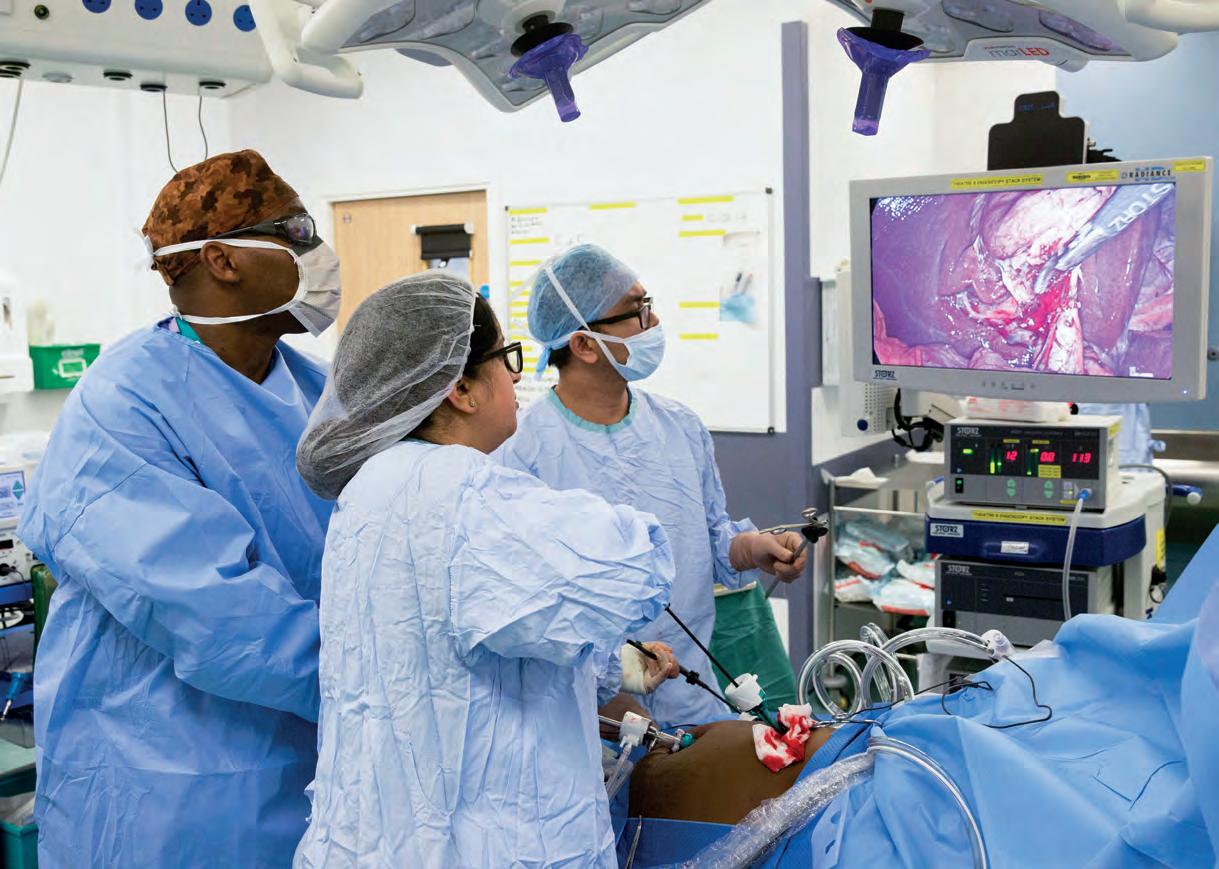
Racial healthcare inequalities and the NHS
Clinical Scientists
Elisa Ly and Sara Majid look at inequality in the health service and ask what action is needed.
The concept of “race” that we have today didn’t always exist. For example, in the 16th Century, the term was used to refer to families or tribes. It was only in the 18th Century, just 300 years ago, that race science and the definition of race that we have today was spawned. Self-proclaimed race scientists were keen to define human categories, to broadly outline “varieties” of human beings, according to the colour of skin and overall appearance. By defining human categories, this unsurprisingly led to a hierarchy of races, with White people placed at the very top.
Horrific surgical experiments were carried out on Black people to try and prove that White people were biologically
better than other races. Many of these experiments were carried out on Black slaves, and without anaesthesia. Invasive gynaecological procedures were carried out on Black women and children without consent. The results of these experiments were used to justify slavery, colonisation, White supremacy, and eugenics.
These experiments and unfounded beliefs about the superiority of White people have fed the medical myths about Black people, still believed by some healthcare professionals today. Incorrect claims such as Black skin is thicker, that nerve endings are less sensitive in Black people, that Black people have a higher pain tolerance, that Black people are physically stronger, and that Black people’s blood coagulates quicker.
IPEM SCOPE 48 WINTER 2022 G F GENERAL FEATURES
IMAGES: MARK THOMAS/SCIENCE PHOTO LIBRARY, ALAMY, SHUTTERSTOCK
This has devastating effects by either consciously or unconsciously altering clinical practice, treatment, and management of Black patients and patients from minoritised backgrounds. This ultimately leads to patient harm, with some having fatal consequences.
Different populations and communities around the world will have genetic variation. This is a scientific fact. However, the problem lies in the fact that false “biological” differences in Black people are being used to justify and perpetuate harmful actions and outcomes. This is a known and documented issue within healthcare – see “Unequal outcomes”, below, for examples.
UNEQUAL OUTCOMES

● Black patients are half as likely to receive pain medication as White patients
● Black women are almost twice as likely to suffer stillbirths
● Black women are four times more likely to die in childbirth
● Black babies are less likely to die when cared for by Black doctors
● The detention rate under the Mental Health Act for ‘Black or Black British’ people is more than four times higher than those in the ‘White’ group
● Patients from minoritised backgrounds receive worse mental healthcare than White patients
● Diagnostic algorithms and practice guidelines containing adjustments for race and ethnicity, show negative effects on patients from minoritised ethnicities
● Pulse oximeters incorrectly monitor oxygen levels in Black patients.
Within the workforce
It is not only patients who experience racerelated inequality within healthcare. The NHS Workforce Race Equality Standard (WRES) report 2021 identified over 20% of the NHS workforce as from Black and Minority Ethnic (BME) backgrounds, yet racism continues to be prevalent. The WRES report highlighted a worsening of the experience of BME staff when compared to White staff in key areas, including discrimination from seniors and sense of equal opportunity. One in six BME staff experienced discrimination personally from a manager. BME staff also endured abuse from patients, their relatives, and the public more frequently than their White colleagues.
White applicants are still 1.61 times more likely to be appointed from shortlisting, showing no improvement in the last six years. BME staff are also 1.14 times more likely to enter the disciplinary process. WRES hope that using detailed demographic analysis at the organisation level will encourage local, regional and national operations to improve diversity through recruitment and promotion. Each organisation should have detailed targets, such as recruitment or leadership development, and the learning shared with similar institutions.
The NHS Race and Health Observatory recently conducted a review of ethnic inequalities in healthcare through the lens of racism. With regard to the NHS workforce, evidence suggested ethnic minority staff were disproportionately affected by the pandemic in terms of access to adequate PPE and negative impact on
FAMOUS SCIENTISTS
Famous scientists throughout history. Let’s take a couple of seconds to think about the names that pop up in your head as you’re reading this.


Maybe you came up with Albert Einstein, Stephen Hawking, Isaac Newton, Marie Curie, Galileo Galilei, Erwin Schrodinger or Alan Turing. Maybe you didn’t, but you probably recognise their names and have some idea of their contributions to our understanding of science, medicine and the world.


Let’s spend a few more seconds to think about this topic. But this time, let’s focus on naming some Black scientists.
How about Alice Bell, Katherine Johnson, Daniel Hale Williams, Charles Drew, James West, Neil DeGrasse Tyson, Madame CJ Walker, or perhaps Patricia Bath?
Chances are that some of you likely found this question more difficult than the last question. This is just an example of implicit biases that we all hold, that is constantly upheld by the media we consume and the current society we live in.
IPEM.AC.UK 49 WINTER 2022 ■ White applicants ■ BAME applicants
of STP
% of successful applicants Specialism (year of entry) Medical Physics (2020) 24 18 12 6 0 Medical Physics (2021)Clinical Engineering (2020)Clinical Engineering (2021)
Success
Applicants Based on Ethnicity
1
mental health. There were numerous incidences of racist abuse from staff and patients, although studies were centred on the nursing profession. Evidence showed racism hampering career progression, development, and ethnic pay gaps for groups identifying as Black, Asian, mixed and other ethnicities.
The report recommends a systematic review of racist experiences for specific professions and settings, and a review of global literature to identify interventions to improve racial inequality in large institutions. Research is needed on how experiences of institutional, structural and interpersonal racism impact mental health and career outcomes, as the latter are likely interlinked. WRES urge to ensure all staff, including those casually employed, are accounted for to build a comprehensive picture of ethnicity within the NHS. NHS England and NHS Improvement are also advised to review recruitment and development procedures to identify the barriers in place for ethnic minority staff.
In the summer of 2020, past and present trainees of the Scientist Training Programme (STP) signed an open letter to the National School of Healthcare Science (NSHCS) asking for better representation of BAME trainees and more action to tackle covert and systemic racism within healthcare science. This led to the founding of the BAME Scientist Trainee Network. The NSHCS responded and has since created an equality, diversity and inclusion committee to discuss these issues. Published ethnicity data for total applications vs. accepted offers are available for the years 2020 and 2021, with only aggregate data available for previous years. Acceptance rates, as shown
EQUALITY AND INCLUSION IS AT
CONVERSATION LIKE NEVER BEFORE
in 1, on the previous page, are consistently higher for White applicants than nonWhite. But what about initial offers, not just acceptance rates? Publishing this data may better reflect the success of both White and non-White applicants.
STP interviewers participate in a voluntary capacity and the school has stated this reflects the diversity of the current workforce. Collection of their ethnicity and diversity data would help understand if the diversity of the current workforce is truly represented and how this compares to the ethnicity of the UK and NHS. Unfortunately, the request to incorporate anti-racism training into the STP curriculum and include trainee reflections on racial discrimination was not met in the syllabus update in 2021. We encourage the school to re-think this, as it is by having open conversations that we can challenge stereotypes and stigmas concerning race.
Only 18% of Heath and Care Professions Council (HCPC) registrants responded to the HCPC Diversity Survey 2021, with 85% of respondents identifying as White. This includes registrants from the 15 health and care professions regulated by the HCPC. The report recognises a need to improve the quantity and quality of diversity data and we welcome the HCPC looking to capture this on a voluntary basis at registration. This need was reiterated in its 2021-2026 EDI strategy. The strategy further identified opportunity for the senior team to
demonstrate clearer, feasible commitment to quality and inclusion through more comprehensive and engaging learning and development, beyond e-learning platforms.
The future of the NHS Equality and inclusion is at the forefront of conversation like never before. Greater diversity in the healthcare workforce is associated with greater patient participation in care, higher patient satisfaction, greater patient adherence to treatment, and greater reach into minoritised communities.
Positive progress has been made but much more can be done to address the inequalities still evident within the NHS, healthcare science and the wider community. More specific research is needed to better understand the racial demographic of healthcare scientists, to help establish practical, tangible policies and practices for racial equality.
By keeping the conversation going, being able to recognise our unconscious biases and advocating for change, we can continue to work towards equality for all. This is vital for the future of the NHS.
IPEM SCOPE 50 WINTER 2022 G F GENERAL FEATURES
THE FOREFRONT OF
Majid
Ly is a Clinical Scientist in Haematological Malignancies at The
NHS
Trust. HOW YOU CAN HELP AS LEADERS AND MANAGERS 1. Attend training – unconscious bias, active allyship, cultural awareness 2. Diverse interview panels 3. Monitoring training opportunities 4. Mentorship 5. Diverse teams 6. Zero tolerance of inappropriate language 7. Equal opportunities to showcase skills at work 8. Practical advice to address specific needs (e.g., prayer room location, halal options at canteen, how to join staff inclusion networks). AS INDIVIDUALS 1. Read WRES reports 2. Actively learning about other’s perspectives 3. Check in with colleagues after witnessing incidents 4. Advocate for others 5. Targeted outreach to schools in disadvantaged areas 6. Review clinical practice, ask if you are contributing to or addressing health inequalities.
Sara
is a Clinical Scientist in Diagnostic Imaging and Radiation Protection at University College London Hospitals NHS Foundation Trust. Elisa
Royal Marsden
Foundation
Adapted with permission (Naima Fredericks, 2021)
Consultant Clinical Scientist
The ingredients for the modern mince pie can be traced to the return of European crusaders from the Holy Land. Middle Eastern methods of cooking, which sometimes combined meats, fruits and spices, were popular at the time. Pies were created from such mixtures of sweet and savoury foods; in Tudor England, shrid pies (as they were known then) were formed from shredded meat, suet and dried fruit. Several authors viewed the pie as being derived from an old Roman custom practised during Saturnalia, where Roman fathers in the Vatican were presented with sweetmeats. Mince pies, at Christmastide, were traditionally shaped in an oblong shape, to resemble a manger and were often topped with a depiction of the Christ Child.
Method
Normal adult volunteers were tested in this trial. Inclusion criteria were adults > 18 years of age, non-smokers and who were not on any medication at the time of trials. Exclusion criteria were pregnant females and subjects with any gustatory malfunction such as dry mouth, loss of appetite, malnutrition, etc.
Participants were unaware of the brand being tested and gave a score from 1–10 on a Likert scale according to taste, texture, visual appearance,
and satiety. The order in which they received each sample was randomised using a computer-generated M-sequence and a washout period of three minutes was allowed between treatments. Data were analysed using a non-parametric one-way fixed effects ANOVA (Kruskal-Wallis). The null hypothesis is zero difference between all brands tested. Data were analysed using Matlab version R2018 for Windows (The Mathwork, Natick, US).
Results
A total of six sample brands were tested on 10 subjects (n = 10) with two males and eight females. The median age of subjects
was 25.5 (range: 22 – 56). Results of the Kruskal-Wallis test are shown in 1. No statistically significant differences were detected (χ2 = 2.54, p = 0.7704).
Conclusion
This study shows that Brand D makes the best mince pies, which explains why mums always go there, although it also generated a huge difference in opinion.
In fact, the variation was so large, it overlapped with all other brands, which explains the null effect for the statistical test. Brand F came a close second, proving that it is not just food they make and whilst Brand B makes exceedingly good cakes, its pies taste very similar to those made by Brands A (where every little helps) and C (where you can live well for less). Brand E was from a less wellknown manufacturer, which served as a placebo.

Comparison study of gustatory receptors’ response to seasonal nutriments:
IPEM.AC.UK 51 WINTER 2022
Azzam F. G. Taktak compares mince pie brands to establish which has the best taste.
E N ENDNOTES
Azzam Taktak is an IPEM Fellow and Consultant Clinical Scientist at Royal Liverpool University Hospital. He is also an Honorary Professor at the University of Liverpool and the University of Manchester.
a double-blind randomised
(WHO
control trial
SELLS THE BEST MINCE PIES?)
1 10 8 6 4 2 0 ABCDEF IMAGE: ISTOCK
CLINICAL RISK MANAGEMENT
We hear from four participants who have undergone an IPEM-commissioned NHS Digital training course.
DCB0129 and DCB0160 are mandatory risk management standards for England, published under section 250 of the Health and Social Care Act 2012. They are applicable for developing (DCB0129) and deploying /operating (DCB0160)
health IT systems, including medical device software. IPEM has recently commissioned NHS Digital to provide clinical risk management training to IPEM members at a discounted rate. The first session of this training was delivered in August 2022 with a second in October 2022.
What did you do?
The course is aimed at providing training for clinical safety officers (CSOs), a term defined by the standards as those who are responsible for ensuring the safety of a health IT system through risk management. Although not
A REPORT
IPEM SCOPE 52 WINTER 2022 E N ENDNOTES
immediately apparent, it was indicated on the course that a registered Clinical Scientist or Engineer can act as a CSO. It covers the central risk management process 1, based on ISO 14971, with practical guidance on how to apply it to health IT systems.
It also explains the different documentation required by the standards:
● Clinical risk management file
● Clinical risk management plan
● Hazard log
● Clinical safety case
● Clinical safety case report
● Safety incident management log.
The course was structured in three parts. Firstly, delegates were required to complete and pass an e-learning course to introduce the key information standards. This training is available for free through the e-learning for health platform, which also is used for IRR/IRMER training. Secondly, there was a set of pre-recorded lectures covering the material in more depth, this was to allow the face-to-face session to be run as a hands-on workshop. Finally, during the workshop there was a brief recap of the lectures with a quiz, followed by a series of group exercises where the attendees worked through the steps of developing and reviewing a clinical safety case and report.
What did you think of it?
Adam Chalkley & Ian Stronach, Clinical Computing and Imaging Sciences Service, University Hospitals Birmingham NHS Foundation Trust
DCB0160 is becoming prevalent in our trust, and we are now likely to require the artefacts specified by these standards when deploying major systems in radiotherapy such as oncology information systems and treatment planning systems. As the process is aligned to ISO 14971, it was familiar, but the course provided useful practical guidance for its application, including tools such as structured what if techniques (SWIFT). It is likely that completing the e-learning would be sufficient for most clinical scientists or engineers involved with system deployment, with the taught course useful for one or two people in a department. The consideration of medical devices in DCB0129/0160 is a relatively new addition, and there are areas where clarity is still required. Part
of the course is based on the premise that technology cannot cause direct harm, and that harm only arises through healthcare staff actions based on information from the health IT system. This is not necessarily the case when considering medical devices, where an IT system may drive equipment that can cause harm directly. Information from DCB0129, shared by the vendor, is meant to feed risk management under DCB0160. It is unclear whether medical device vendors, who have followed ISO 14971, are aware of this English requirement.
Josh Kirby, Radiotherapy Physics, Northern Centre for Cancer Care, Newcastle Hospitals NHS Foundation Trust
The course was very helpful in providing information about the DCB standards and demonstrating the very large scope that they apply to. The majority of the content was covered in the pre-course learning, which then facilitated more useful discussions on the day of the workshop. The workshop will have been particularly useful for anyone practically involved in processes relating to either DCB0129 or DCB160 and the related documentation. There does appear to be a discrepancy between requiring the CSO to be actively involved in the risk assessment process at an individual project level as well as having influence at a high level within the organisation. Further work is needed to explore the practicalities of this and how clinical scientists and other MPCE staff fit into these processes as part of a larger NHS organisation, but this course was a useful
stepping stone for informing our staff group of these mandatory standards.
Claire Tarbert, MDU, Department o Clinical Physics and Bioengineering, NHS Greater Glasgow and Clyde While DCB0160 and DCB0129 aren’t yet mandatory in Scotland, there is an increasing awareness of the hazards around healthcare software, and some Scottish boards are starting to implement the standards voluntarily. The NHS Digital course is well designed and caters for people who have no prior experience of risk management. However, the pre-requisites for the workshop (e-learning, pre-recorded lectures) are extensive, and if you are already familiar with ISO 14971, the e-learning might not feel like time well spent. The opportunity to go through a software-specific example in the workshop and discuss the results was really useful.
It’s clear from the course that the scope of the DCB standards is large (all healthcare software), and they require a significant amount of documentation to be generated and maintained. With the technical skillset that medical physics and clinical engineering staff have and our experience of quality management systems, we might be able to offer a valuable contribution not just in our local departments, but more widely within our trusts and health boards in applying the standards. This could potentially involve becoming clinical safety officers and it could be an opportunity to better develop our relationship with our e-health teams.
1 Clinical Risk Management Process, reproduced from DCB0160 Information from NHS Digital, licensed under the current version of the Open Government Licence
Scope Definition (4.2) Clinical Hazard Identification (4.3) Clinical Risk Estimation (4.4) Initial Clinical Risk Evaluation (5.1) Control Option Analysis (6.1) Clinical Risk Benefit Analysis (6.2) Control Measure Implementation (6.3) Completeness Evaluation (6.4) Deployment (7.1) Post-deployment Monitoring (7.2) Maintenance (7.3) Decomission (7.4) Risk Management Risk Analysis Risk Evaluation Risk Control IMAGE: GETTY IMAGES IPEM.AC.UK 53 WINTER 2022
Handbook of Radiotherapy Physics – Theory & Practice –Second Edition


The treatment of cancer by ionizing radiation – radiotherapy (RT) –has undergone continuous development since the early years of the twentieth century. This book, in two volumes (each containing the contents and index of the complete work) represents a comprehensive updating of the first edition (published in 2007).
High-quality RT outcomes depend on close collaboration by radiation oncologists, therapy radiographers (aka radiation technologists), dosimetrists, physicists and, not infrequently, RT equipment engineers. Whilst this book is primarily written by physicists for physicists, some of the material should be useful, even essential, to anyone whose work involves radiotherapy, including those who train such professionals.
The book is organised into eleven “parts”, covering different topics, plus Part L -
data tables – now including the stopping power and ranges of protons (up to 300 MeV kinetic energy) in elements, compounds and mixtures of medical interest. A total of 62 scientists – eminent specialists (from Europe, North America and elsewhere) – have produced the 61 chapters, including colleagues who did not contribute to the first edition.
Parts A through C cover the fundamentals of the relevant physics, radiobiology and technology. Parts D through H give the practical information necessary for the support of external-beam RT: dose measurements, clinicalbeam properties, the computation of dose distributions in patient anatomy, treatment
(aka dose) planning and quality assurance. RT delivered with radionuclides is described in Part I (brachytherapy) and Part J (unsealed sources aka “molecular radiotherapy”). Part K covers the radiation protection framework, with an emphasis on the legislation in the UK.
We editors hope that our readers will learn as much from reading the book as we have by bringing the 2007 edition up to date. May they be inspired to continue the development of the scientific and technical basis of radiotherapy for the benefit of cancer patients worldwide.


Handbook of Radiotherapy Physics
CRC Press/Taylor & Francis Group (Boca Raton, London, New York) 2022.
IPEM.AC.UK 54 WINTER 2022
BOOK PITCH ENDNOTES E N BOOK PITCH
BOOK IS PRIMARILY WRITTEN BY PHYSICISTS FOR PHYSICISTS
THIS
Philip Mayles, Alan Nahum and Jean-Claude Rosenwald outline the ideas behind their new second edition.


+44(0)
enquiry@osl.uk.com
T-105 • • T-300 • • •
1743 462694
www.osl.uk.com
Physics in Medicine & Biology
iopscience.org/pmb


Physics in Medicine & Biology (PMB) covers the development and application of theoretical, computational and experimental physics to medicine, physiology and biology.


Editor-in-Chief
To find out more about submitting, visit iopscience.org/pmb or e-mail pmb@ioppublishing.org.
RETURN ADDRESS: IPEM Fairmount House 230 Tadcaster Road York YO24 1ES UK
JOURNAL IMPACT FACTOR
