Axillary artery – Arteria axillaris
4.6
The axillary artery is a direct continuation of the subclavian artery. It gives off branches in the axillary fossa and then continues as the brachial artery. It supplies the muscles of the shoulder joint, muscles bordering the axillary fossa, deltoid, lateral thoracic wall including its muscles and the mammary gland. The axillary artery can be divided into three parts according to its relationship with the pectoralis minor. Course
1 Suprapectoral part (pars suprapectoralis) – originates from the subclavian artery when it’s crossing the lateral margin of the 1st rib 2 Retropectoral part (pars retropectoralis) – descends behind the tendon of the pectoralis minor 3 Infrapectoral part (pars infrapectoralis) – is located between the inferior margin of the pectoralis minor and the inferior part of the teres minor and latissimus dorsi (at the level of the surgical neck of the humerus)
Branches and areas supplied
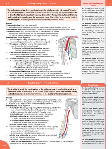
• 1 Superior thoracic artery (arteria thoracis superior) – supplies the pectoral muscles and the mammary gland • 2 Thoraco-acromial artery (arteria thoracoacromialis)
2 1 3
– runs through the clavipectoral triangle 4 2.1 Acromial and deltoid branch, pectoral branches 6 (ramus acromialis, deltoideus et rami pectorales) 5 3 Lateral thoracic artery (arteria thoracica lateralis) 4.1 – descends on the serratus anterior, which it supplies 4.2 3.1 Lateral mammary branches (rami mammarii laterales) 4 Subscapular artery (arteria subscapularis) 4.1 Circumflex scapular artery (arteria circumflexa scapulae) – passes through the omotricipital foramen to the infraspinous fossa where it supplies the posterior scapular muscles 4.2 Thoracodorsal artery (arteria thoracodorsalis) – passes over the latissimus dorsi, which it supplies 5 Anterior circumflex humeral artery (arteria circumflexa humeri anterior) – a smaller artery located on the anterior surface of the surgical neck of the humerus 6 Posterior circumflex humeral artery (arteria circumflexa humeri posterior) – a larger artery that passes through the humerotricipital foramen to supply the deltoid
• •
• •
• •
4.7
Brachial artery – Arteria brachialis
The brachial artery is the continuation of the axillary artery. It supplies the whole arm and elbow joint. It terminates in the cubital fossa where it bifurcates into the ulnary and radial arteries. Its collaterals terminate in the cubital articular anastomosis. Course
1. originates from the axillary artery, close to the inferior margin of the teres major and latissimus dorsi, at the level of the surgical neck of the humerus 2. descends on the medial surface of the arm to the cubital fossa
Branches and areas supplied
•
1 Profunda brachii artery / deep artery of arm (arteria profunda brachii) – runs between the lateral and medial heads of the triceps brachii and then passes in the radial canal with the radial nerve 1.1 Deltoid branch (ramus deltoideus) – supplies the deltoid 1.2 Middle collateral artery (arteria collateralis media) – enters the medial head of the triceps brachii 1.3 Radial collateral artery (arteria collateralis radialis) 1 – terminates in the cubital articular anastomosis 3 2 Humeral nutrient artery (arteria nutritia humeri) 3 Superior ulnar collateral artery (arteria collateralis ulnaris superior) – descends on the anterior surface in the medial intermuscular 4 septum of the arm with the ulnar nerve – terminates in the cubital articular anastomosis 5 4 Inferior ulnar collateral artery (arteria collateralis ulnaris inferior) – arises just above the cubital fossa – enters the cubital articular anastomosis 5 Cubital articular anastomosis (rete articulare cubiti) – a vascular plexus of the elbow joint – formed by the brachial, radial, and ulnar arteries
•
• •
284
Heart and blood vessels Arteries
The lateral thoracic artery descends on the surface of the serratus anterior with the long thoracic nerve. The thoracodorsal artery runs on the inner surface of the latissimus dorsi with the thoracodorsal nerve. The posterior circumflex humeral artery can be damaged in a fracture of the surgical neck of the humerus. The radial artery is palpable in the radial foveola (the “anatomical snuff-box”). The princeps pollicis artery is an inaccurate but widespread term, usually synonymous with the first palmar metacarpal artery. The radialis indicis artery is synonymous with the radial proper palmar digital artery. It most commonly arises from the first palmar metacarpal artery. The superficial palmar arch is completely formed only in 27 % of cases. It is unclosed in a majority of cases. The arteria comitans nervi mediani (median artery) accompanies the median nerve and can be variably enlarged. The artery supports the arterial supply of the palm.
Clinical notes The thoracodorsal artery serves as a nutritive vessel for muscle and myocutaneous flaps in plastic and reconstructive surgeries. When measuring blood pressure, the stethoscope is placed over the brachial artery, medially to the biceps brachii tendon in the cubital fossa. Pulse is most usually palpated by compressing the radial artery against the radius in the distal part of the forearm, a few centimetres proximal to the wrist. The radial artery serves as an approach for diagnostic and therapeutic coronary catheterisation. The ulnar artery is less commonly used The radial artery can be used to create an arterio-venous fistula (anastomosis) for haemodialysis. The most common type is the radiocephalic fistula. The radial artery can be also used as a graft in coronary artery bypass surgery. Its diameter is 2.0–3.2 mm.