Uterus – Uterus
3.3
Reproductive system
The uterus is a pear-shaped organ located in the lesser pelvis between the urinary bladder and rectum. Its mucosa undergoes cyclic transformation, synchronised with the ovarian cycle. This ensures that the uterus is ready for implantation and the subsequent development of the fertilised ovum. It is positioned in anteversion and anteflexion and is almost completely covered by peritoneum. Due to its position, spermatozoa can easily reach the uterine cavity and it provides sufficient space for a growing embryo/foetus. External structure
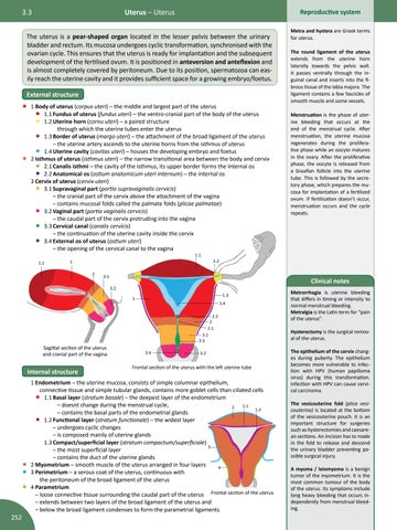
• 1 Body of uterus (corpus uteri) – the middle and largest part of the uterus • 1.1 Fundus of uterus (fundus uteri) – the ventro-cranial part of the body of the uterus • 1.2 Uterine horn (cornu uteri) – a paired structure through which the uterine tubes enter the uterus • 1.3 Border of uterus (margo uteri) – the attachment of the broad ligament of the uterus – the uterine artery ascends to the uterine horns from the isthmus of uterus • 1.4 Uterine cavity (cavitas uteri) – houses the developing embryo and foetus • 2 Isthmus of uterus (isthmus uteri) – the narrow transitional area between the body and cervix • 2.1 Canalis isthmi – the cavity of the isthmus, its upper border forms the internal os • 2.2 Anatomical os (ostium anatomicum uteri internum) – the internal os 3 Cervix of uterus (cervix uteri) • 3.1 Supravaginal part (portio supravaginalis cervicis) – the cranial part of the cervix above the attachment of the vagina – contains mucosal folds called the palmate folds (plicae palmatae) • 3.2 Vaginal part (portio vaginalis cervicis) – the caudal part of the cervix protruding into the vagina • 3.3 Cervical canal (canalis cervicis) – the continuation of the uterine cavity inside the cervix • 3.4 External os of uterus (ostium uteri) – the opening of the cervical canal to the vagina 1.1
1.1
1 2
1.3
1
1.4 2.2 2 2.1 3.1 3.3 3.4
3.2
Frontal section of the uterus with the left uterine tube
1 Endometrium – the uterine mucosa, consists of simple columnar epithelium, connective tissue and simple tubular glands, contains more goblet cells than ciliated cells • 1.1 Basal layer (stratum basale) – the deepest layer of the endometrium
– doesnt change during the menstrual cycle, 1.1 2 1.2 – contains the basal parts of the endometrial glands 1.2 Functional layer (stratum functionale) – the widest layer – undergoes cyclic changes – is composed mainly of uterine glands 1.3 Compact/superficial layer (stratum compactum/superficiale) 3 – the most superficial layer – contains the duct of the uterine glands 2 Myometrium – smooth muscle of the uterus arranged in four layers 4 3 Perimetrium – a serous coat of the uterus, continuous with the peritoneum of the broad ligament of the uterus 4 Parametrium – loose connective tissue surrounding the caudal part of the uterus Frontal section of the uterus – extends between two layers of the broad ligament of the uterus and – below the broad ligament condenses to form the parametrial ligaments
•
• • •
252
Menstruation is the phase of uterine bleeding that occurs at the end of the menstrual cycle. After menstruation, the uterine mucosa regenerates during the proliferative phase while an oocyte matures in the ovary. After the proliferative phase, the oocyte is released from a Graafian follicle into the uterine tube. This is followed by the secretory phase, which prepares the mucosa for implantation of a fertilized ovum. If fertilisation doesn’t occur, menstruation occurs and the cycle repeats.
Clinical notes
3.2
Internal structure
The round ligament of the uterus extends from the uterine horn laterally towards the pelvic wall. It passes ventrally through the inguinal canal and inserts into the fibrous tissue of the labia majora. The ligament contains a few fascicles of smooth muscle and some vessels.
1.2
3.1
Sagittal section of the uterus and cranial part of the vagina
Metra and hystera are Greek terms for uterus.
Metrorrhagia is uterine bleeding that differs in timing or intensity to normal menstrual bleeding. Metralgia is the Latin term for “pain of the uterus”. Hysterectomy is the surgical removal of the uterus. The epithelium of the cervix changes during puberty. The epithelium becomes more vulnerable to infection with HPV (human papilloma virus) during this transformation. Infection with HPV can cause cervical carcinoma. The vesicouterine fold (plica vesicouterina) is located at the bottom of the vesicouterine pouch. It is an important structure for surgeries such as hysterectomies and caesarean sections. An incision has to made in the fold to release and descend the urinary bladder preventing possible surgical injury. A myoma / leiomyoma is a benign tumor of the myometrium. It is the most common tumour of the body of the uterus. Its symptoms include long heavy bleeding that occurs independently from menstrual bleeding.