14
Hypophysis / Pituitary gland – Hypophysis / Glandula pituitaria
3
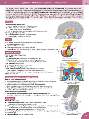
The pituitary gland is a small gland located in the hypophyseal fossa of the sphenoid bone (sella turcica). The pituitary stalk connects the pituitary gland to the hypothalamus. The hypothalamus controls the activity of the pituitary gland. The hypophysis and hypothalamus form the hypothalamohypophysial system/axis, which is responsible for the majority of hormonal regulation and is also closely related to the autonomic nervous system. The anterior part of the pituitary gland is called adenohypophysis and is derived from the roof of the stomodeum (ectoderm). The posterior part, termed neurohypophysis, represents a pouch of the diencephalon at the base of the third ventricle (derived from the neuroectoderm). Structure 1 Adenohypophysis (anterior lobe) 1.1 Pars distalis – the major part of the anterior lobe 1.2 Pars intermedia – a strip of hypophyseal tissue at the border with the posterior lobe 1.3 Pars tuberalis – a layer surrounding the stalk of the posterior lobe 2 Neurohypophysis (posterior lobe) 2.1 Infundibulum – the stalk of the posterior lobe 2.2 Neural lobe – the posterior lobe itself
• • • • •
Syntopy
• 3 Cranially: hypothalamus (median eminence, tuber cinereum) • 4 Ventrocranially: optic chiasm 4 • 5 Ventrocaudally: sphenoid sinus • 6 Dorsally: dorsum sellae, basilar artery and pons • 7 Laterally: cavernous sinus (with 3 , 4 , 5 and 6 cranial nerves and internal carotid artery) rd
th
th
3 1.3 2.1
th
Histological structure Anterior lobe (adenohypophysis)
1 Pars distalis 1.1 Chromophobe cells – stem cells or hormone secreting cells 1.2 Chromaffin cells – somatotrophs, mammotrophs, gonadotrophs, thyreotrophs, corticotrophs (named according to the target gland) 2 Pars intermedia – corticotrophs 3 Pars tuberalis – gonadotrophs or thyreotrophs
Posterior lobe (neurohypophysis)
1 Axons of neuroendocrine neurons – the cell bodies are positioned in the supraoptic and paraventricular nuclei of the hypothalamus – the hypothalamohypophysial tract transports secretory granules to the hypophysis 2 Pituicytes – glial cells
Functional connections with the hypothalamus Anterior lobe (adenohypophysis)
1.1 6
1.2
2.2
Close-up of a sagittal section of hypothalamus and hypophysis 3 4 1.3 1.1
7 5
1. hormones are secreted in the arcuate nucleus (parvocellular part) of the hypothalamus and stimulate or inhibit cells of the adenohypophysis 2. hormones are carried via axonal transport to the median eminence where there are released into capillaries during a process called neurosecretion 3. the portal system of the hypophysis transmits hormones to the chromaffin cells 4. secretion of hormones into the blood stream
Frontal section of the hypophysis and cavernous sinus
Posterior lobe (neurohypophysis)
1. neuroendocrine secretion from axons of the hypothalamohypophysial tract (supraoptic and paraventricular nucleus) to the drainage area of the inferior hypophysial artery
Blood supply Arterial blood supply:
1
• 1 Superior hypophysial artery (a branch of the internal carotid artery) 3 – supplies the infundibulum and median eminence • 2 Portal system of the hypophysis – originates from the spiral arteries in the pituitary stalk and continues to the second capillary bed in the adenohypophysis • 3 Trabecular artery – supplies the pars distalis of the anterior lobe • 4 Inferior hypophysial artery (branch of the internal carotid artery) – supplies the posterior lobe Venous drainage:
• 5 Hypophysial veins (into the cavernous sinus)
2 4
5
Scheme of the hypothalamohypophysial system (sagittal section)
523