1.4
13
Organ of hearing and balance – Organum vestibulocochleare
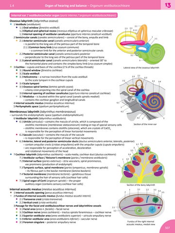
Internal ear / vestibulocochelar organ (auris interna / organum vestibulocochleare) Osseous labyrinth (labyrinthus osseus)
• 1 Vestibule (vestibulum) • 1.1 Oval window (fenestra vestibuli) 1.2 Elliptical and spherical recess (recessus ellipticus et sphericus maculae cribrosae) 1.3 Internal opening of vestibular canaliculus (apertura interna canaliculi vestibuli) 2 Semicircular canals (canales semicirculares) – consist of the bony, ampulla and limb • 2.1 Anterior semicircular canal (canalis semicircularis anterior)
– is parallel to the long axis of the petrous part of the temporal bone 2.1.1 Common bony limb (crus osseum commune) – a common limb for the anterior and posterior semicircular canals 2.2 Posterior semicircular canal (canalis semicircularis posterior – perpendicular to the long axis of the petrous part of the temporal bone 2.3 Lateral semicircular canal (canalis semicircularis lateralis) – oriented 30° to the horizontal plane and contains the simplex bony limb (crus osseum simplex) 3 Cochlea – cupula and basis of the cochlea (2 ¾ of the cochlea threads) 3.1 Round window (fenestra cochleae) 3.2 Scala vestibuli 3.3 Helicotrema – a narrow transition from the scala vestibuli to the scala tympani in the cochlear cupula 3.4 Scala tympani 3.5 Osseous spiral lamina (lamina spiralis ossea) – a bony crest projecting into the spiral canal of the cochlea 3.6 Internal opening of cochlear canaliculus (apertura interna canaliculi cochleae) 3.7 Modiolus – is located within the spiral canal (canalis spiralis modioli) – contains the cochlear ganglion and longitudinal canals 4 Internal acoustic meatus (meatus acusticus internus) 5 Perilymphatic space (spatium perilymphaticum)
2.3
2.1 2.1.1 1
• • • • • • • • •
Membranous labyrinth (labyrinthus membranaceus)
2.2
1.1
3.1 3
Lateral view of the osseous labyrinth 2.3
2.1
6.3
2.2
3.3
3.4
6.1 1 6.2
– surrounds the endolymphatic space (spatium endolymphaticum) 3.2 7 6 Vestibular labyrinth (labyrinthus vestibularis) 6.1 Utricle (utriculus) – contains the macula of utricle, which is composed of the Section of the inner ear otolithic membrane (membranae statoconiorum) resting on top of special sensory cells – otolithic membranes contains otoliths (otoconia), which are crystals of CaCO3 is responsible for the perception of linear horizontal movements 6.2 Saccule (sacculus) – contains the macula of the saccule – is responsible for the perception of linear vertical movements 6.3 Anterior, lateral and posterior semicircular ducts (ductus semicircularis anterior, lateralis, posterior) 3.7 3.3 – contain ampullar crests (cristae ampullares) with the ampullar cupula (cupula ampullaris) 3 – are responsible for perception of acceleration, deceleration 3.5 and rotational movements of the head 3.4 7 Cochlear labyrinth (labyrinthus cochlearis) – scala media, cochlear duct (ductus cochlearis) 7.1 Vestibular surface / Reissner’s membrane (paries / membrana vestibularis) 7 3.2 7.2 External surface (paries externus) – stria vascularis, spiral prominence, vas prominens (production of endolymph) 7.5 7.3 Tympanic surface, spiral membrane (paries tympanicus, membrana spiralis) – its fibrous part is the basilar membrane (lamina basilaris) 7.4 Tectorial membrane (membrana tectoria) – gelatinous tissue surrounding the hair of sensory cells (cochlear hair cells) 7.5 Spiral organ of Corti (organum spirale) – the proper auditory organ (contains sensory cochlear hair cells) Section of the bony labyrinth
3
• • • • •
Internal acoustic meatus (meatus acusticus internus)
• 1 Internal acoustic opening (porus acusticus internus) • 2 Fundus of internal acoustic meatus (fundus meatus acustici interni) • 2.1 Transverse crest (crista transversa) • 2.2 Vertical crest (crista verticalis) 3 Passage for the facial and vestibulocochlear nerves and labyrinthine vessels 2.1 • 3.1 Facial area (area n. facialis) – facial nerve 3.2 3.2 Cochlear nerve (area cochlearis) – tractus spiralis foraminosus – cochlear nerve • • 3.3 Superior vestibular area (area vestibularis superior) – utriculo-ampullary nerve • 3.4 Inferior vestibular area (area vestibularis inferior) – saccular nerve • 3.5 Foramen singulare – posterior ampullary nerve
2
1
3.1
2.2
3.3
Fundus of the right internal acoustic meatus, medial view
3.4
3.5
507