Improving Time to Diagnosis
mechanical support, or the need for palliation.2,3,18–21 The temperature of the extremities as assessed by palpation may also used as an indicator of cardiac output. Cool peripheries are present in up to 24 % of unselected patients.19 The Forrester classification system based upon whether patients have adequate circulation (warm or cold) alongside the presence or absence of congestion (wet or dry) is no longer included in current guidelines but provides a useful guide to applied haemodynamics.29,30 Assessment of the heart rate and rhythm will help to elicit important arrhythmias (e.g. atrial fibrillation), which may have triggered the presentation and will influence management. Other important ancillary signs, which indicate underlying structural heart disease, include cardiac murmurs. The most prevalent features of congestion are peripheral oedema (65–68 %) and bibasal crepitations or rales (64– 80 %), which in patients presenting to the emergency department with undifferentiated dyspnoea offer sensitivity and specificity of 51 and 76 % and 60 and 78 %, respectively.2,3,18–21 Measurement of the jugular venous pressure has been an integral part of bedside cardiology since its inception. In expert hands the value of jugular venous distension as an indicator of elevated right atrial pressure and its correlation with raised pulmonary capillary wedge pressure has been elegantly demonstrated in advanced chronic HF with excellent specificity.31,32 Yet even in this context the practical utility of this sign has not been universal, while in undifferentiated patients with acute dyspnoea sensitivity falls to as low as 39 %, perhaps reflecting in part a more general lack of confidence among the newer generation of clinicians in eliciting such signs.33–36 Similarly the third heart sound, a low pitched sound best heard over the left ventricular apex in conjunction with the rapid filing phase of ventricular diastole demonstrated excellent specificity and was an independent predictor of mortality in a large cohort of AHF patients in Japan.37 However, a lack of familiarity in the detection of this sign and poor inter-observer variability has been documented elsewhere, reflected in its low sensitivity (13–40 %) in patients with AHF presenting to the emergency department, even when augmented by acoustic cardiography (computerised measurement of the heart sounds with a synchronous electrocardiogram [ECG] trace using specialised electrodes placed at V3–V4).34,38–40
Special Considerations Alongside an ageing population the demographics of HF patients are changing, e.g. changing cardiovascular risk profile (reduction in incidence of myocardial infarction) and developments in cardiac care (e.g. primary percutaneous coronary intervention). Some contemporary epidemiological series indicate the mean age at diagnosis may be as high as 80 years old.41 Patients in this category warrant special consideration and particular vigilance from the assessing clinician. Presentation may be subtle and more gradual, perhaps marked by atypical symptoms (e.g. anorexia and weight loss), with co-morbid conditions (e.g. musculoskeletal disease) acting as confounders to potentially decrease sensitivity.42 In contrast to their younger counterparts, elderly patients (>85 years) with AHF are more likely to be female and have a preserved EF.43 Atrial fibrillation, hypertension, anaemia, renal dysfunction and cerebrovascular disease are all more common – while diabetes and coronary artery disease are conversely less frequent – perhaps implying that patients with these conditions are more likely to present earlier.44 The expected typical examination findings are also less frequent compared with younger (<65 years of age) patients.45
CARDIAC FAILURE REVIEW
Sharma_FINAL.indd 71
Table 2: Clinical Assessment in Acute Heart Failure – History Clinical Features Prevalence (%)
Sensitivity (%) Specificity (%)
Background
Lowest
Highest
Heart failure
72
88
60
90
Myocardial
22
31
40
87
44
57
52
70
Hypertension
60
73
60
56
Diabetes
27
45
28
87
Atrial fibrillation
31
40
NR
NR
COPD
9
31
34
57
Dyspnoea
61
77
84
34
Orthopnoea
27
59
50
77
Paroxysmal
15
53
41
84
infarction Coronary artery disease
Symptoms
nocturnal dyspnoea Prevalence figures compiled from the following international registries of patients presenting to hospital with acute heart failure: ADHERE,2 OPTIMIZE,3 EFICA,18 ATTEND,19 IMPACT-HF,20 and EHFSII.21 Sensitivities and specificities adapted from a separate metaanalysis by Wang et al.34 – a synthesis of data from 22 observational series of patients presenting with undifferentiated dyspnoea. COPD = chronic obstructive pulmonary disease.
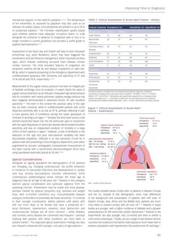
Figure 1: Clinical Assessment in Acute Heart Failure – Examination
Jugular venous distension Prevalence 39–52 % Sensitiviy 39% Specificity 78 %
Third heart sound Prevalence 11–41 % Sensitivity 13 % Specificity 99 %
Haemodynamic disturbance SBP < 90 mmHg 5–10 % SBP 90–140 mmHg 42–46 % SBP > 140 mmHg 44–50 % Peripheral oedema Prevalence 27–68 % Sensitivity 51 % Specificity 76 %
Pulmonary crepitations Prevalence 64–71 % Sensitivity 60 % Specificity 78 % SBP = systolic blood pressure.
The studies detailed above mostly refer to patients in Western Europe and the US. Outside of this demographic some main differences in the background and presentation of patients with AHF exist. In Eastern Europe, Asia, Africa and the Middle East, patients are much more likely to present acutely with de novo HF.46–50 Patients in Saudi Arabia are younger, with a higher incidence of diabetes and a greater preponderance for left ventricular systolic dysfunction.50 Patients in the Asian-Pacific are also younger, less co-morbid and tend to exhibit a more severe phenotype.49 Finally, across a range of sub-Saharan African countries the incidence of ischaemic heart disease is much lower than in western populations with hypertension the predominant HF aetiology.48
71
19/10/2015 14:06