Cardiac or Other Implantable Electronic Devices and Sleep-disordered Breathing
excessive daytime sleepiness.10,11 CSA is characterised by repetitive cessation of ventilation during sleep resulting from a loss of ventilatory drive. A central apnoea is a 10-second pause in ventilation with no associated respiratory effort. CSA is present when a patient has five or more central apnoeas or hypopnoeas per hour.10,12
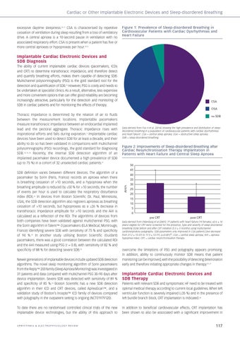
Figure 1: Prevalence of Sleep-disordered Breathing in Cardiovascular Patients with Cardiac Dysrhythmias and Heart Failure
Implantable Cardiac Electronic Devices and SDB Diagnosis The ability of current implantable cardiac devices (pacemakers, ICDs and CRT) to determine transthoracic impedance, and therefore detect and quantify breathing efforts, makes them capable of detecting SDB. Multichannel polysomnography (PSG) is the gold standard tool for the detection and quantification of SDB.13 However, PSG is costly and needs to be undertaken at specialist clinics. As a result, alternative, less expensive and more convenient options that can offer good reliability are becoming increasingly attractive, particularly for the detection and monitoring of SDB in cardiac patients and for monitoring the effects of therapy.
CSA OSA
ability to do so has been validated in comparisons with multichannel polysomnography (PSG) recordings, the gold standard for diagnosing SDB.7,8,14–20 Recently, the internal SDB detection algorithm of an implanted pacemaker device documented a high prevalence of SDB (up to 75 %) in a cohort of 32 unselected cardiac patients.21 SDB definition varies between different devices. The algorithm of a pacemaker by Sorin (Paris, France) records an apnoea when there is breathing cessation of >10 seconds, and a hypopnoea when the breathing amplitude is reduced by ≥50 % for >10 seconds; the number of events per hour is used to calculate the respiratory disturbance index (RDI).22 In devices from Boston Scientific (St. Paul, Minnesota, USA), the SDB detection algorithm also registers apnoeas as breathing cessation of >10 seconds, but hypopnoeas as a ≥26 % decrease in transthoracic impedance amplitude for >10 seconds and the AHI is calculated as a reflection of the RDI. The algorithms of devices from both companies have been validated against multichannel PSG, with the Sorin algorithm in Talent™-3 pacemakers (ELA Medical, Montrouge, France) identifying severe SDB with sensitivity of 75 % and specificity of 94 %.22 In another study utilising Boston Scientific (Guidant) pacemakers, there was a good correlation between the calculated RDI and the AHI measured using PSG (r = 0.8), with sensitivity of 82 % and specificity of 88 % for detecting severe SDB.23 Newer generations of implantable devices include updated SDB detection algorithms. The novel sleep monitoring algorithm of Sorin pacemakers from the Reply™ 200 family (Sleep Apnoea Monitoring) was investigated in 31 patients and data compared with multichannel PSG 30–90 days after device implantation. Severe SDB was detected with sensitivity of 89 % and specificity of 85 %.24 Boston Scientific has a new SDB detection algorithm in their ICD and CRT devices, called ApneaScan™, and a validation study of Boston’s Incepta™ ICD family of devices compared with polygraphy in the outpatient setting is ongoing (NCT01979120). To date there are no randomised controlled clinical trials of the new implantable device technologies, but the ability of this approach to
ARRHYTHMIA & ELECTROPHYSIOLOGY REVIEW
Fox_FINAL.indd 117
no SDB
Data derived from Fox H et al. [2014] showing the high prevalence and distribution of sleepdisordered breathing in a population of cardiovascular patients with cardiac dysrhythmias and heart failure9. CSA = central sleep apnoea; OSA = obstructive sleep apnoea; SDB = sleep-disordered breathing.
Figure 2: Improvements of Sleep-disordered Breathing after Cardiac Resynchronisation Therapy Implantation in Patients with Heart Failure and Central Sleep Apnoea 50 45 40 35 AHI/h
Thoracic impedance is determined by the relation of air to fluids between the measurement locations. Implantable pacemakers measure transthoracic impedance between an endocardial implanted lead and the pectoral aggregate. Thoracic impedance rises with inspirational efforts and falls during expiration.4 Implantable cardiac devices have been used to detect SDB for at least a decade, and their
30 25 20 15 10 5 0 pre CRT
post CRT
Data derived from Oldenburg et al [2007]; 77 patients with heart failure (19 females; 62.6 ± 10 years) eligible for CRT were screened for the presence, type and severity of sleep-disordered breathing (SDB) before and after CRT initiation (5.3 ± 3 months) using multichannel cardiorespiratory polygraphy. SDB parameters only improved in CSA patients [AHI decrease from 31.2 ± 15.5/h to 17.3 ± 13.7/h, p<0.001]26; CSA = central sleep apnoea; AHI = apnoea hypopnea index; CRT = cardiac resynchronisation therapy.
overcome the limitations of PSG and polygraphy appears promising. In addition, ability to continuously monitor SDB means that patient monitoring can be improved, with the possibility of detecting deterioration early and therefore initiating appropriate changes in therapy.25–27
Implantable Cardiac Electronic Devices and SDB Therapy Patients with relevant SDB and symptomatic HF need to be treated with optimal medical therapy according to current local guidelines. When left ventricular function is severely impaired (≤35 %) and in the presence of left bundle branch block, CRT implantation is indicated.28 In addition to beneficial cardiovascular effects, CRT implantation has been shown to also be associated with a significant improvement in
117
15/08/2014 12:17