40
Society spotlight
Operating on the cutting edge
Virtual planning and 3D modelling technology is helping surgeons at Chris O’Brien Lifehouse perform intricate operations with tremendous results. When 46-year-old mother-of-two Tara Flannery first noticed a lesion on her upper gum, she never could have imagined the road that lay ahead. “I received a call saying I needed to meet with the surgical team at the Lifehouse Centre because my lesion was something more sinister – cancer,” she said. After months of uncertainty and multiple biopsies, Tara was diagnosed with a squamous cell cancer. It was already surrounding her teeth and her medical team recommended the complete removal of her upper jaw. Surgically, it was far from the easiest option but it would give her the best chance of a cure. On its own, the removal of Tara’s cancer would leave significant cosmetic deformities and make it difficult, if not impossible, for her to chew and swallow. But the surgical team at Chris O’Brien Lifehouse utilised the collective expertise of head and neck ablative and reconstructive surgeons, maxillofacial surgeons, and prosthodontists from Westmead Oral Restorative Sciences to map out the removal and complete reconstruction of Tara’s jaw. Associate Professor Carsten Palme, Director of Head and Neck Surgery at Lifehouse, was one of Tara’s surgeons. He said that while medical teams are good at getting cancers out, historically they have not measured up when it comes to rehabilitation. “Dental rehabilitation is crucial to restoring a patient’s appearance and function and allowing them to successfully integrate back into work and society,” Associate Professor Palme said. Lifehouse surgeons have expertise in virtual surgical planning, allowing them to comprehensively map out a patient’s
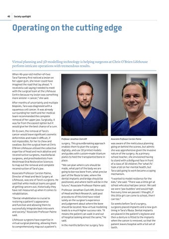
Professor Jonathan Clark AM
Associate Professor Carsten Palme
surgery. This groundbreaking approach enables them to plan the surgery digitally, and use 3D printed models and guides with custom-made titanium plates to hold the transplanted bone in place.
was aware of the meticulous planning going on behind the scenes, but admits she was apprehensive given the invasive nature of the surgery. As a primary school teacher, she envisioned having to stand with a disfigured face in front of a class of 30 children.The ‘what ifs’ took a toll on her mental health, but Tara said going to work became a coping mechanism.
“We can plan where cuts should be made, what part of the body we are going to borrow bone from, what precise part of the fibula to take, where the dental implants and bridge should be positioned, and where teeth will be in the future,” Associate Professor Palme said. Professor Jonathan Clark AM, Director of Head and Neck Research, said past procedures of this kind have relied solely on the surgeon’s experience and judgement about where the bone should be located. Now virtual modelling leads to a much higher success rate. “It means the patient can walk in and out of hospital looking almost the same,” he said. In the months before her surgery Tara
“I wanted to model resilience for the kids,” she said. “There was a little girl at school who also had jaw cancer. We said we were ‘jaw buddies’ and would high five every time we passed. I thought, if this little girl can come to school, then I can too.” Six weeks before Tara’s surgery, surgeons placed implants and a new gum lining in Tara’s fibula. Dental implants are placed in the patient’s leg bone and then a denture is fitted to the implants when the cancer is removed, so that the patient leaves hospital with a full set of teeth.