Knowledge
Vet Insight
Hallmarq was founded 20 years ago by Dr Nick Bolas, who wanted to develop a diagnostic imaging system that would assist vets to accurately diagnose a horse’s injury. Nick and his team of imaging experts worked with local vets in the South-East of the UK, to develop the world’s first Standing MRI system for horses. Since then, Hallmarq has continued to improve equine MRI technology and has also gone on to develop a vet-specific, small animal MRI system, using expertise from across the company
Photography by Hallmarq Veterinary Imaging
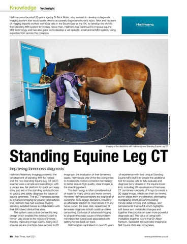
Imaging of the distal limb with Hallmarq’s new Standing Equine Leg CT
Standing Equine Leg CT
Improving lameness diagnosis Hallmarq Veterinary Imaging pioneered the development of standing MRI for horses and the new Standing Equine Leg CT (slCT) scanner uses a simple and safe design, with a unique low, flat platform for quick and easy entry and exit of the standing sedated horse to quickly and safely diagnose the cause of equine lameness. The slCT increases access to advanced imaging for equine vet practices and Hallmarq has had success imaging standing sedated horses in collaboration with their UK-based clinical trial sites. The system uses a dual-concentric ring design which enables the detector plate to remain very close to the region of interest, thereby improving image quality. Using slCT ensures equine practices have access to 3D
58
Polo Times, April 2021
imaging in the evaluation of their lameness cases. Hallmarq is one of the few companies to incorporate motion correction technology to better ensure high quality, clear images in the standing patient. This technology is often considered out of reach for many clinics and horse owners. However, Hallmarq considers the total cost of ownership in its design decisions, providing an affordable solution to most clinics. For any horse owner, the treat, rest, repeat loop of lameness diagnosis is both costly and time consuming. Early use of advanced imaging to pinpoint the exact cause of the problem minimises the overall cost associated with getting horses back on track. Hallmarq has capitalised on over 20 years
of experience with their unique Standing Equine MRI (sMRI) to create this additional tool for equine vets to fully evaluate and diagnose bony disease in the equine lower limb, including 3D visualisation of fractures. CT combines hundreds of X-rays to create a 3D digital image, which can then be viewed as thin slices from any direction, eliminating overlapping structures and revealing minute detail in bone and cartilage. slCT complements their sMRI which highlights soft tissue and metabolic changes and together they provide an even more powerful diagnostic aid. The value of using both modalities together is one that Dr Alison Fairburn, Specialist in Diagnostic Imaging at Bell Equine Vets also recognises,
www.polotimes.co.uk