New Laboratory at New Bolton Center New Laboratory at New Bolton Center
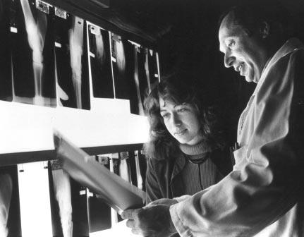
Adedication ceremony was held on June 17 at New Bolton Center, the large animal facility of the University of Pennsylvania School of Veterinary Medicine, to mark the completion of The Richard S. Reynolds, Jr. Comparative Orthopedic Research Laboratory.
The new laboratory is named as a tribute to Richard S. Reynolds, Jr., a former University Trustee. Housed in a 2,800 square-foot addition to the C. Mahlon Kline
Orthopedic and Rehabilitation Center, it includes a mechanical testing facility, a computer and microscope viewing room, a bone morphology unit and an orthopedic engineering and machine shop.
43 (continued on page 3)
1
University of PennsylvaniaSummer 1998 Newsmagazine of the School of Veterinary Medicine
43
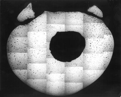
Microradiograph of a cross section of a cannon bone.
From the Dean
Too often we read reports of international comparisons showing that US school children are poor in mathematics and science. Inevitably, this sets off a national debate about the quality and dedication of science teachers in this country. It has always struck me as curious that in spite of these discouraging results public support for science in the United States is markedly greater than anywhere else. This country leads the world in the quality and magnitude of its scientific research program; our science is the envy of every other country on earth. I was therefore interested to read a recent publication by the National Science Foundation entitled Science and Engineering Indicators, 1998, showing that an astonishing 70% of Americans claim they are interested in science and among the adult population, understanding of science is higher than in many other countries.
Could it be that our science teachers are doing a better job than the international comparisons lead us to believe? I would like to think so and would like to see the comparisons extended to look at long term retention of scientific information by school children and young adults in different countries. However, I believe there are additional factors at work. The National Science Foundation report goes on to show that the American people are vastly more positive about the impact of science and technology than those in Europe and Japan. In these countries advances in transgenesis, the experimental modification of individual genes in plants and animals pioneered by the School’s distinguished faculty, Ralph Brinster, are surrounded by suspicion and fear. The Swiss even held a referendum on whether to permit the creation of transgenic plants and animals in their country despite the fact that they have a large pharmaceutical industry and transgenic techniques are enormously valuable to medicine and agriculture.
In the United States, I believe our news media also play a role. They are replete with scandal and gloomy stories about ethnic cleansing, drug offences, abuses of
First International Feline Genetic Disease Conference
More than 120 scientists from the US, Canada and Europe met at VHUP the end of June for the First International Feline Genetic Disease Conference, organized by Dr. Urs Giger, Charlotte Newton Sheppard Professor of Medicine and, chief, Section of Medical Genetics. Two days were devoted to the presentation of scientific papers on feline hereditary disorders and the feline gene map focusing on the future of feline health. The third day offered a series of presentations on the same topics for cat breeders and veterinary practitioners. This session drew more than 180 participants. The more than 70 presentations during the three days stimulated a lot of discussion and interaction between scientists, breeders and practitioners. The conference and the breeder/practitioner session were underwritten by the Ralston Purina Company and the Winn Feline Foundation.
managed health care, handgun violence and so on. But in the past few years we have become accustomed to much more positive and illuminating news reports of scientific advances, crops with improved resistance to disease, the identification of genes that cause heart disease, neurologic disease or cancer, about new vaccines and about the prospects for gene therapy and improved health. By and large, Americans appear to be interested and enthused by the news of these discoveries and are increasingly supportive of the molecular biology revolution in science. This bodes well for America’s leadership in research and technology and for our capacity to embrace change as we enter the 21st century.
We have reason to be especially grateful for the support of an enlightened American populace for the School’s research budget increased by a remarkable 40% in the academic year just ended. This is a wonderful barometer of the success of the School and it creates an environment of opportunity and discovery that energizes students and faculty alike.
Alan M. Kelly
The Gilbert S. Kahn Dean of Veterinary Medicine
New Name for Center
Dean Alan Kelly announced that the School’s Center for Equine Sports Medicine has been renamed the Mark and Lila Allam Center for Equine Sports Medicine in honor of former dean Dr. Mark W. Allam and his wife, Lila G. Allam. Dr. and Mrs. Allam died this spring within weeks of each other.
“Mark and Lila were an integral part of the School for many decades,” said Dr. Kelly. “They were instrumental in the “birth” of New Bolton Center and in its development. Their dedication to the idea of creating at New Bolton one of the outstanding teaching and research facilities for horses and other large animals was truly inspirational. It is fitting that their names are linked now with the School’s Center for Equine Sports Medicine, an interdisciplinary center to advance equine health and fitness.”
2
IN THIS ISSUE New Laboratory at New Bolton Center. . . . . . . . . cover, 3 From the Dean. . . . . . . . . . . . . . . . . . . . . 2 Avian Influenza Vaccine. . . . . . . . . . . . . 4 Cystinuria Test. . . . . . . . . . . . . . . . . . . . . 4 Alumni Award of Merit. . . . . . . . . . . . . . 5 Dr. Reid Retires. . . . . . . . . . . . . . . . . . . . 5 Very Much a Doctor. . . . . . . . . . . . . . . . . 7 Rosettes & Ribbons. . . . . . . . . . . . . . . . . 8 Feline Symposium. . . . . . . . . . . . . . . . . . 10 Student Government Teaching Awards. . . . . . . . . . . . . . . . . 15 113th Commencement. . . . . . . . . . . . . . . 16 Animal Crackers. . . . . . . . . . . . . . . . . . . 18 Canine Symposium. . . . . . . . . . . . . . . . . 20 1998 Penn Annual Conference. . . . . . . . 27 Special Gifts. . . . . . . . . . . . . . . . . . . . . . . 28 A Legacy. . . . . . . . . . . . . . . . . . . . . . . . . 28 Scholarships. . . . . . . . . . . . . . . . . . . . . . . 30 VHUP Pet Memorial Program. . . . . . . . . 31
New Laboratory at New Bolton Center
(continued from cover)
The work in the mechanical testing facility focuses on an Instrom 1331 dynamic materials test system, used to apply stress to bone and thus to determine fatigue levels in bone and in various materials used for fixation of bone in treating fractures.
The computer and microscope viewing room is the center for all computer facilities in the C. Mahlon Kline Center and will be especially utilized for the histology and mechanical testing programs of bone.
threats from loss and injury of the racehorse:
•The development of bone remodeling techniques and training regimens that show promise in reducing the incidence of bucked shins in young Thoroughbred racehorses. These training regimens are being implemented at training facilities throughout the country.
•A designed and patented external skeletal fixation device that can be used to save the lives of horses that have catastrophic breakdown injuries, including fractures that occur during racing.
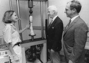
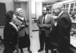
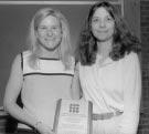
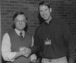
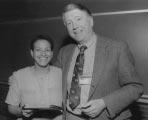
Dr. Corinne Sweeney, associate professor of medicine, explains equipment in the new laboratory to Robert Kane from State Senator Bell’s office and to State Representative Chris Ross.
The bone morphology unit, made possible by gifts in fond memory of Mrs. Joan Ferguson Pew, a former member of the School’s Board of Overseers, provides special information on the microscopic structure of bone in both the healthy state and in fractures. The laboratory is used to prepare calcified and noncalcified bone sections and is equipped for microradiography and photomicrography.
The orthopedic engineering and machine shop contains a milling machine and lathe along with other machines needed for construction of new sophisticated devices for the fixation of equine fractures.
Since its founding in 1981 the Comparative Orthopedic Laboratory (CORL) has been under the direction of Dr. David M. Nunamaker, Jacques Jenny Professor of Orthopedic Surgery. Despite its make-shift space in the Kline Center, CORL has had remarkable success in limiting traditional
•The development of several revolutionary techniques to improve the surgical treatment of fractures including:
- plate-luting, a method that improves the fatigue resistance of internal fixations using plates and screws up to 200%.
- tension band wiring, pin and wire, and cerclage wiring procedures that allow fixation of elbow fractures without interference with the growth plate in young animals.
- similar wiring techniques for suc-
cessful treatment of mid-body and base sesamoid fractures.
- new surgical approaches to fracture fixation that decrease surgery time and reduce the infection rate of patients.
CORL, one of five research laboratories in the Mark and Lila Allam Center for Equine Sports Medicine, continues the School’s long tradition in comparative medicine, advancing not only veterinary medicine but also human medicine.
The new facility became a reality because of the generosity of many people, among them the Richard S. Reynolds Foundation; Mr. Robert H. Crompton III; Mrs. Kathleen Crompton; Mrs. Georgiana Ducas; Doris Duke; the Estate of Louise B. Barclay; The Hunt Foundation; Mr. and Mrs. Robert P. Levy; Mr. Henry S. McNeil, Jr.; Mrs. J. Maxwell Moran; David M. Nunamaker, V’68; Mrs. Roberta Odell; 1993 Pennsylvania Hunt Cup; Ms. Joan E. Pew; Mrs. E. Miles Valentine; Mrs. Charlotte C. Weber; and Mr. and Mrs. George Strawbridge, Jr. The mechanical testing facility was made possible by a gift from the Thoroughbred Retirement Foundation, Inc.; the computer and microscope viewing room was made possible by the 1994 An Evening in Old Saratoga.
3
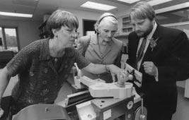
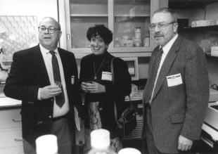
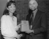
Judy Lloyd, research assistant, and Mrs. Georgiana Ducas and Mr. Glenn Pew look at histology equipment in the new laboratory.
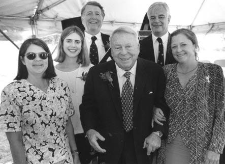
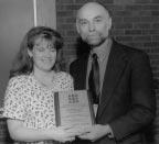
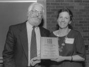
Attending the dedication were members of the Reynolds family, (l to r) Mrs. Julie Swords, Ms. Julia Swords, Mr. David Reynolds, Mrs. Dorothy Brotherton, shown here with Dr. Kelly and Dr. Nunamaker.
Avian Influenza Vaccination
Sherrill Davison, V.M.D., Phillip Scott, Ph.D., and Robert J. Eckroade, D.V.M.
Avian Influenza (AI) is a viral respiratory disease of many species of domestic and wild birds. Historically, in commercial poultry flocks, turkeys are most commonly affected due to the practice of range rearing and comingling with wild birds. While only six outbreaks of AI have occurred in commercial chickens in the United States prior to the current 1997 outbreak, when they occur they can be devastating. The 1983-1984 AI outbreak in Pennsylvania resulted in the depopulation of 17 million birds with a cost to the federal government of $60 million.
The recent AI outbreak in Pennsylvania began in December 1996 when a live bird market dealer’s flock in Lebanon County was found to be positive for nonpathogenic AI (H7N2). Subsequent to that, a flock of
commercial layers in the same vicinity was diagnosed with nonpathogenic AI (H7N2). Then in April 1997, a flock of commercial layers was diagnosed with AI in Lancaster County. A quarantine was placed on poultry facilities in a five mile radius of the index flock in Lancaster County. To date, a total of 19 commercial layer flocks, two commercial layer pullet flocks, and a commercial meat turkey flock have been diagnosed with non-pathogenic AI (H7N2) viral infection.
Control of avian influenza includes depopulation, quarantine and vaccination. Research and field experience (Mexico and the United States) has shown that vaccination for avian influenza can decrease the clinical signs of avian influenza, decrease the viral shed from known infected birds and therefore decrease the potential transmission of the disease to surrounding flocks.
Avian influenza vaccination has not
Identification of gene defect leads to cystinuria test
The gene defect for cystinuria in Newfoundlands has been identified by Dr. Paula Henthorn, associate professor of medical genetics, and colleagues in the Section of Medical Genetics at Penn’s School of Veterinary Medicine. The team, which includes Drs. Urs Giger and Jung Long Lin, has developed a test to identify carriers, affected, and normal dogs for the disease in Newfoundlands. Cystinuria, which also affects many other breeds, is an autosomal recessive trait.
The molecular test is the sixth such test the Penn group has developed. Newfoundland breeders can now screen their breeding stock for this disease to reduce the number of affected animals. If all breeding animals are screened, the disease can be eliminated from the Newfoundland population in the relatively short period of one to two generations.
Cystinuria is caused by excessive urinary excretion of cystine and other amino acids due to a defective transport system for these substances in the kidney. It leads to crystal and eventual
stone (bladder and kidney stones) formation in the urinary tract and is particularly problematic in male dogs.
Dr. Henthorn and her colleagues are now working to identify the gene defects causing cystinuria in other affected breeds so that additional tests can be developed. Unfortunately, in genetic diseases, the gene defect for a disease and its location vary from breed to breed, requiring much painstaking research to develop tests for each breed.
Dr. Henthorn’s work on cystinuria is supported by grants from the National Institutes of Health and the AKC Canine Health Foundation. The test for cystinuria in Newfoundlands is one of the many tests for canine and feline genetic diseases available through the Josephine Deubler Genetic Disease Testing Laboratory in the Section of Medical Genetics, School of Veterinary Medicine, University of Pennsylvania. For additional information, contact Dr. Urs Giger at 215-898-3375(phone), 215-573-2162 (fax) or via e-mail at<penngen@vet.upenn.edu>.
been approved for use in the United States in chickens unless the virus is highly pathogenic. The use of vaccine in Pennsylvania during the 1997 avian influenza outbreak was denied because the virus was classified as nonpathogenic. Currently, the only available vaccine for use in commercial poultry is a killed product. Additional vaccines under development are the fowl pox viral vectored vaccine and sub-unit protein vaccines.
Major advances in our understanding of vaccines have occurred due to basic molecular studies, and are currently being applied to the improvement of vaccines against many human diseases. One of the most exciting advances in the development of new vaccines has been the discovery that injection of DNA-encoding viral antigens that are known to be protective — can induce protection that is equivalent to, or superior to, that obtained following immunization with killed whole organisms, purified proteins, or subunit vaccines.
There are several notable advantages to using DNA vaccines over conventional protein antigens, some of which include: 1) the ease of manufacturing the vaccine; 2) the stability of the vaccine; 3) providing long-term antigen expression that continuously stimulates the immune response; 4) the lack of a requirement for a traditional adjuvant; 5) the ability of DNA vaccines to stimulate both strong antibody responses, T cell responses and the generation of cytotoxic T cells; and 6) the ability to co-deliver the vaccine with plasmid-DNA-encoding cytokines to enhance the immune response. Of particular interest has been the inclusion of cytokines, one of the most important of which is interleukin-12 (IL-12) which, when administered in a vaccine, dramatically improves the efficacy. Unfortunately, these advances have been slow to be applied to food animals, where they could make a major impact in the agricultural industry. Our research will focus on the development of a molecularly defined avian influenza vaccine that incorporates IL-12, and compare the efficacy of such a vaccine with a commercially available killed vaccine. Development of a DNA vaccine will provide the basic information and tools necessary to offer the poultry industry of Pennsylvania the most effective vaccine available against avian influenza.
4
Alumni Award of Merit
“The Veterinary Medical Alumni Society of the University of Pennsylvania Salutes Daniel D. Bleicher, V.M.D., Class of 1953—For your contribution to veterinary medicine through the publishing of scholarly articles that promote professional inquiry and the good name of your alma mater in publications of professional note.
For your recognition of the historical merits of your profession by participating in the creation of the Pennsylvania Veterinary Medical Historical Society.
For your longstanding commitment and dedication to the School of Veterinary Medicine in your roles as past President of the Veterinary Medical Alumni Society, Alumni Member of the Long Range Planning Committee and Chairperson of the Alumni Faculty Teaching Award Committee.
For your commitment to your community through your receipt of the Governor’s Award for Community Crime Prevention, presented by the Commonwealth of Pennsylvania.”
Dr. Reid Retires
by Dr. Midge Leitch, V’73
For those of us educated at Penn during the second half of this century, an era ended Friday night, May 15th, when Charles F. Reid, D.V.M., professor of radiology, officially retired. A member of the triumvirate which included Charles W. Raker, V.M.D. and Loren H. Evans, D.V.M.,
“The Veterinary Medical Alumni Society of the University of Pennsylvania Salutes Fred Fernich, V.M.D., Class of 1963—For your efforts to uphold a higher level of veterinary medicine as an Inspector for the Veterinary Facility Accreditation Program administered by the New York State Veterinary Medical Society.
For your commitment to the community through your volunteer work with the Kiwanis Club.
For your longstanding commitment and dedication to the University of Pennsylvania School of Veterinary Medicine in your roles as Class Agent Co-Chairman, Class Agent, Veterinary Medical Alumni Society Executive Board Member and Alumni Liaison Committee Member.
For your devotion to the School as both an alumnus and parent of a Veterinary School graduate, you have set the highest standards for alumni to follow.”
“The Veterinary Medical Alumni Society of the University of Pennsylvania Salutes Gordon W. Robinson, V.M.D., Class of 1963—For your extensive dedication to your profession and the welfare of animals through your work with the American Society for the Prevention of Cruelty to Animals as both the Head of the Department of Surgery and Director of the Bergh Memorial Animal Hospital.
For your efforts to uphold a higher level of veterinary medicine and education through the American Animal Hospital Association in your roles as Chairman of the Education Committee, and Area Director for New York and New Jersey, and as past President, Treasurer and a member of the Executive Board of the Veterinary Medical Association of New York City.
For your contribution to your country as a career officer in the New Jersey National Guard, retiring with the rank of Colonel after 37 years of service.”
Charlie was the first veterinary radiologist who developed a special interest in horses and who made an effort to connect the x-rays with both the clinical condition of and competitive expectations for the horse. He was a vital member of the team which created the clinic at New Bolton Center, and made it the premier equine facility in the world, where innovation was the password.
Charlie came to Penn in 1963 and oversaw the creation of the Radiology Suite, the development of what we now consider to be the ‘standard views’ of the large animal radiographic study, supported Dr. Mike Ross’ efforts to develop scintigraphy at New Bolton Center, and fought the good fight against western (read UC Davis) nomenclature, which
(continued on page 6)
5
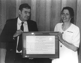
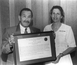
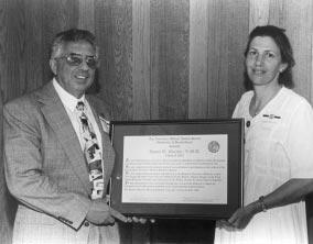
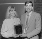
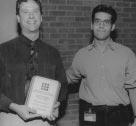
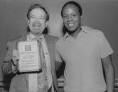
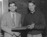
The Veterinary Medical Alumni Society presented the Alumni Award of Merit to Dr. Daniel Bleicher, V’53, Dr. Fred Fernich, V’63 and Dr. Gordon W. Robinson, V’63. VMAS President, Dr. Suzanne Smith, V’82, presented the award certificates on Alumni Day, May17.
Dr. Reid Retires
(continued from page 5)
undoubtedly helped provide a great foundation for the well known phrase ‘at Penn we’.
He has trained untold numbers of fledgling radiologists who now populate the American College of Veterinary Radiologists and have gone on to help develop the Sections of Radiology in many of the veterinary schools both in the United States and abroad. Foreign radiologists from as diverse locations as The Netherlands and Israel have spent valuable time under Charlie’s tutelage and remembered those times and experiences in written tributes which were shared with the two hundred people who attended the official retirement dinner.
From Dr. Barney Kerr, Nassau, The Bahamas: ‘The news of your retirement sent me spinning down memory lane. I can still hear you storming into Radiology every morning with laughter echoing in your wake... The rivalry and agitation that often erupted around the job. You enjoyed the confidence of the most cynical, experienced horse people in every corner of the globe that I ever visited. You were about 1% slave driver and 99% marshmallow. I will never forget the countless hours you spent teaching and encouraging me. Your enthusiasm was infectious and Radiology was the most enjoyable place in the world to me.”
From Drs. Nadine Oakley and John Simms, Shippensburg, Pennsylvania: “We have fond memories of days spent in large animal radiology sessions endlessly repeating ‘normal, normal, normal, normal.....OOPS!’ in an attempt to read films with the master overseeing this whole process. You challenged us unmercifully, yet made it fun and taught us to laugh at ourselves. We all learned so much, but more importantly, we learned to think for and believe in ourselves.
Your contagious smile, great sense of humor, and your ability not to take things too seriously influenced all whom you taught.... You will be sorely missed by those of us out here in the trenches.”
From a friend in Unionville, Pennsylvania, whom Charlie counseled: “...you said I was smarter then that and should set my goals much higher. At the time, I wasn’t sure if I, a high school drop-out, could even handle algebra. Your words stuck with me and somehow helped me discover the confidence and discipline I needed to pursue a greater challenge.
Massachusetts came to roast and toast Charlie. And inspired by the object of their obvious respect and affections, rose to unsuspected heights of both emotion and humor. Charlie, however, as usual, had the last word and the last laugh and managed once again to remind us of why he has played such an influential role in so many lives.
For many of us, he was not only one of the most dynamic teachers we encountered, but also an untiring and often entertaining source of personal advice! Because teaching was obviously his first love, the committee responsible for organizing his retirement celebration, which consisted of Lisa Suslak-Brown, V.M.D., Jill Beech, V.M.D., Dean Richardson, D.V.M., Carole Johnson Brady, and me, developed the idea of creating the Charles F. Reid Scholarship Fund in his honor. The interest from this perpetual fund will assist veterinary students during the pursuit of their degree. Contributions to the fund can be made at any time by checks payable to the Trustees of the University of Pennsylvania, earmarked for the Charles F. Reid Scholarship Fund, and sent to New Bolton Center, Development Office, 382 W. Street Road, Kennett Square, PA 19438-1692. And of course your donations are fully tax deductible!
You are able to give people a sense of their own potential and, at the same time, keep the inspiration based firmly in reality by serving as an example of real achievement in your own life and work.”
From Dr. Scott Palmer, New Jersey Equine Clinic: “The influence of a teacher extends throughout our profession like ripples caused by a pebble thrown onto the quiet surface of a still pool. Your career, by any measure, has been a splash!”
Master of Ceremonies Dean Richardson was encouraged by all who witnessed his performance to pursue a career in comedy, in spite of his rather prodigious talents as an orthopedic surgeon. Colleagues, including both Deans Kelly and Marshak, and friends from as far as Florida, California, and
The diversity of the group which collected to honor Charlie mirrored the breadth of his interest in people and their pursuits. His contributions to the School of Veterinary Medicine are too numerous to mention and the list of devoted friends, students, and colleagues too long to recite. The commemoration of his career with a perpetual scholarship fund seems the only appropriate token of our gratitude for the concern and effort he made on so many individuals’ behalf. From professional horsemen to veterinarians to millionaire investors to fellow neighbors, he helped us all with the same even hand. Ever convinced of his own perspective, he encouraged us all to have the courage of our convictions. We thank you and we salute you and we rest assured that retirement will not deprive us of your presence and influence!
6
Dr. Reid teaching.
Very Much a Doctor
By Dr. Marne Platt V’93
Ientered veterinary school planning, as I had since the age of six, to be an equine veterinarian. By my fourth year, however, small questions were tickling the back of my mind; so many fascinating diseases beckoned in small animal practice! Unable to decide, I joined a mixed practice in Califon, NJ. The current practice owners, then senior associates, are both Penn grads (Debbie Cronin, V’80 and MaryBeth Hanorski, V’87) who helped me use my training to pursue a diagnosis and an understanding of the disease, instead of just treating symptoms. The practice owner at the time, Dr. John Kenney, is a Cornell grad who loved to pick on our VMD’s but shared our search for knowledge. Starting practice at Califon Animal Hospital showed me that the theory I had learned at Penn really could be integrated into everyday practice, which never really became “everyday”.
After two years, personal reasons dictated that I had to move and give up my beloved horse work. I joined the small animal practice of Dr. Michael Tuder. There I was able to fly solo as much as I wanted, or get a second opinion from one of the other six doctors on staff. I also
Deubler Laboratory Dedicated
The Josephine Deubler Genetic Disease Testing Laboratory was dedicated following the Canine Symposium on January 31. Contributions by many individuals and clubs made the establishment and opening of this laboratory possible. Funds are still being raised to complete several projects within the laboratory.
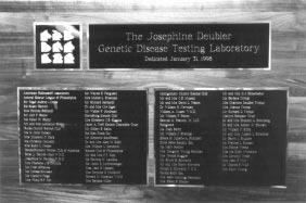
Guests toured the new laboratory, located on the fourth floor of VHUP in the Section of Medical Genetics. The Deubler Laboratory offers DNA, biochemical and immunologic tests for canine and feline genetic diseases. For information on sending samples for testing please contact 215-898-3375 (phone) or 215-573-2162(fax).
further honed my surgical skills (who says a Penn grad can’t spay a cat in under 15 minutes?) Unfortunately, two years later my immune system had enough. After realizing that I was taking 10-12 antihistamines a day, I visited an MD. My worst fears were confirmed: I am now allergic to most mammals. It seemed that a lifetime of practice was not my destiny.
After scraping my heart off the floor and making the fateful call to Mom and Dad, I set about finding something new to do. After considering pursuing a Ph.D. in pubic health or some related topic, I finally accepted my current position, Regional Technical Manager for Novartis Animal Health.
My job description is one of the most varied around. I am responsible for the technical training of approximately 15 sales representatives who cover Maine to Maryland, Michigan and Illinois. I provide in-clinic seminars for my colleagues and their staff. I answer calls on our Professional Services phone lines about our products, SENTINEL™, INTERCEPTOR®, and PROGRAM®, and their varied uses. I am the company liaison for five veterinary schools, including Penn, so I spend time at each of them with students, clinicians, researchers and admin-
istrators. I analyze research for its strengths, weaknesses, and various interpretations. Here my Penn education is a real plus: having been taught to analyze every result, question every conclusion, and make my own decisions on the validity of studies, this aspect of my job is a most enjoyable challenge.
I travel an average of three to four days a week, maintaining a strong network of contacts with colleagues and friends. Special projects may involve analyzing raw data (those statistics courses come in handy here) or creating presentations. In each aspect of this position, my Penn training boosts my performance.
My many extracurricular activities while at Penn also contribute to my success. Editing the yearbook, working on Development Office Phone-a-thons, participating in the Student Rights and Responsibilities Committee, SCAVMA, and the Colic Team, all taught me teamwork and how to survive in a pressure cooker.
My training and education at Penn have helped to make my new career as fulfilling as I ever expected when I set off to “fix horses, Daddy!” I am Very Much a Doctor!
7
Left: Mr. and Mrs. Lawrence Brown and Mr.Daniel Shoemaker tour the laboratory.
Above: Mrs. Connie Vanacore, Dr. Deubler, Dr.Urs Giger, chief, Section of Medical Genetics, and Dr. Joseph Slick in the new laboratory.
Dr. Amanda Fine, V’97, has been named a Luce Fellow by The Henry Luce Foundation. Dr. Fine will spend one year at the Mongolian Veterinary Research Institute in Ulan Bator, Mongolia.
She will be working on livestock diseases and public health issues in the field and in the laboratory. Mongolia is a major exporter of cashmere wool. Much of the population is nomadic and the livestock, goats, sheep, cattle, horses and yak, moves from pasture to pasture. Dr. Fine is only the second veterinarian to be awarded a Luce Fellowship.
Dr. Raymond Sweeney, V’82, associate professor of medicine, received the Norden Distinguished Teaching Award.
The School of Veterinary Medicine’s site “Veterinary Service At Oncolink” on Penn’s Medical School’s Oncolink web site won The Best of Internet Award. Congratulations to all in VHUP’s Oncology Service.

The Marshak Dairy received a “Dairy of Distinction Award” from the Northeast Dairy Farm Beautification Program.
Dr. Arnold Kornblatt, V’78, is the regional veterinarian for the Mate Bibyamin District in Israel and product manager for Biogal-Galed Laboratories at Kibbutz Galed in Megiddo, Israel. He and colleagues have developed testing kits for canine ehrlichia, bovine leptospirosis, and avian psittacosis.
Dr. Sue McDonnell, assistant professor in reproduction, presented the Fred Pierce Lecture at a meeting of Canadian horse breeders in Deer, Alberta. The lecture was established by the Canadian SPCA and is presented by a speaker honored for research contributions to the welfare of horses. The American Hanoverarian Society presented Dr. McDonnell with its first Annual Research Award at the Society’s 1998 Annual Meeting. Dr. McDonnell was recognized for her pioneering work in horse behavior and she presented a lecture on the subject.
She also traveled to Helsinki, Finland, in March to present a program to the Finnish Equine Veterinary Association.
Dr. Stuart Meyers, assistant professor of large animal reproduction, received funding for the second year from the Grayson Jockey Club Research Foundation for his study “Evaluation of Stallion Fertility Based on Sperm Cellular Function: A Prospective Study.” He also received a two-year grant from the Pennsylvania Department of Agriculture for the study “Assessment of Sperm Function in Preserved Boar Semen.” Both studies seek to apply new techniques in cell and molecular biology to large animal male infertility.
Dr. Andre Ziegler, staff veterinarian in the Laboratory of Avian Medicine and Pathology, received the Pennsylvania Game Commission’s Sport Ethics Award for his assistance with a diagnostic case submitted to the laboratory. The case involved duck intoxication with diazinone that resulted in mortality of ducks in a local river.
Dr. Barbara Davis, V’84, was a faculty member at the International Symposium and Histopathology Seminar on the Reproductive Tract of Laboratory Animals in Nara, Japan in April. She presented two papers and led three seminars on toxicologic pathology. Dr. Davis is veterinary pathologist at the National Institute of Environmental Health Sciences, Raleigh, NC.
Dr. Robert Eckroade, associate professor of poultry pathology, presented a talk at the Ontario Poultry Health Conference in Kitchnener, Ontario and a gave a talk at the Pfizer Seminar in Quebec, Canada. He also spoke at the Pacific Egg and Poultry Association meeting in Monterey, CA and at the Western Poultry Disease Conference in Sacramento, CA in March. Dr. Eckroade received a grant from the Pennsylvania Animal Health Commission to study Salmonella enteritidis.
Dr. Sherrill Davison, V’83, assistant professor of poultry pathology, received a grant from the Pennsylvania Animal Health Commission to study E. coli in
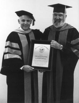
Dr. James Buchanan, professor of cardiology, was honored by Michigan State University and named a Distinguished Alumnus. Dr. Buchanan, a member of the Class of 1960 of MSU’s College of Veterinary Medicine, was honored for his role as a pioneer in veterinary cardiology, for his pioneering heart surgery techniques and for his accomplishments as a teacher, researcher, clinician and surgeon. Dr. Buchanan came to Penn 38 years ago.
8
Dr. James Buchanan receives MSU’s Distinguished Alumnus Award from Dr. Lonnie J. King, Jr., dean of MSU’s College of Veterinary Medicine.
poultry. Dr. Davison also gave presentations on avian influenza and laryngotracheitis at the New England Poultry Health seminars and gave a presentation on avian influenza at the PADL’s Diagnostic Conference.
Dr. Fred Rude, V’58, received the American Animal Hospital Special Recognition Award for his outstanding contributions as an AAHA leader and for his dedication and commitment to AAHA student members in the Northeast Region.
Dr. James Orsini, associate professor of surgery, was the Coughlin Visiting Professor at the College of Veterinary Medicine, University of Tennessee in January. He is the co-author, with Dr. Thomas Divers, of Manual of Equine Emergencies: Treatment and Procedures, which was published by W.B. Saunders and Co.
Dr. Michael Woltz, V’78, was honored by the Greenburgh Nature Center, Westchester County, NY for his decadelong volunteer service to the organization and its live animal museum.
Dr. Kathryn Michel, assistant professor of nutrition, is now a diplomate of the American College of Veterinary Clinical Nutritionists. Dr. Lilian Aronson, V’92, assistant professor of surgery, Dr. Daniel Brockman, assistant professor or surgery, and Dr. Elizabeth Hammer, lecturer in sports medicine, are diplomates of the American College of Veterinary Surgeons. Dr. Charles Vite, post-doctoral fellow in pathobiology, is a diplomate of the American College of Veterinary Internal Medicine in the specialty of neurology Dr. Rebecka Hess, lecturer in medicine, and Dr. Nicola Mason, lecturer in medicine, are diplomates of the American College of Veterinary Internal Medicine.
Dr. E. Neil Moore, professor of physiology, was visiting professor at Michigan State University. He also gave the Buchanan Lecture to the Schools of Veterinary Medicine, Medicine, and Osteopathic Medicine there.
Dr. Colin Johnstone and the School received a $25,000 grant from the Merck Co. Foundation, on behalf of Merial, for the development of the “Merial Electronic Book on Parasites and Parasitic Diseases of Domestic Animals.” The work will present lectures in veterinary parasitology, illustrated by slides. It will
be in English and Spanish and transferred to the Internet as a teaching tool.
Ken Mullin, director of medical records at VHUP, won an “Honorable Mention for Black and White Photography” in the annual competition sponsored by the Dog Writers of America. His wife, Dr. Nina Beyer, V’87, also won an “Honorable Mention” in the same competition for an article in AKC Afield.
Dr. Carla Drozdowicz, V’85, research director in the Preclinical Research and Development Group at Hoffman-La Roche Inc. received the Tribute to Women in Industry Award for her leadership. The award is sponsored by the YMCA of Bergen County
Dr. Peter Schantz, V’65, received the first United States Public Health Service Commissioned Officer Veterinarian-ofthe-Year Award The award formally recognizes individuals whose contributions have led to the advancement of public health and veterinary medicine. Dr. Schantz is internationally recognized for his work on the diagnosis, treatment, epidemiology, and control of zoonotic parasitic diseases. He was instrumental in opening opportunities for veterinarians with the Centers for Disease Control.
Dr. Frederick Fregin, V’64, director of the Virginia-Maryland Regional College of Veterinary Medicine’s Marion duPont Scott Equine Medical Center has been named to the Loudon County Rural Economic Task Force.
Barbara Davis, the School’s director of financial aid, is a baseball expert for Total Sports and a weekly commentator on WHYY on the Phillies, who are doing nicely
Dr. John Whitehead, V’52, was honored by the Veterinary Medical Association of New York City with its Distinguished Life Service Award.
Dr. Eric Tulleners, holder of the Lawrence Baker Sheppard Professorship in Surgery, has been promoted to professor of surgery Dr. Mark Saunders, V’81, has been promoted to associate professor of radiology. Dr. Sydney
Evans, V’77, has been promoted to associate professor of radiology Dr. David Holt has been promoted to associate professor of surgery. Dr. Erika Holzbaur has been promoted to associate professor of biochemistry
Dr. Eileen Mera, V’86, received the Woman of Distinction Award from the Great Valley Girl Scout Council. Dr Mera, a partner in the Wright Veterinary Medical Center in Bethlehem, PA, was honored for her mentorship of Girl Scouts.
Dr. Kirk Gelatt, V’65, professor of ophthalmology at the University of Florida College of Veterinary Medicine, received the American Kennel Club’s annual award for Career Achievement in Canine Research.
Dr. Kevin Byrne, lecturer in dermatology, received his Masters of Science degree in Veterinary Medicine from the University of Illinois. He presented a paper at the Annual Meeting of the American Academy of Dermatology and the American College of Dermatology. In June he participated in a round table discussion “Latest Advances in Allergy Therapy — Diagnostics and Treatment in Food Allergy,” held in Orlando, Fl.
Dr. Urs Giger, Charlotte Newton Sheppard Professor of Medicine, was elected president of the Association of Veterinary Hematology and Transfusion Medicine.
Dr. James Serpell, Marie A. Moore Associate Professor of Humane Ethics and Animal Welfare, participated in the National Bioethics Institute at Oregon State University in August. Dr. Serpell also had his grant renewed by the Provost’s Interdisciplinary Seminar Fund for the seminar series Human Relations with Animals and the Natural World.
Catch up with happenings and events at the School of Veterinary Medicine and read the Bellwether on line by looking at the School’s website at:<www.vet.upenn.edu>
9
Feline Symposium
The 21st Annual Feline Symposium was held on Saturday, April 4, 1998. In light of the enormous emotional investment owners make in their feline companions, the seminar focused on the strong symbiosis between cats and people, as well as the critical care and preventive measures designed ultimately to prolong that bond. The 22nd Annual Feline Symposium will be held on March 6, 1999.
History of the Cat
The unprecedented popularity felids today enjoy as companion animals is the latest phase in a centuries-old lovehate relationship with mankind. Dr. James A. Serpell, Marie A. Moore Associate Professor of Humane Ethics and Animal Welfare at the School, chronicled the domestication of cats throughout the millenia.
Archaeological, genetic and behavioral evidence suggest that the domestic cat (Felis catus) is descended from the African wildcat (Felis libyca). There are also etymological reasons for this assertion: The English word “cat,” the French “chat,” the German “katze,” the fourth-century Latin “cattus” and the modern Arabic “quttah” likely derive from the Nubian word for cat — “kadiz.”
The oldest known bony remains of African wildcats date back to 60007000 B.C. They were excavated from Jericho, and from Cyprus where they coincide with the earliest human settlements on this Mediterranean island. Whether these early cats were domesticated, however, is unclear, said Dr. Serpell. The prevailing theory is that cats likely achieved domestic status in Egypt sometime during the third millenium B.C. Egyptian iconography from 1600 B.C. and onward frequently portrays cats in domestic vignettes; cats were illustrated sitting under their owners’ chairs, playing with other animals and even helping people to hunt birds among the papyrus swamps of the Nile delta.
How did cats slink so gracefully into the company of humans? “The process that led to the cat’s domestication is shrouded in a certain amount of mystery,” said Dr. Serpell. Experts believe a mutualistic association based on the need for rodent control in early Egyptian settlements — which thrived on grain cultivation and storage — was responsible.
Much like the celestial ascent of cows in India, domesticity led to divine worship for cats. Deities with feline heads and human bodies populated Egyptian spiritual imagery. The cat was linked to the mother goddess Isis and to the goddess Bastet, who symbolized fertility, fecundity and motherhood. According to ancient writings, the temple built in deference to Bastet was inhabited by thousands of cats who were fed and cared for by the priesthood.
So deified were cats in ancient Egypt that the death of one sent its human family into a state of mourning manifested by shaved eyebrows as a sign of respect. The dead cat was embalmed and buried in a sacred repository, provided adequate funds were available. Owing to their status as a protected species in Egypt, causing the death of a cat — even accidentally — was a capital offense. The Egyptians restricted the export of cats, thereby retarding their spread to neighboring Mediterranean countries.
The earliest known representation of cats in Greece is on a marble block dating back to about 500 B.C. At the time, cats were regarded as novelties in Greece and Italy, where rodent control was relegated to ferrets. The earliest reference to cats in India dates back to about 200 B.C., and cats probably colonized the Orient soon thereafter. The Romans propagated the spread of the domestic cat — which owes much of its colonizing ability to its facile adjustment to shipboard life — to northern Europe and other outposts of the Roman Empire; by about the middle of the fourth century A.D., domestic cats were present in Britain.
Modern studies have concluded that
current gene frequencies for feline coat color variation throughout the world correspond to early colonization patterns. For example, the sex-linked, orange coat-color mutant, which appears to have originated in Asia Minor, is today quite prevalent throughout the Near East, northern Africa, southern Italy, Germany, France, northern England and Scandinavia. This, said Dr. Serpell, may reflect the movement of cats on Viking trade ships in the eighth and ninth centuries A.D.
By about 1200 A.D., the cat experienced a calamitous change in fortune. “The gradual extinction of pagan gods and goddesses and the rise and spread of Christianity produced very dramatic changes in attitudes to cats throughout Europe,” Dr. Serpell explained. Cats were rapidly transformed from benevolent symbols of femininity to malevolent agents of the devil. At the time, nearly all the major heretical sects were accused of worshiping the devil in the form of a black cat. Up until the eighteenth century, cats were caught up in the wave of persecution of witches, of whom cats were viewed as demonic companions. On feast days throughout Europe, cats were captured and tortured to death as symbolic means of driving out the devil.
“By associating the cat with the devil and bad luck,” Dr. Serpell alleged, “the Church provided the superstitious masses of Europe with a kind of universal scapegoat, something they could blame for all the hardships of life.”
The metaphoric link between cats and women, particularly the threatening aspects of female sexuality, was also responsible for this rancor toward cats. Dr. Serpell referred to monstrous vampire cats of Japanese folklore that assumed the forms of women in order to “suck the blood and vitality from unsuspecting men.”
This malice did not forestall the spread of cats to virtually every corner of the world. In 1986, the cat overtook the dog as the most popular companion animal. Today, the cat’s popularity is unprecedented, comparable only with
10
its heyday in ancient Egypt. Yet it is a wavering acceptance. In a modern survey of American attitudes toward cats, 17.4 percent of respondents expressed some animosity toward cats (versus 2.6 percent who reported disliking dogs).
Over the years, cats have aroused antipathy due likely to their somewhat ambivalent relationships with people. “The cat leads sort of a double life,” Dr. Serpell asserted in a National Geographic story (The Human-Cat Connection, June 1997). “It likes to enjoy the fruits of domesticity. It likes to lead its own wild life too. It resists conforming to human standards.”
What It Means to Be Owned by a Cat — An Owner’s Perspective
Cooly aloof, cats seem to have an almost calculating way of soliciting their owners’ indulgence. Cat breeder Janet Wolf gave her personal account of the feline mystique that — in most households — renders the owner subservient to the cat.
“The question is not only who owns who,” Ms. Wolf said, “but who’s the boss.” She remembers how one of her cats “taught” her father to give her treats by slinking past his legs. She related how her other cat taps her husband’s arm each morning in order to procure her share of his donut.
Why all the fuss over a pet? Ms. Wolf answered this question with a flurry of revealing numbers. She said studies have shown that pet owners have significantly greater psychological health than people who do not own pets. In one study, cat owners over 60 years of age reported having greater life satisfaction and less loneliness, anxiety and depression one year after adopting a cat than did non-cat-owners.
“Cats buffer any sense of social isolation, provide companionship, can be icebreakers and social facilitators and can also be a source of social and tactile contact. They also offer stress reduction. Think of how relaxing it is to have a cat just purring on your lap.”
All this coddling, she suggested, is the price one pays to win the affection of an animal that knows how to play “hard to get.” Ms. Wolf, who has been breeding Birmans since 1987, introduced her cat family, members of whom each have their designated living quarters in her house. She said the accommodations she makes for them include buying their favorite foods, constructing cat “trees” to give her pets vertical space for climbing, hiring cat sitters when she goes away on vacation, even using only kitty-safe ornaments to decorate her Christmas tree.
“When we bought our first Birman, I don’t think we ever envisioned becoming so involved with our cats. But after thinking this through,” she conceded, “I am convinced without a doubt that I am owned by my cats.”
Losing a Best Friend — Copingwith the Death of a Cat
Love is species-blind. In no way, perhaps, is this more apparent than in the manner in which a pet owner grieves the loss of a beloved animal. Mrs. Kathleen Dunn, M.S.W. social worker at VHUP, profiled the petowner relationship and explained the mourning process owners undergo when pets die.
Pets are humanized in modern American society, said Mrs. Dunn. Examples abound of owners who talk to and kiss their pets, even celebrate their pets’ birthdays. “Through a process called ‘attachment and bonding,’ a very special relationship develops.”
Among the accolades Mrs. Dunn has heard owners shower on their pets: “The fun the animal gives” and “No matter what happens at work, my cat is always there for me when I get home.” “‘Unconditional love’ is also a term that’s used a lot,” she added.
When she first joined the VHUP patient-care team, Mrs. Dunn figured she would be working mostly with the lonely elderly. To her surprise, she soon found that the people who needed
her services ran the age gamut.
She also learned that many of them — men and women alike — related to their pets as though the animals were their children. “Because this bond is so deep, the animal becomes a person to you, part of the family — sort of a perennial two-year-old. And if the relationship is threatened by death or illness, it’s like a child dying. It doesn’t matter that what died was ‘only’ an animal. A loss is a loss is a loss.”
She recounted the case of an owner whose cat was stolen. Days later, the distraught woman was still dismantling the household in search of the missing cat. “She told me it felt as though she’d lost a child.”
In her Pet Bereavement Support Group at VHUP, one of few such groups in the country, Mrs. Dunn sees many pet owners who are troubled in their mourning. It is not unusual for an owner to report seeing or hearing the deceased pet. Someone recently told her she thought she saw her dog — who died two years ago — walking down the street. She attributes this phenomenon to the fact that owners spend so much time caring for their pets that their presence becomes “ingrained” in their lives.
The grieving process for the deceased pet involves distinct stages of mourning, including guilt, which, she said, is the toughest to reconcile. Empathetic veterinarians can be particularly instrumental in helping owners deal with guilt, as this emotion is often linked to medical misperceptions, perhaps the notion that the animal died because the owner forgot to administer a pill, e.t.c. During this sorrowful time, owners may experience difficulty eating, sleeping and concentrating. The loss of their pet can even trigger the memory of a previous loss.
One of the most valuable coping strategies for dealing with pet loss is talking, particularly with someone who has also experienced the death of a pet and understands the emotional bond,
(continued on page 12)
11
FELINE SYMPOSIUM FELINE SYMPOSIUM
Feline Symposium
said Mrs. Dunn, whose support group meets every other week. During the bereavement period, particularly the first two weeks following the pet’s death — which are typically the most difficult to endure, it is important for one to resume one’s schedule and get adequate exercise and nutrition. Other therapeutic options include reading about pet loss, writing about one’s pet and joining a support group. Mrs. Dunn added that surviving animals, who also may be lamenting the loss of their buddy, should be comforted and given loving attention.
Feline Oral Health: Disease and Preventing It
Cats are skilled hunters and, as such, supreme carnivores. Yet domestic felines lack true grit — dietetically speaking, because most commercial diets are deficient in the abrasive “toothbrushing action” of bones. Dr. William Rosenblad, resident in dental medicine at VHUP, discussed dental health in cats.
The importance of oral health in cats and dogs, said Dr. Rosenblad, cannot be overstated. “Because they use their mouths to eat, groom and communicate, their oral health has that much more importance.”
Two oral conditions to which cats are prone are resorptive lesions and ulcerative stomatitis. Both diseases begin with plaque, a gummy layer of bacteria and their by-products that coats the tooth, and subsequent periodontal disease. Within 24 hours of adherence to the enamel, plaque begins to mineralize into calculus, or tartar, which can be mechanically chipped away. The outer (buccal) surfaces of the upper teeth are predisposed to plaque formation because they lack both the abrasive, shearing forces that exist between the inside (lingual) surfaces of the upper teeth and the buccal surfaces of the lower teeth, as well as close contact with the tongue, which washes the lingual surfaces of the teeth with saliva.
(continued from page 11)
Periodontal disease is probably the most prevalent health problem in cats and dogs. In fact, Dr. Rosenblad estimates, 75-90 percent of all adult cats have periodontal disease, and it is more prevalent in purebreds. Periodontal disease, which is manifest as either periodontitis or the more mild gingivitis, results when plaque and tartar build up under the gum line.
Gingivitis is a reversible condition apparent as a reddened, inflamed gum margin. Periodontitis is a comparatively more deep-seated infection affecting the structures (periodontal ligaments and bony tissue) that support the tooth within the alveolus, or tooth socket. The inciting bacteria, which travel inside the well-vascularized gingiva, can readily reach the bloodstream, leading to serious systemic ramifications. Furthermore, periodontitis, which in severe cases involves pus accumulation and oral tissue necrosis, can stress the immune system and additionally compromise cats suffering from concomitant systemic illnesses like renal and liver disease, and diabetes.
Periodontal disease is of greater consequence to cats than dogs because the former have a thinner, and therefore more vulnerable, band of gingiva attached to their enamel. Gingival recession leads to bone resorption or “cervical line (neck) lesions.”
“This is a progressive problem,” said Dr. Rosenblad. “This isn’t important for only the affected tooth because this is basically an infection of the bone itself.”
Neck lesions, in which the tooth below the crown is eaten away, are the feline equivalents to cavities in people. These lesions are extremely painful due to both resorption into the innervated pulp canal and associated gingival inflammation.
The canine teeth are more likely than the molars or premolars to undergo root resorption. These cats may present with the affected canine tooth appearing longer than the contralateral normal tooth. The reason for this, explained Dr. Rosenblad, is that when the root re-
sorbs, the socket becomes inflamed and the crown is gradually extruded.
Neck lesions may be apparent by visual inspection of the oral cavity, but their presence can be confirmed radiographically. For cats showing clinical signs, such as refusal to eat, the affected teeth are typically extracted.
The second major feline oral disease, ulcerative stomatitis, involves generalized oral inflammation caused by an excessive immune response to plaque bacteria. Inflammation can be quite severe and culminate in tissue necrosis. Signs include drooling and anorexia. The acute stage of this illness is managed with antibiotics and antiinflammatory agents. Subsequent treatment may also include extractions, dental scaling and polishing, plaque retardants and antiviral agents. Ulcerative stomatitis must be differentiated from other causes of oral ulceration in cats, like kidney disease, oral eosinophilic granulomas and squamous cell carcinoma.
Dental prophylaxis is valuable both diagnostically and therapeutically. The extra-oral structures, including the head, eyes, ears, throat and lymph nodes, are typically examined. The intra-oral structures, such as the teeth, tongue and palate, are checked and a periodontal exam is performed under anesthesia. Each tooth is probed to detect crevices suggestive of neck lesions. Radiographs are made to locate resorptive lesions. Following any necessary tooth extractions, scaling and root cleaning are completed. Finally, the teeth are polished to smooth out any roughened surfaces to which bacteria can adhere. Prophylactic antibiotics are given to debilitated cats, such as those with heart or kidney disease, as well as those with severe oral disease.
When it comes to feline oral disease, prevention is an accessible goal. “This is one of the ways we can keep our cats happy,” said Dr. Rosenblad. He advises most owners to brush their cat’s teeth at home (buccal surface of upper teeth using a brush with bristles) and to offer them abrasive food substances such as kibble (i.e. Hill’s TD dental diet) and tartar-control chews.
12
Feline Renal Transplantation
Kidney transplantation is a sophisticated procedure for changing the delicate blood “filter” that fails so many cats. Dr. Lillian R. Aronson, assistant professor of surgery at VHUP, described the transplantation technique, which is now available at VHUP, and reviewed patient selection criteria, post-operative care and prognosis for transplant patients.
Renal transplants in animals date back to the early 1900s. The first feline kidney transplant at a university hospital took place at the University of California, Davis in 1984. The patient, a Persian cat named Queenie, lived with normal kidney function for two years following surgery, eventually succumbing to heart failure.
One of the most important aspects of a successful renal transplant program, said Dr. Aronson, is careful patient selection. “Renal transplantation is an excellent treatment option for some cats, but it’s not for every cat.”
The ideal candidate is the cat in very early decompensated kidney failure. This status is gauged by body weight, which declines in debilitated renal failure patients. An acceptable candidate has up to a 10-20 percent weight loss. The recipient must also be free of other diseases, such as FeLV, FIV, heart disease, diabetes and a history of inflammatory bowel disease. Bloodwork and urine tests are performed on the potential recipient, as are EKG and chest/abdominal radiographs and ultrasound. If there is suspicion that a dormant medical condition like a urinary tract infection will be unmasked by the administration of the immunosuppressive drugs that maintain the cat following transplant, a two-week trial of these drugs is performed prior to surgery. Age is not a consideration for this surgery, said Dr. Aronson, whose transplant recipients have ranged in age from 2-16 years.
The kidney donor should be a healthy, young, FeLV/FIV-negative
adult cat, ideally the same size or slightly larger than the recipient. The cat should also be blood-crossmatch compatible with the recipient, as antigens present on red blood cells are also present on the endothelium of graft blood vessels. Blood and urine analyses are performed, as is excretory urography to assure that the donor has two normal-shaped, well-vascularized kidneys. The other major condition is that the donor cat, which comes from the Pennsylvania S.P.C.A., must be adopted by the owner of the recipient cat.
“This has been a very positive part of the program,” said Dr. Aronson. “Owners love it. They feel they are saving the life of the cat that saved their cat’s life.”
Unilateral kidney removal does not clinically compromise the donor, she added. In a recent study, about 20 donor cats were followed post-surgery. Only two of these cats showed mild changes in urine-concentrating ability and minor increases in serum creatinine; they remained clinically normal.
Presurgical preparation is crucial to the success of renal transplantation. The recipient is diuresed with a balanced electrolyte solution and fed a protein-restricted diet. Anemia, a serious byproduct of renal failure, is corrected with either whole-blood transfusion or erythropoietin administration. Betablockers are given if blood pressure is dangerously elevated, and a glycerin suppository is administered to both the donor and recipient the night before surgery to empty their colons.
To decrease the likelihood of organ rejection, immunosuppressive drugs are started. The recipient is given cyclosporine orally 1-2 days before surgery. Prednisone is administered orally starting the day of the procedure.
The transplant procedure involves two surgical teams working on both cats simultaneously. The donor cat is brought to surgery first and opened along the ventral midline. Using loupes that provide magnification for vascular dissection, the left and right kidneys are examined for vascular pedicles consisting of suitable vessels. The left kidney is preferred because of its slightly
FELINE SYMPOSIUM FELINE SYMPOSIUM
longer vein. The vessels and ureter are carefully dissected out and cleaned.
Once the recipient is opened and prepared to receive the kidney, donor nephrectomy is performed and the renal vessels of the transplanted kidney are anastomosed to the recipient’s aorta and caudal vena cava. The ureter is sutured into the bladder and, to prevent torsion of the vascular pedicle, the transplanted kidney is sutured to the adjacent abdominal wall.
The native kidneys are biopsied but usually left in situ as a reserve should the donor kidney fail. Most transplanted kidneys are functioning well by 72 hours after surgery, at which time dramatic clinical improvement is generally evident; if the transplanted kidney fails to function normally, re-transplantation is an option for most patients.
Post-operative handling and stress should be minimized. IV fluids, gastrotomy tube feedings, antibiotics and cyclosporine are administered. Cyclosporine levels are checked regularly. The cat is discharged once graft function is determined to be stable. Intravenous cyclosporine is sent to the referring veterinarian’s hospital for use in an emergency rejection episode. Bloodwork at the referring hospital to monitor cyclosporine levels and creatinine must be performed weekly until drug levels are stable, and then every 23 months thereafter.
Renal transplant surgery mandates extensive owner commitment and expense. The procedure costs $4-5,000 if no complications develop. For the average-sized cat, the cost of cyclosporine, which must be given for the remainder of the recipient’s life, runs $.30-2.40 per day. And there are no guarantees: 70 percent of patients survive the surgery and are home for at least one year with normal kidney function; the mean postoperative survival time is 26 months.
“Owners really need to understand the risks,” cautioned Dr. Aronson. “You’re taking a cat with an underlying fatal disease and putting him through a big procedure he may not survive.”
(continued on page 14)
13
Feline Symposium (continued from page 13)
Caring for the Critically-Ill Cat
Contrary to popular folklore, cats do not have nine lives. Fortunately, sophisticated emergency and intensive care measures are available to save critically decompensated cats. Dr. Deborah C. Mandell, lecturer in emergency medicine at VHUP, provided an overview of the four major body systems and discussed critical care management procedures for insults to those systems in cats.
Feline critical care is problematic for several reasons. First of all, Dr. Mandell said “Cats are notorious for compensating very well in response to certain diseases. So by the time they’re showing signs, they’re already in a very advanced state of disease.”
To worsen matters, critically-ill cats can become easily stressed and tolerate little manipulation, rendering medical workup and treatment somewhat precarious. The four critical organ systems, which must be rapidly assessed in cats presenting for emergency care, are the respiratory, cardiovascular, renal and neurologic systems.
The most common causes of respiratory distress in cats are asthma, heart disease and pleural space disease. A thorough physical exam and radiographs are essential in distinguishing between these.
On physical exam, respiratory competency is evaluated by checking mucous membrane color, which should be pink, and respiratory rate (normal feline respiratory rate is 15-36 breaths per minute) and effort. For a cat in severe respiratory distress, which can be manifest by and/or nostril flaring openmouth breathing, the exam is temporarily suspended. Oxygen is administered through either a flow-by tube held near the nostrils; an oxygen mask if the cat is placid and/or comatose; or placement in an oxygen cage, which supplies air comprised of as much as 60 percent oxygen.
The next stop on the physiologic route is the cardiovascular system,
which distributes inspired oxygen throughout the body. Cardiovascular health is reflected in mucous membrane color and capillary refill, time pulse rate (normal feline pulse rate is 160-220 beats per minute) and quality, and heart beat-peripheral pulse synchrony. A common feline problem that can lead to cardiovascular disease is chronic renal failure. Cats do not show signs of renal failure until 75-80 percent of kidney function is lost. At this point, toxins accumulate in the blood and serum creatinine and blood urea nitrogen (BUN) rise, leading to anorexia, nausea and vomiting. Sequelae of this vicious process include hypovolemia and anemia, both of which compromise cardiovascular status.
These patients are infused with intravenous fluids at high rates, and often prescribed regular subcutaneous fluids to be administered at home for the remainder of life. H-2 blockers are administered to prevent stomach ulcers. Phosphate binders are given to reduce serum phosphorus, which becomes elevated in animals with renal failure and leads to nausea and anorexia. Chronic renal failure is progressive and ultimately incompatible with life.
The renal system can be impacted by problems other than primary kidney dysfunction. “It’s not just whether a cat is able to produce urine, but also whether the cat is able to excrete urine,” Dr. Mandell explained.
Feline urethral obstruction is a lifethreatening emergency caused by mineral crystals or mucus plugs that clog the distal urethra and block urine flow and potassium excretion. Rising serum potassium levels can slow or stop the heart. Signs of feline urethral obstruction, to which male cats are predisposed, include straining to urinate, frequent trips to the litterbox, vocalizing and vomiting. Treatment involves sedating the cat and mechanically dislodging the obstruction. Intravenous fluids are also administered at high rates, as is medication to decrease potassium, and transfusion in the infrequent cases involving substantial blood loss.
The fourth emergency system, the
FELINE SYMPOSIUM FELINE SYMPOSIUM
neurologic system, is assessed by surveying mentation and gait. Impaired mentation may be manifest by decreased responsiveness, depression or stupor. The most common feline neurologic gait abnormality is hindlimb paralysis. This is typically caused by emboli that lodge in the distal aorta, severing blood flow to the hind legs. A cat that presents with this painful condition, which usually occurs secondarily to myocardial disease, usually has cold paralyzed hindlimbs with absent pulses. The prognosis for this disease is poor.
Other critical conditions peculiar to cats arise from idiosyncracies of feline metabolism. Obesity in cats is a risk factor for diabetes and hepatic lipidosis. Hepatic lipidosis, which is life-threatening, can develop in cats when they refrain from eating for days to weeks. Fat infiltrates and enlarges the liver, and the animal becomes icteric. The intensive therapy these patients require includes intravenous fluids, nutrition and treatment of the underlying cause of anorexia.
Alternate feeding mechanisms for anorectic cats include nasogastric tubes; PEG (percutaneous and endoscopicallyplaced gastrotomy) tubes, which is implanted through the body wall into the stomach; jejunostomy tubes, which is inserted through the body wall into the jejunum, thereby bypassing the stomach (used in vomiting cats); and total parenteral nutrition (TPN), which is given intravenously to cats that cannot tolerate food. Force feeding is another option, but is not recommended in cats because it can result in aspiration or development of food aversion.
Cats have few options for pain relief due to their inability to process certain substances. Because they cannot metabolize acetaminophen, Tylenol is lethal to cats. Other non-steroidal antiinflammatory drugs (NSAIDS) like ibuprophen, can cause acute renal failure and gastric ulcers in cats. Gastric lavage is usually effective in patients that are presented for emergency treatment within four hours following such toxin ingestion. J.C.
14
Student Government Teaching Awards
The annual Student Government Teaching Awards Ceremony was held April 24, 1998 in Room B101 at VHUP. Each class selects a favorite teacher. Awards are also presented to outstanding veterinary technicians who work with the veterinary and the nursing students. Technicians also honor outstanding students and the interns and residents too present an award. This ceremony is the occasion where the dean announces the recipients for the Dean’s Award for Leadership in Education.
Dr. Raymond Sweeney, associate professor of medicine, receives the Norden Award for Distinguished Teaching from Kenneth Basal, V’99, president of the Veterinary Medical Student Government.
Dr. Peter Dodson, professor of anatomy, receives the Class of 2001 Teaching Award from Elsa Campos, class president.
Dr. Mattie Hendrick, associate professor of pathology, receives the Dean’s Award for Leadership in Education from Dean Alan Kelly. The other recipient of the award is Dr. Colin Johnstone, associate professor of parasitology.
Dr. Suzanne Smith, president of the Veterinary Medical Alumni Society, presents the VMAS Commendation Award to Dr. Robert Washabau, associate professor of medicine.
The Jules and Lucy Silver Animal Bedside Manner Award was presented to Dr. Coleen Brady and Dr. Melissa Noakes, interns at VHUP.
Dr. Dorothy Brown, lecturer in surgery, receives the Resident’s Award for Outstanding Teaching from Dr.Colin Harvey, professor of surgery and dentistry and vice-chair, Clinical Studies, Philadelphia.
Dr. Jennifer Baez, resident in medicine, receives the Interns’ Mentor Award from Dr. Harvey.
Kristin Dance, V’98, receives the Senior Student Patient Care Award (NBC) from Jane Cohen, orthopedic nurse at VHUP.
Laura Snyder, V’98, and Lisa Ziemer, V’98, are the Senior Student Patient Care Award recipients for VHUP. They are shown with Jane Cohen.
Dr. James Lok, associate professor of parasitology, receives the Class of 2000 Teaching Award from Omar Farias-Llovet, class president.
Dr. Colleen Brady, intern, receives the Class of 1998 Intern Teaching Award from Jeffrey Berman.

Dr. Victoria Nelson, resident in surgery, receives the Class of 1998 Resident Teaching Award from Jeffrey Berman.
Dr. James Orsini, associate professor of surgery, receives the Class of 1999 Teaching Award from Emily Graves, class president.
Dr. Rebecca Hess, lecturer in medicine, receives the Class of 1998 Faculty Teaching Award from Jeffrey Berman, class president.
Dr. Charles Newton receives the VMSG Commendation Award from Kenneth Bixler.
Dr. Raymond Sweeney presents the William B. Boucher Award for Outstanding Teaching at New Bolton Center by a House Officer to Dr. Jane Axon, resident in medicine.
Mark Your Calendar
Penn Annual Conference
January 27 and 28, 1999 Adams Mark Hotel, Philadelphia
29th Annual Canine Symposium
Saturday, January 30, 1999
All Day, VHUP, Philadelphia
22nd Annual Feline Symposium
Saturday, March 6, 1999
All Day, VHUP, Philadelphia
15
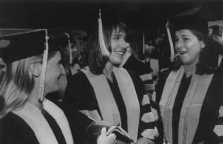
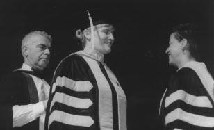
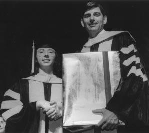
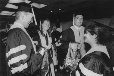
113th Commencement
The 98 graduates of the Class of 1998 bring the total of Penn’s veterinary graduates to 5,422. Of these, 3,897 are men and 1,525 are women. The Class of 1998 is composed of 70 women and 26 men.
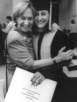
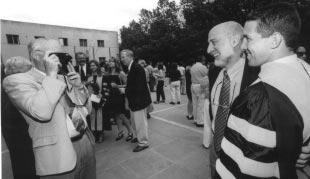
The Commencement exercises were held on May 18 at the Zellerbach Theater in the Annenberg Center. Parents, spouses, children, and other family members filled the auditorium to capacity and cheered for their special graduate.
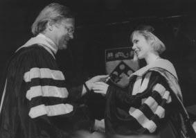
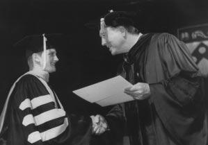
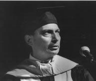
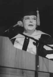
The Commencement address was given by Mary Beth Leiminger, D.V.M., a companion animal veterinarian and immediate past president of the American Veterinary Medical Association. Dean Alan M. Kelly presented the diplomas, assisted by Drs. Mattie J. Hendrich, Colin Johnstone, and Charles D. Newton. Dr. Raymond Sweeney assisted with the awarding of prizes. The Class Flag was presented to Dr. Jeffrey Berman V’98, class president, by Dr. Suzanne Smith
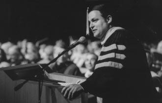
V’82, president of the Veterinary Medical Alumni Society and Dr. Harvey Bendix, V’75, president of the Pennsylvania Veterinary Medical Association, administered the Veterinarians oath. Once the newly minted V.M.D.s and faculty marched out of the auditorium, the informal festivities began on the plaza with much hugging and picture taking. Itwas a proud and happy day for everyone.
Class of 1998
David Adam Castrillo
Deborah Christine Anderman
Sally Anne Crellin Aschenbrand
Lisa Beth Barnett
Steven Jeremy Bensinger***
Felicia Lynn Berkowitz*
Jeffrey Ian Berman
Sara Elizabeth Bernat
Heather Elizabeth Bixler
Derek Sean Boen
Amy Diane Bowman
Edwin Dale Bracken, Jr.
Janet Keith Burke
William Whiteley Bush IV**
Natalie Domenica Campbell
Morgan T. Cavanaugh*
Kimberly Ann Christy
Susan Reed Cooke
Jennifer Tuttle Cromwell
Kristin Foster Dance**
Kerri Lee Davis**
EdytheAnn DeMaria**
Ingrid den Outer-Bergin
Jennifer Laurie Derstine*
Christina Joy Dolan
Mark Conrad Doran
Michael Howard Dunn
Elizabeth A.H. Ewaskiewicz**
Michael Scott Fanelli
Robert Flahive
Martha Anne Franklin
Jennifer Lynn Fry
Thomas Narinder Garg
Melissa A. George
Joan Phyllis Capuzzi Giresi
Margaret Wistar Gober
Chris Gretzinger
Jeevraj Singh Grewal
Bethany Jane Grohs
Barbra Michelle Hart
Gregory Saylor Heins*
Laura Margaret Aggson Holland
Julia Winters Irwin
Victoria Johnson
Courtney Alexandra Jones*
Erin Mary Jordan
Mark Albert Kapolka
Kathy Jude Kazmierski
Amy Lynn Kidd
Dorothy Abigail Kielkopf
Elizabeth Ann Krug
Hamilton Lincoln III
R. Thomas Livezey
Amy Elizabeth Lloyd
Carolyn M. Lyon
Katherine Cole MacGillivray
Robert Fraser MacGregor
Sean Abraham Maguire
Joann Cecile Martineau
David Dominic Matunis
Zachary Paul Matzkin
David Vinton McCrork
Claire Selawski McNesby*
Christy Joy Meola
Susan Elizabeth Monteforte
George Austin Motley
Karen Amy Moulin
Cynthia Christine Nass
Molly Ayres Northrop***
Sandra Lee Orben
Heather Peikes***
Sarah Anna Pesillo***
Karen Sloan Phillips
Erica Lee Pickman
Esteban Pokorny
Donna L. Riddle
Kimberly Ann Scalise
Beth Ann Shane
Lisa Ann Sherman
Roberta Ann Smith
Laura Jean Snyder
Heidi Anne Sproul
Kimberly Joy Sprouse
Jessica Ellen Stehr*
Lauren Merrill Stein
Jane Carolyn Teichner
Deanna Karen Tobin
Anson Joji Tsugawa
Elaine Kay Tucker
Lisa Ann Twardus
Shelly Susan Wagner
Debbie Leigh Wardius
Chick Winston Christopher Weisse***
Amy Marie Woodford
Lisa Suzanne Ziemer
Kathryn Elizabeth Zingle
***Summa Cum Laude
**Magna Cum Laude
*Cum Laude
16
Award Recipients
Leonard Pearson Prize
Steven Jeremy Bensinger
J.B. Lippincott Prize
Steven Jeremy Bensinger
1930 Class Prize in Surgery
Zachary Paul Matzkin
Auxiliary to the American Veterinary Medical Association Prize
Jeffrey Ian Berman
Auxiliary to the Pennsylvania Veterinary Medical Association Prize-Small Animal Award
Chick Winston Christopher Weisse
Auxiliary to the Pennsylvania Veterinary Medical Association Prize-Large Animal Award
Katherine Cole MacGillivray
1956 Class Medal for Achievement in Pathology
Sarah Anna Pesillo
James Hazlitt Jones Prize in Biochemistry
Heather Peikes
American Animal Hospital Association Award
Sarah Anna Pesillo
Merck Awards
Small Animal Award
Mark Albert Kapolka
Large Animal Award
EdytheAnn DeMaria
George M. Palmer Prize
Barbra Michelle Hart
Everingham Prize for Cardiology
Zachary Paul Matzkin
Large Animal Surgery Prize
Jeevraj Singh Grewal
Large Animal Medicine Prize
Elizabeth A.H. Ewaskiewicz
Morris L. Ziskind Prize in Food Animal Medicine
Lisa Ann Sherman
Morris L. Ziskind Prize in Public Health
Molly Ayres Northrop
Hill’s Award R. Thomas Livezey
Pharmacia & Upjohn Awards
Small Animal Award
Heather Peikes
Large Animal Award
Kristin Foster Dance
Faculty/SCAVMA Prize
William Whiteley Bush IV
American College of Veterinary Surgeons Prizes
Small Animal Surgery Prize
Janet Keith Burke
Large Animal Surgery Prize
Elizabeth A.H. Ewaskiewicz
American Association of Feline Practitioners
Award
Lisa Suzanne Ziemer
Field Service Prize
Barbra Michelle Hart
E.L. Stubbs Award in Avian Medicine
Kathryn Elizabeth Zingle
Anatomy Prize
Molly Ayres Northrop
American College of Veterinary Radiology Award
Sarah Anna Pesillo
Iams/VECCS Award for Excellence in Veterinary
Emergency and Critical Care Medicine
Jeffrey Ian Berman
Charles F. Reid Award
David Adam Castrillo
17
Dog Bites
An average of 12 people die each year from dog bites and as many as one million require medical attention. The Insurance Information Institute reports that insurance companies paid a record $1 billion for dog bite liability claims in 1996 alone. Medical treatment for dog attacks costs society $102.4 million annually.
The American Veterinary Medical Association and State Farm Insurance Companies have initiated a campaign to raise awareness about dog bites and promote responsible pet ownership. The brochure published explains that any dog, if provoked, has the potential to bite and gives tips on how to prevent dog bites. These include: socialization so the dog feels at ease around people and other animals, training the dog to obey basic commands such as “stay”, “no”, and “come”, and not allowing the dog to run free. Copies of the brochure may be obtained by calling 887-254-FIDO (3436).
The AVMA supports dangerous animal legislation by state, county, or municipal governments, provided the legislation does not refer to specific breeds.
Those planning to purchase a puppy for the first time should carefully consider the original purpose of a breed (hunting, herding, guarding, etc.), and temperament. One of the reasons it is better to buy from a breeder is that it provides the opportunity to see littermates, parents, and other adult animals.
Dog bites are the number one health problem of children. It is important to teach children how to correctly interact with dogs and not to approach strange dogs. Dogs can be wonderful companions, but it is the responsibility of the owners to make them “good citizens”.
Health Concerns
The Morris Animal Foundation has released results of a survey of pet owners. Dog owners identified cancer, skin disease, and dental problems as conditions most affecting their animal’s health. Other concerns were nutrition, hereditary disease, and external parasites.
Among cat owners, urinary problems were the main concern, followed by dental problems, cancer, and feline leukemia.
Cancer was the leading diseaserelated cause of death in dogs and cats.
Most Popular AKC Breeds
Total registrations are the basis of the AKC’s “Top Ten” ranking. The Labrador retriever with 158,399 registrations continues to be the most popular breed. Rottweilers remain in second place, although their total registrations are declining. Chow Chows and Dalmatians are recent fad breeds whose registration numbers have been declining rapidly. In 1997, German shepherds ranked third, golden retrievers fourth, and poodles fifth. German shepherds have been in the top ten for over seventy years.
The other breeds leading in registrations are beagles, dachshunds, cocker spaniels, Yorkshire terriers, and Pomeranians.
The miniature schnauzer was the most popular terrier breed in 1997, followed by West Highland white and Scottish terriers.
A total of 1,307,362 dogs were registered by AKC in 1997.
White German ShepherdDogs
Breed standards are under the control of breed clubs and some have disqualifi-
cations which prevent competition at AKC championship shows. There have been white German shepherds since breed was established over a hundred years ago. They can be registered with AKC as purebred, but cannot be shown as that color is listed as a disqualification in the standard.
These dogs are not albinos, they have dark eyerims, nose, and pads. The white color is a recessive gene, both parents must carry the gene to produce a white puppy. It is said that the heredity deafness found in some white animals is not linked to this gene. It also is said that white is the wrong color for the breed’s work in the armed forces and other areas. The white shepherds seem to lack the exaggerated rear angulation and sloping toplines which are seen in present day show dogs.
Recently, the United Kennel Club announced that it is considering registering a new breed — white shepherds. These dogs will compete at UKC shows, but unless the German Shepherd Dog Club of America changes its breed standard, they cannot compete at AKC shows.
Creating a new breed is not without precedent. AKC recognizes varieties with breeds. Not too long ago, Norfolk and Norwich terriers were separated by ear carriage and wire and smooth fox terriers became separate breeds based on coat texture.
Book Reviews
The Irrepressible Toy Dog by Darlene Arden (Howell Book House/Macmillan USA, $17.95 hardcover).
Small dogs have special needs which may be quite different from those of larger dogs. These distinctions are covered, from the new-born through puppyhood, going to a new home, housebreaking, and learning house man-
18
ners. Health care and behavior problems are discussed and there are short profiles of the various toy breeds.
The importance of socializing from the 21st to the 45th days is stressed. Generally, toy dogs are too fragile for small children, but children can learn how to behave gently with them. Grooming should start at five to six weeks and should include teeth cleaning (toy dogs are notorious for dental problems). It’s unusual for a breeder to send a puppy off before the age of three months because, as a rule, toy dogs take longer to develop than a large breed puppy. A crate is important as a safe haven and an aid in housebreaking. Traveling with a crate can convince a hotelier to make an exception to a No Dogs policy.
Other bits of information in the book are that it is not unusual to see toy dogs become geriatric at around 11 to 12 years of age — when many giant breeds may have come to the end of their lives... Collapsing trachea, considered to be an inherited condition, is a problem in a number of toy breeds. Coughing is one of the signs and the owner smoking may contribute to the problem..... Traction alopecia is caused by the topknot in some breeds as the elastic or barrette
used to hold the topknot in place pulls the skin and hair and eventually causes permanent hair loss in the area. The recommendation is not keeping the hair in a topknot all of the time... Not every breed is right for every person. Every dog has considerations such as activity level, temperament, coat care and special health concerns that could be either negatives or positives for you. The pug dates back to 400 B.C. originating in China where it was depicted as the Foo dog. Dutch traders brought pugs to Holland, where they became favorites of William of Orange when a pug saved the prince’s life because its barking alerted to invading Spaniards. William’s tomb contains a carving of the monarch with his pug.
Much more useful and interesting information about toy breeds can be found in the book.
The 1998 Cat Fanciers Association Yearbook (Cat Fanciers Assn., P.O. Box 1005, Manasquan, NJ 08736 — $39.00 postpaid — hardcover).
This large volume is of particular interest because of many color photographs of the different breeds of cats in the advertisements. While the breed may
not be identified, there is an address for obtaining more information. There is a large section of black and white photographs of CFA Grand Champions and Grand Premiers and an interesting wellillustrated article on Abyssinians in America. Another article is on the American shorthair and covers early breed history. A few excerpts — “there is definite proof that several shorthair cats were brought to America on the Mayflower..... During the San Francisco Gold Rush of 1849, miners paid approximately $50 each for top quality proven rodent hunting cats from the ship S.S. Ohio..... Shorthairs sold for $50 to $100 a piece during the San Francisco rat plague of 1884..... As American cats became more plentiful, farmers and miners began to select kittens that appealed to them based on flashy colors.....”
The comprehensive index covers many articles published in earlier editions of the Yearbook. I have been assured that copies of these articles can be obtained from the office of the CFA.
Much CFA business is covered along with show results. However, the book may have much of interest for anyone who likes to read about cats.
Dr. Josephine Deubler, V’38, had the ultimate dog show judging assignment. She judged the best in show competition at the Westminster Kennel Club show at Madison Square Garden, New York, in February. The School hosted a reception in her honor, though Josephine could not be there. Her stand-in was a lifelike, life-size photo cut out which was a great success.
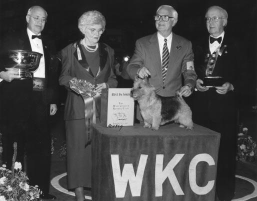
19
Canine Symposium
Cancer in Dogs
The 28th Annual Canine Symposium was held January 31, 1998 at VHUP. The event was organized with the help of the School’s Mari Lowe Center for Comparative Oncology and featured a series of presentations on Cancer in Dogs.
Mari Lowe Center for Comparative Oncology
The words cancer and growth are brutally intertwined. But they are also connected in several positive ways, said Dr. Narayan G. Avadhani, professor of biochemistry at the School. In recent years, cancer has met tremendous growth in research, knowledge, technology and treatment modalities. Dr. Avadhani presented Penn’s Mari Lowe Center for Comparative Oncology, which conducts extensive cancer research and clinical programs.
Cancer is a genetic disease, Dr. Avadhani explained. Yet cancer is inherited in only five percent of cases. The rest spontaneously arise following a primary DNA lesion that occurs during the life of an individual. “It is this 95 percent of the population that’s going to be extremely important for understanding cancer in both humans and animals.
“Animals are exposed to the same environment and some of the same putative causal factors that we are,” he said.
With these issues in mind, the Center, which is supported primarily through individual and foundation contributions, has created a state-of-the-art clinical oncology service at VHUP and today trains veterinary oncologists, conducts multidisciplinary research and supports outside cancer research. Since its establishment four years ago, said Dr. Avadhani, who heads the Center, the number of canine cancer patients at VHUP has more than doubled to some 1,500 cases annually.
J.C.
Cancer Terminology andSymptoms
What exactly is cancer? The language used to discuss cancer and its treatment is composed of many words that are not part of everyday conversation. Dr. Kim Cronin introduced some of the basic terminology that veterinary cancer specialists use and also discussed the early warning signs of cancer to help the symposium audience prepare for the lectures that would follow.
The early warning signs of cancer in small animals are similar to the seven warning signs of cancer in people used by the American Cancer Society. The signs range from: an abnormal swelling that persists or continues to grow, a sore that does not heal , weight loss, bleeding or a discharge from any body opening, reluctance to exercise, a loss of stamina or difficulty breathing, urinating or defecating. Even subtle changes, such as sleeping more, not playing as much and lack of desire to interact may indicate the need for a veterinary evaluation to determine if additional tests are necessary.
The term neoplasia means new growth and is used interchangeably with cancer. It takes about a billion cells, or thirty doublings, before we can actually see a tumor. When rapidly dividing cancer cells invade normal tissue the cancer becomes malignant. Malignant cancers can metastasize — spread to other parts of the body. During metastasis the cancer cells move through the blood stream or lymphatic vessels and may settle in tissues distant from their origin. The location where the tumor grew before metastasizing is called the primary site. Benign tumors are not usually invasive and do not metastasize.
The lungs and lymph nodes are the most common sites for tumor metastasis, however, there are many types of cancer that selectively spread to other parts of the body. The way a tumor grows and spreads is called its biologic behavior. The biologic behavior of a tumor determines what tests will be performed, how
the animal will be treated therapeutically and what the probable outcome will be. There are many unique terms used to describe the procedures involved in the clinical workup of an animal with cancer. An aspiration is when a needle attached to a syringe is inserted into the tumor and the plunger on the syringe is drawn back to suction a few cells from the mass for microscopic analysis. Cytology refers to the examination of the cells collected by aspirate. A biopsy involves the surgical removal of a small piece of tissue and examining it under the microscope. Grading of tumors is accomplished by characterizing the tumor cells using the microscope. Tumors are assigned into high, intermediate and low grades based on microscopic evaluation. High grade tumors tend to be very aggressive, they spread early and are difficult to treat. Low grade tumors are exactly the opposite. Staging is the evaluation of the extent of the tumor and the prognosis is the expected outcome for that tumor. A protocol is the overall treatment plan, the drugs used, how often as well as the number of times repeated. Finally, when the bone marrow is no longer able to produce normal cells the patient loses the ability to fight infection and sepsis results. Sepsis is an overwhelming systemic infection. Recognizing the early warning signs of cancer may lead to early detection and significantly increases the likelihood of successful treatment.
M.R.
Cancer Detection through Imaging Studies
Imaging techniques are indispensable in the management of cancer in domestic animals. Dr. Jeffrey A. Wortman, associate professor of radiology at the School, discussed the applications of various imaging modalities in diagnosing cancers, assessing prognoses, planning treatments and monitoring disease response.
Diagnostic imaging techniques, such as radiography — or “x-rays,” are used for cancer screening, detection, staging
20
and surveillance. Screening tests are employed to detect cancer in a population prior to the onset of clinical signs; unlike in people, in whom screening tests like routine mammography are often performed, these tests are infrequently done in animals. Imaging studies are routinely used to detect the presence of neoplasia in clinically-affected animals. These studies have variable sensitivities and specificities for diagnosing different tumor types; additional tests like tissue biopsy are often added to augment imaging studies.
Imaging tests also serve in staging of cancers. Once the diagnosis is confirmed by histopathology, such imaging diagnostics as thoracic radiography can then be used to detect tumor metastasis from primary sites, such as bone; these “mets” show up as radiopaque (white) nodules. This information is important in affixing prognosis and planning treatments like surgery, radiation therapy and chemotherapy. “If the cancer has spread,” said Dr. Wortman, “this indicates that it’s aggressive and will be more difficult to treat and cure.”
Follow-up imaging studies can then be incorporated into disease surveillance to chronicle disease regression, progression, relapse, and treatment complications.
The imaging methods that are particularly efficacious in veterinary oncology include radiography +/- contrast (i.e. iodinated agents) ultrasound, x-ray computed tomography (CT), magnetic resonance imaging (MRI) and nuclear scintigraphy (scans). The choice of imaging study, Dr. Wortman explained, “depends on many things, like the patient history and (tentative) diagnosis, which will give us some idea of the nature of the cancer and its biological behavior.” The choice also depends on what information is being sought, availability and quality of the imaging technology and expertise of the veterinarian interpreting the study.
Ultrasonography, an imaging technique in which deep structures of the body are visualized by recording the reflections of sound waves directed into the tissues, enables one to view masses that may or may not be externally pal-
pable. It can also be used to safely direct biopsy instruments to internal sites so tissue samples can be obtained for pathology. Likewise, a CT scan or an MRI may elucidate a brain tumor that would not be apparent on skull radiographs.The availability of imaging modalities in veterinary medicine is variable. Radiography, including contrast techniques, is ubiquitous throughout private veterinary practice, said Dr. Wortman, and ultrasound is steadily becoming more accessible. But more sophisticated studies, like CT and MRI, are less accessible to animal patients.
Whatever imaging test(s) one selects to screen for, diagnose, stage or monitor cancer, Dr. Wortman advised, one should maximize the fitness of the test by using proper technique. In radiography this includes correct body positioning and film exposure, and use of contrast agents where appropriate.
“You need to be aware of the limitations of a specific test and try to expand on the test in order to accurately make the diagnosis.”
J.C.
Breed Related Cancers
Many cancers have been found to have a breed predilection. Dr. Michael H. Goldschmidt, professor of pathology at the School and head of the Surgical Pathology Service of the Laboratory of Pathology and Toxicology, showed examples of specific cancers in the dog and the breeds most commonly affected.
Dr. Goldschmidt cited a study conducted in the 1960’s in the USA that estimated tumor incidence at about 1,100 cases per 100,000 dogs per year. The Laboratory of Pathology had developed an extensive computerized database of canine cases; in the decade preceding 1995 over 130,000 cases in dogs were submitted to the Surgical Pathology Service for diagnosis. Most tumors in these dogs were from the skin, oral cavity, mammary gland and male genitalia. Because of the large size of the database it
was often possible to calculate the breed risk for commonly diagnosed tumors.
An example of this was mammary tumors. Benign mammary tumors, seen clinically as well encapsulated masses of varying size and consistency on palpation, were found to have a higher predilection for small breeds of dogs such as the miniature poodle, Yorkshire terrier, Chihuahua and Maltese. Malignant mammary tumors, seen clinically as invasive, inflamed, ulcerated masses with lymphatic invasion in some cases, had a higher predilection in the English setter, Chihuahua, miniature poodle and Afghan hound. Many breeds at decreased risk for developing mammary tumors, such as the golden retriever, Rottweiler and boxer were at high risk for developing soft tissue and mesenchymal tumors.
The incidence and types of tumors are breed related and probably controlled by a single or multiple genes. An example given was the German shepherd which is predisposed to developing nodular dermatofibrosis, a syndrome that is comprised of a three tumor; multiple dermal fibromas (multiple firm masses in the skin), renal cystadenocarcinomas (malignant cystic epithelial tumors throughout the kidneys), and in females uterine leiomyomas (benign tumors of the smooth muscle).
Dr. Goldschmidt also illustrated the increased and decreased breed risks associated with two specific skin tumors, including infundibular keratinizing acanthoma, to which the Norwegian elkhound is highly predisposed with a relative risk of 29, and trichoblastoma, to which the Kerry blue terrier is predisposed with a relative risk of 12.
Other specific tumors with high breed risks were also discussed. These included:
•Trichoepithelioma: basset hound
•Pilomaticoma: Kerry blue terrier
•Ceruminous adenoma: cocker spaniel
•Anal sac gland carcinoma: English cocker spaniel
•Subungual squamous cell carcinoma: giant schnauzer and Gordon setter
•Subungual malignant melanoma: Scottish terrier
(continued on page 22)
21
CANINE SYMPOSIUM CANINE SYMPOSIUM
Canine Symposium
•Melanocytoma: Vizsla and miniature schnauzer
•Liposarcoma: Shetland sheepdog
•Mast cell tumor: boxer and pug
•Cutaneous plasmacytoma: cocker spaniel
Dr. Goldschmidt concluded that by selecting for the specific external features of a dog, its phenotype, we are selecting specific genes, which make that breed more or less susceptible to developing a specific cancer.
J.C.
Cancer Treatment Options
When it comes to treating cancer, quantity and quality should go hand-inhand. Dr. Karin Sorenmo, assistant professor of oncology at the School, said that cancer therapy is best governed by attempts to prolong life within the realm of maintaining reasonable quality of existence. “We try to maintain the highest degree of function and take as few risks for serious complications as possible” when planning cancer treatment protocols, she said.
Several factors influence cancer treatment decisions. Most important are the tumor type, biological behavior and staging. Malignant tumors are characterized by local invasion +/- distant metastasis. Highly malignant (high grade) tumors often have metastasized by the time the patient’s cancer is diagnosed. For highgrade tumors, aggressive treatment combining surgery and chemotherapy is often warranted even if metastases are not yet detectable radiographically. The goal then is not typically to cure, but rather to prolong survival by slowing disease progression and to provide palliation. On the other hand, tumors that are locally invasive but less likely to spread offer better prognoses and are often treated effectively with surgery and/ or radiation therapy.
The patient’s overall health status plays a major role in therapy choices. Concurrent diseases should be attended to and the patient evaluated for its ability to tolerate cancer treatment. Life expect-
(continued from page 21)
ancy should be taken into consideration as well; for a slow-growing tumor in an older dog, for example, the treatment drawbacks may outpace the potential benefits.
Owner factors also impact medical decisions because treating cancer requires the owner’s money, commitment and emotional resilience.
The three principal cancer treatment modalities are surgery, radiation therapy and chemotherapy. Surgery has two functions in cancer management: to obtain tissue biopsies for definitive diagnosis and to excise lesions completely. Biopsy provides information about the mitotic index and vascularity of the tumor, both of which are increased in rapidly-growing masses. Local lymph nodes can also be biopsied to detect evidence of tumor spreading.
Tumor excision is performed for both curative and — in the case of painful tumors like osteosarcomas — palliative reasons. Tumors are resected with wide margins; the objective is to obtain “clean” or cancer-free borders, thereby minimizing the likelihood of local recurrence. Once the lesion is excised, the borders are examined histologically for the presence of neoplastic cells.
Radiation therapy is a suitable alternative to radical surgery for tumors in certain locations, such as the head or legs. Radiation is also important for adjunctive treatment of tumors that are not completely resectable, as well as for locally-invasive tumors like nasal carcinomas, for which surgery alone is usually not curative. As an adjunct to surgery, radiation therapy is effective in sterilizing the borders. If the borders are clean, the main concern then becomes distant metastasis.
Chemotherapy is often combined with surgery or radiation therapy to treat highgrade tumors that are likely to metastasize. In addition, it’s the treatment of choice for multicentric cancers like leukemia or lymphosarcoma. Chemotherapy strikes rapidly-dividing cells, which, in addition to cancer cells, include normal cells of the bone marrow, oral mucosa and G.I. tract. Many owners fear potential chemotherapy side effects. However,
those seen in people, such as hair loss and nausea, are not typically experienced by dogs. Because most breeds do not have continuously-growing hair, the hair follicular cells are not affected by chemotherapy. More importantly, the objective for most canine cancer patients is to prolong life rather than establish a cure. Therefore, lower doses and fewer drug combinations are used.
“If we treat aggressively and cause serious complications in these normal tissues, all the while knowing we are not going to cure the animal,” Dr. Sorenmo explained, “then we probably haven’t done that animal a favor.”
Veterinarians administer the same chemotherapy drugs used in human oncology. The drugs are given orally, intravenously or subcutaneously. Extensive owner cooperation is necessary in complying with strict and sometimes rigorous treatment schedules. But the returns can be well worth the effort. Dr. Sorenmo pointed out that chemotherapy extends the average lymphosarcoma survival time from just two months (untreated) to a year; ten to 15 percent of these cases never relapse. For osteosarcoma, following surgery with chemotherapy doubles the survival time over amputation alone.
In designing a cancer treatment protocol utilizing any or all of the aforementioned options, one must first define the treatment goal based on a realistic prognosis. This determines acceptable levels of owner resources spent and patient discomfort tolerated. Dr. Sorenmo cautioned: “The treatment should not be worse than the disease itself.”
J.C.
Quality of Life Issues for CanineCancer Patients
“Quality of life concerns are important to veterinarians who want their patients to feel good, they are important to pet owners because pets are often considered family members, and quality of life is certainly important to our patients” ex-
22
plained Dr. Lillian Duda, lecturer in radiation oncology.
Once a diagnosis of cancer is made, the decision to pursue treatment hinges on the determination of the quality of life that can be expected for the animal from that point onward. The veterinarian can help the pet owner feel comfortable talking about the many issues involved, and they must work together toward a common goal. It is something that should also be discussed between family members and friends because the matter is both personal and emotionally charged.
The main goal in cancer treatment is to secure a high quality of life for as long as possible. A pet may be kept alive in the hospital using supportive measures such as oxygen and intravenous fluids, but, it may not be acceptable to do so if there is no hope of improving the pet enough that it can go home. Quality of life issues are more easily overlooked and more difficult to assess than quantity of life issues. There must be a balance between both. The veterinarian can help by remaining objective and providing information about the cancer and the effects of treatments on the animal. However, the veterinarian depends on the owner to report how their pet is feeling and behaving.
The side effects of cancer therapy are numerous and vary greatly between patients. In general, it is not acceptable to expect the animal to suffer side effects from the treatment without a good chance of a better life. The Animal Medical Center in New York City has developed a scale to assess the overall quality of life for dogs undergoing cancer treatment. They measure the dog’s ability to carry out normal daily activities using five parameters. Eating is one of the parameters that is measured. As their quality of life begins to decrease, dogs will often show changes in their eating habits or may have a general loss of appetite, some other parameters are alertness, body conditions, and activity level. This scoring method makes the difficult task of assessing the dog’s condition more objective and provides useful medical information. Studies have shown that indi-
viduals scoring closer to normal tend to do better overall.
Quality of life means something different to each pet owner. People are asked to come up with and write down their personal minimum level of acceptable quality of life for their pet before it receives treatment. This is done to help them anticipate, and thereby prepare for some of the important decisions they may need to make. If the unanimous goal is to prolong a quality life and to minimize suffering then euthanasia is sometimes the only option remaining. Euthanasia is a humane and responsible decision — a final therapy allowing the pet to be released from pain and suffering.
M.R..
Nutritional Needs of CanineCancer Patients
Animals with serious chronic illness are often malnourished. One of the primary reasons this occurs is because their appetites are impaired by the illness and oftentimes the only food they accept — table food — is nutritionally inadequate. This may not be critical for short periods of time, but, when treatments extend for weeks or even months there can be serious effects on the animal’s health related to malnourishment. “I like to tell people it is their job to find something their dog will eat adequate amounts of consistently and I can build a balanced diet around it,” said Dr. Kathryn Michel, assistant professor of nutrition. Cancer not only affects the patient’s quality of life, but, can also impair their ability to tolerate cancer treatments. Meeting the unique needs of patients undergoing cancer therapy is augmented by providing adequate nutrition, however, dietary therapy in this context is considered supportive rather than primary care.
Dr. Michel reviewed the six basic classes of nutrients. “I know that we don’t normally think of water as a nutrient but really it is the most essential nutrient there is. An animal will die of dehydration in a much shorter time than it will die from starvation.” After water,
CANINE SYMPOSIUM CANINE SYMPOSIUM
the body must have nutrients that provide energy such as carbohydrates, fats and protein. Protein not only supplies calories, it is also the source of amino acids which the body uses to make new proteins. Micronutrients are also derived from the minerals and vitamins in the diet. A dog with cancer will require water, adequate calories, protein, minerals and vitamins, however, cancer causes changes in normal metabolism that are not overcome by supplying calories and nutrients. At this time the specific nutritional requirements for dogs with cancer have not been established.
Cancer therapies that reduce the chances of deleterious side effects are always sought, but, they are not always completely successful. Some patients may have nausea, vomiting or diarrhea as a result of their treatment. If an animal associates side effects such as nausea with the act of eating they may stop eating for that reason. This phenomenon is termed a learned food aversion and presents quite a challenge when we try to feed these animals. An important part of nursing a dog or cat through an illness is coaxed feeding. It is very important not to push food on an animal that does not want to eat. It is best to wait until the pet is comfortable and not stressed before presenting food. Do not attempt feeding right after changing bandages or giving medications. It may be best to feed them cold food directly from the refrigerator, as it will not have much taste or aroma. Drugs may be used to reduce nausea as a short term approach if the dogs are having gastrointestinal side effects. Appetite stimulants are most useful in convalescent animals to “jump-start” their appetites, but, really have very limited application. They are not intended for long term use. If the dog shows some interest in food try feeding novel food items or offer food in a different setting or at different times. You can also divide the day’s food up into a number of small meals instead of one or two large ones.
Some patients may benefit from nutritional support. These patients often show signs of malnourishment and have not responded well to coaxed feeding. It may
(continued on page 24)
23
Canine Symposium
be necessary to feed these dogs using a nasoesophageal or esophageal tube. In critical patients intravenous feeding is given to help animals through a short time period until the chemotherapy takes effect. This is not complete nutritional support, nor is it meant to be used as life support. It is providing adequate nutritional care for a limited time to cancer patients who are unable to nourish themselves during the treatment of their disease.
M.R.
Canine Osteosarcoma
Osteosarcoma is the most common primary bone tumor in dogs, accounting for some 85 percent of canine bone tumors. VHUP clinical specialists Drs. Kim Cronin and Amy Kapatkin explained the disease process of — and treatments for — osteosarcoma.
The average osteosarcoma patient is seven or eight years of age, although dogs as young as six months old have been diagnosed with this cancer. It typically strikes large- and giant-breed dogs like great Danes, golden retrievers and German shepherds. Associated with high amounts of stress on weight-bearing limbs, osteosarcoma is almost 500 times as likely to affect dogs over 35 kg than dogs weighing less than 10 kg.
Osteosarcomas tend to anchor themselves in areas of increased bone remodeling, said Dr. Cronin, lecturer in oncology. “Every time you have cell damage or increased turnover, the DNA is more likely to make a mistake when coding for new cells, which can lead to tumor formation.” So naturally, previous fractures and chronic bone infections are predisposing factors. These tumors are more likely to occur in the limbs, particularly the forelimbs, which bear most of the body weight; other bones, such as the ribs and skull, can also be affected.
Osteosarcoma is both locally invasive and metastatic. It infiltrates the bone and weakens it. It then spreads throughout the body. The chief presenting signs for osteosarcoma are lameness and pain
from page 21)
(which may be intermittent), limb swellings and pathologic fractures at the tumor site.
Because osteosarcoma shares common clinical signs with other conditions like degenerative joint disease (arthritis), infectious diseases and other tumors of the bone (i.e. fibrosarcoma, chondrosarcoma, hemangiosarcoma and synovial cell tumor), a thorough physical exam, radiographs and biopsy are used to make the final diagnosis. Chest radiographs should be performed in dogs diagnosed with osteosarcoma. According to Dr. Cronin, over 90 percent of afflicted dogs have pulmonary metastasis at the time of diagnosis, although lung nodules may not yet be visible radiographically.
Because of its high metastatic potential, osteosarcoma carries a guarded prognosis. Most patients eventually die of metastasis to the lungs or other organs. The average survival time from the date of diagnosis is 8-12 months. Only 20 percent of patients are alive two years after diagnosis. Without treatment, most patients succumb to the disease within a couple of months.
Treatment is aimed at removing neoplastic sites and preventing further seeding. If the tumor is on a leg, amputation is the usual course. “We must be very aggressive in our initial approach so we don’t get regrowth,” said Dr. Kapatkin, assistant professor of orthopedics and neurosurgery. The leg is typically disarticulated at the coxofemoral (hip) or scapulohumeral (shoulder) joint. “Amputation is very cosmetic and most of our patients can ambulate well on three legs and are incredibly happy,” said Dr. Kapatkin.
Limb sparing is the other surgical approach. Here, the tumor is resected en bloc with 3-5 cm margins, and the gap is filled with bone graft or donor bone. Potential complications include infection, implant failure and tumor recurrence. However, the survival rate is the same for limb-sparing as for amputation.
Surgery can be performed also to excise metastases. Prolonged survival rates have been achieved in cases where three or fewer nodules were present and survival until metastasis was diagnosed ra-
diographically was 300+ days, Dr. Kapatkin said.
Radiation therapy can be potent in destroying neoplastic cells at the primary tumor site and chemotherapy may be employed to prevent or delay metastasis. Several drugs — including carboplatin, cisplatin and doxorubicin, are part of the osteosarcoma chemotherapy protocol, which is typically initiated 10-14 days after surgery. Immunotherapy, which activates the immune system to combat cancer cells, is part of newer treatment protocols.
J.C.
Canine Brain Tumors
Brain tumors in dogs are not infrequent. Nor are they untreatable. Drs. Charles Vite and Amy Kapatkin clarified the key issues revolving around brain tumors in dogs and their surgical biopsy.
According to Dr. Vite, post-doctoral fellow in neurology at VHUP, brain tumors are a common cause of neurological dysfunction in dogs over five years of age. Primary brain tumors arise from neuroepithelial (i.e. astrocytoma, oligodendrocytoma, choroid plexus neoplasm and ependymal tumors) and meningeal tissue (meningioma). Brain tumors can also arise secondarily from surrounding tissues like the nasal cavity or bone (i.e. nasal carcinoma, chondroma and osteosarcoma), or originate from hematogenous metastasis from tumors elsewhere in the body (i.e. malignant melanoma, hemangiosarcoma and lymphosarcoma).
Clinical signs of a brain tumor depend on the location of the mass. Tumors of the cerebrum and thalamus may result in seizures and depression; tumors of the cerebellum and brain stem typically result in incoordination and cranial nerve deficits. “The clinical signs tell us what part of the brain is affected,” said Dr. Vite.
A thorough neurological exam should be performed. Once the location of the lesion is determined based on the neurologic signs, the lesion is then character-
24
(continued
ized as either neoplastic, inflammatory, cerebrovascular or degenerative. Because nothing is pathognomonic for a brain tumor except biopsy results, one must compile a list of differential diagnoses that can account for the clinical signs. The factors that go into ranking these differential diagnoses are patient signalment (age, breed and sex), chronicity and progression of signs, evidence of focal or multifocal neurologic disease, and presence of other systemic disease.
For example, said Dr. Vite, the differential diagnoses for a six-year-old golden retriever presenting with a sudden onset of generalized seizures include: intracranial tumor, encephalitis, metabolic disease and idiopathic epilepsy. Add to this the dog’s history of increasing mental dullness, and idiopathic epilepsy moves further down on the list. Throw in the fact that the dog is blind in the right eye and has right-sided postural deficits and the clinical picture is now most closely referable to either a brain tumor or encephalitis.
A cerebrospinal fluid (CSF) tap, titers to known organisms capable of causing encephalitis, and magnetic resonance imaging (MRI) of the brain can aid in distinguishing between these two possible causes. However, Dr. Vite added, definitive diagnosis of a brain tumor requires biopsy of the mass.
Once a brain tumor is tentatively diagnosed, surgical feasibility is then determined. Surgical success depends on the type of tumor present and its location, said Dr. Kapatkin, assistant professor of orthopedics and neurosurgery. Most meningiomas are treatable with surgery and adjunctive therapy, increasing a patients’s lifespan; pituitary tumors, on the other hand, are not easily treated. The cerebrum is the best location to have a tumor, she added. “We can remove a whole half of the cerebrum and you probably wouldn’t notice a big difference in your pet.”
The three main surgical approaches are the lateral, transfrontal and suboccipital entries. The lateral — or parietal — approach involves making a curved incision and elevating the muscles over the parietal bone. A hole is then drilled in
the bone and the cerebrum is accessed. In the transfrontal approach, the frontal sinus is penetrated and the frontal bone is removed using a bone saw. The tumor is removed from the prefrontal area and the bone is replaced. The suboccipital approach, used to enter the cerebellum or brainstem, involves a midline incision made at the caudal aspect of the skull. Care must be taken to avoid potentially lethal laceration of the sinuses in this region. A fourth approach, the ventral approach, is made through the oral cavity. Used mainly to access pituitary microadenomas, this approach carries high risk of infection. Treatment of pituitary tumors may incorporate radiation as a substitute for — or adjunct to — surgery.
In addition to sophisticated surgical techniques, Dr. Kapatkin credits advanced post-operative care with the high success rate for brain surgery. Intensive care, including fluids and ventilator therapy, is provided after surgery to prevent or rapidly respond to potential complications, such as seizures, surges in intracranial pressure, and intracranial hemorrhage. Fortunately, most patients suffer no complications. In fact, she said, “Seventy-five percent of patients we do brain surgery on literally eat dinner the same day of their surgery.”
J.C.
Mammary Cancer
Tumors of the canine mammary gland are common. They comprise 52% of all tumors in the intact female dog and occur most often between 10 and 11 years of age. The breeds most highly represented are spaniels, terriers, German shepherds as well as toy and miniature poodles. Mammary gland tumors are often associated with early estrogen exposure. “The risk for tumor development is associated with estrogen exposure during the first few years of life and that has a significant importance when it comes to preventing these tumors” said Dr. Karin Sorenmo, assistant professor of oncology. When a dog is spayed before the
CANINE SYMPOSIUM CANINE SYMPOSIUM
first estrus, she has a 0.5% chance of developing breast cancer later in life. If she is spayed before the second estrus there is an 8% chance of developing breast cancer, and spaying after the third estrus — or any estrus thereafter — increases the likelihood of mammary cancer to 26%. In dogs that were spayed in addition to having the tumor removed the median survival was 659 days. Whereas, dogs that were left intact after their treatment surgery had a median survival of only 198 days.
The relationship between estrogen and mammary cancer in canines is also important because there are similarities with estrogen and breast cancer in women. Women with breast cancer are usually middle aged to older — the same age distribution seen in dogs. The most common types of tumors in dogs are the same as those found in women. Canine estrogen positive tumors respond to hormonal therapy by removing the ovaries. Estrogen positive tumors in women respond to Tamoxifen which is an antiestrogen. Both dogs with mammary gland tumors and women with breast cancer can be treated very effectively if the tumors are small and there is no evidence of metastasis. And both canine and human patients with large primary tumors or metastatic tumors are at high risk for dying from the disease. “I think that dogs can provide some very interesting and valuable models for this disease in women” said Dr. Sorenmo.
It has been reported that about half of the dogs with mammary gland tumors will actually have multiple masses. The caudal glands are affected more frequently. Mammary tumors can feel firm, soft or thickened and vary widely in size. They may be ulcerated, inflamed or edematous and one cannot determine if they are malignant or benign from these signs. “The good thing is that even though this is a very common type of tumor in the intact female dog, half are likely to be benign.” To obtain a diagnosis, a wide-margin excisional is performed if the surgical margins are clean this is also the treatment of choice for the primary tumor. However, it is necessary
(continued on page 26)
25
Canine Symposium
to first evaluate the patient’s general health through blood analysis and to look for possible systemic spread of the cancer with chest radiographs. If there is lymph node involvement then the risk for developing distant metastasis in the lungs is much higher.
Once the tumor type has been determined from the biopsy, and the extent of disease has been determined by one work-up, it is possible to suggest an outcome as well as an appropriate treatment. Several good retrospective studies indicate that survival rates are higher for dogs with small tumors. Tumors that are made up of well differentiated cells are likely to have a better prognosis than tumors that are anaplastic and have a high mitotic index. Invasive tumors can be more likely to metastasize than encapsulated ones. In general, tumors that have metastasized, have a much poorer prognosis.
The best treatment option is surgical resection. If the tumor is small it can be effectively treated with a lumpectomy or a mastectomy. A large tumor or tumors in multiple glands require a broad approach — regional or radical mastectomy — in order to remove all the malignant tissues at the primary site. It is thought that the progression of these tumors is dependent upon the presence of estrogen and an ovariohysterectomy — removal of the ovaries — makes it less likely that the tumor will continue to grow. Chemotherapy is indicated in dogs that have multiple negative prognostic factors and lymph node metastasis. More work needs to be undertaken in this area because there are no controlled studies to document the effectiveness of adjuvant chemotherapy after surgery in dogs with high risk mammary gland tumors.
The owner has a significant role in preventing canine mammary gland tumors through early detection. All tumors start out small and often appear insidiously. Therefore, it is important for the owner to either examine their dog’s glands or provide for regular veterinary check ups. There is one retrospective study from the University of Pennsylvania that found 95% of dogs with mammary gland tumors were likely to be
(continued from page 21)
overweight during their first year of life. By feeding a balanced diet, spaying early and providing regular examination of the mammary glands, the pet owner may significantly reduce the chances of tumor development. All tumors should be removed and biopsied because early treatment is crucial for a good outcome. Do not watch and wait.
M.R.
From the Laboratory Bench tothe Patient’s Bedside
New research in angiogenesis — or neovascularization — and its regulation has led to the discovery that inhibitors of this process are potent and promising anti-tumor drugs. “While this work is in its early stages we are very hopeful that we can make a contribution to this growing field” said Dr. Andrei Tikhonenko, assistant professor of pathology.
In the normal adult body there is very little angiogenesis apart from wound healing and menstrual cycling. When angiogenesis occurs, a new blood vessel sprouts from the pre-existing one. The membrane surrounding the original vessel dissociates and the endothelial cells migrate and undergo self proliferation. Cancer biologists and doctors have known for years that almost all malignant tumors are heavily vascularized. The tumor is penetrated by a tightly knit network of small blood capillaries which supply blood and feed the tumor with oxygen and nutrients. If angiogenesis can be controlled then it may be possible to cut off the blood supply to the tumor and prevent its growth. This concept is attractive because tumor cells are capable of rapidly accumulating mutation and becoming resistant to chemotherapy and other therapies.
Within the last three to four years some of the molecules which are responsible for endothelial cell growth were identified. Most cells in the body — including tumor cells — secrete both inhibitors and activators of angiogenesis. However, normal tissues secrete
more repressors than activators while tumor cells secrete either more activators or less repressors and turn the angiogenic “switch” on. Some of the molecules that have been identified are: Vascular Endothelial Growth Factor — the most potent activator of angiogenesis — as well as some inhibitors such as Thrombospondin I. There are other even more potent inhibitors such as some members of the Interleukin family and Angiostatin, and the most recent addition to the list is a protein called Endostatin.
In a recent study published in Nature magazine, a purified form of Endostatin — a very potent inhibitor of angiogenesis — was used. Tumors treated with Endostatin regressed very rapidly. The tumor cells remained, but, in the absence of blood vasculature they did not grow and slowly regressed. When the therapy was discontinued the tumor cells regrouped and formed another tumor. If Endostatin therapy was given again, the tumor regressed again but grew back as soon as therapy was stopped. Surprisingly, after twelve to fourteen cycles the ability of the tumor to grow back was lost. Still, the problem is that Endostatin therapy is too costly for prolonged use.
One type of tumor may be more sensitive to Endostatin — tumors of the endothelial cells themselves. In canines this tumor is often known as hemangiosarcoma. Hemangiosarcoma is a very malignant tumor and is capable of metastasis and dissemination. This tumor is often fatal for the dog. It most commonly affects the spleen or the right atrium of the heart, particularly in German shepherds. Hemangiosarcomas are comprised of chaotically arranged endothelial cells and may not respond to all of the inhibitors of angiogenesis in the same way that normal endothelial cells do. “We are poised to test the numerous inhibitors of angiogenesis and measure their ability to inhibit the growth and viability of hemangiosarcoma cells,” explained Dr. Tikhonenko. M.R.
26
1998 Penn Annual Conference
The 1998 Penn Annual Conference, held in late January, drew 750 veterinarians and 200 veterinary technicians for two days of lectures at the Adams Mark Hotel in Philadelphia. The 1999 edition of the nation’s oldest continuing education event for veterinarians is scheduled for January 27 and 28.
Many vendors participated and had booths in the exhibition area. Patrons and sponsors were honored with a plaque, presented by Dr. Charles Newton, associate dean.
JOIN US FOR THE AMERICAN GOLD CUP
September 10-13 at the Devon Show Grounds, Devon, PA
Four days of Olympic caliber show jumping, culminating in the Gold Cup competition on Sunday, Sep. 13 at 2:30PM.
Saturday, Sept. 12 is Family Day with activities for children, including an art show, a celebrity dog show and a M*A*S*H tent where injured, stuffed animals are brought back to health by Penn’s veterinary students. Children under 12 who bring a drawing or painting of their pet receive free admission that day.
A reserved seat, including admission to the show, is $5 for Thursday, $10 each Friday and Saturday and $20 on Sunday. Admission and a reserved for children under 12 is $5 on Sunday. General admission for any day without a reserved seatis$5.
Planned Giving Programs for the School of Veterinary Medicine
Over the years, the School of Veterinary Medicine has received generous support from donors who have made planned gifts through the Planned Giving Programs of the University of Pennsylvania.
Planned gifts are flexible, tax-advantaged arrangements that enable donors to make substantial gifts in ways that complement their personal financial planning. They can be designed to generate life-long income, obtain significant income tax deductions and reduce or eliminate estate taxes. Planned gifts can also be a means of converting lowyielding assets into a higher income stream at a reduced capital gains cost.
The Office of Planned Giving Programs of the University of Pennsylvania offers a variety of life income arrangements including: Charitable Remainder Trusts and
Charitable Lead Trusts. The Office of Planned Giving Programs can also help donors tailor bequests and structure gifts of life insurance and other assets for the benefit of the School of Veterinary Medicine.
Participation in any of Penn’s Planned Giving Programs also bestows the benefits of membership in The Charles Custis Harrison Society. Those benefits include annual luncheons, seminars and the University’s planned giving newsletter, Partner’s in Penn’s Future.
The Office of Planned Giving Programs is always willing to meet with donors and their financial advisors to design the most advantageous ways of giving to the School of Veterinary Medicine. For more information, please contact the Office of Planned Giving Programs at 1-800-223-8236.
27
The Iams Company representatives accept the Sponsor plaque.
The DVM Pharmaceuticals, Inc. representatives accept the Sponsor plaque.
The Hill’s Pet Nutrition representatives accept the Sponsor plaque.
Dr. Marne Platt, V’93, representative of Novartis Animal Health, accepts the Patron plaque.
Dr. Marlene Gumbs, V’83, scientific franchise manager for Mars-Kal KanWaltham, representing Waltham, a sponsor.
The A.J. Buck and Son, Inc. representative accepts the Patron plaque.
Special Gifts
The following have contributed to the Imaging/ Heart Station Building at New Bolton Center in memory of Mrs. Almira Jackson Scott:
Mr. and Mrs. Thomas H. Beddall
Mrs. Edith H. Overly
Paoli Memorial Hospital Auxiliary/Radnor
Three Day Event
Ms. Diane S. Quillman
Radnor Hunt
Dr. Charles W. Raker
Mr. and Mrs. James K. Robinson, Jr.
Ms. Edith Rosato
Mr. and Mrs. Fred R. Seiter
Ms. Caroline M.C. Shipley
Mr. and Mrs. David S.J. Smith
Mrs. George Strawbridge
Mrs. Anne F. Thorington
Ms. Dolores R. Widzgowiski
The Following are gifts made to New Bolton Center in memory or honor of those listed:
Mrs. Harry H.S. Phillips, Jr. in memory of Mr. Alexander C. Stokes
Mr. Michael J. Renehan in memory of Ms. Hilary Segal
Mrs. George Strawbridge in memory of Mr. W. Burling Cocks
The Following are gifts made to New Bolton Center in memory of a beloved animal listed:
Ms. Doris Broomall in memory of REDS
Ms. Susan W. Sensor in memory of JAKE
Ms. Trish Swigart in memory of OBNOB
The following are gifts made to New Bolton Center in honor of the person or animal listed: Thomas & Nephele Wing Domenicich Foundation, Inc. in honor of FLAMINGO
Ms. Jane B. Nigra in gratitude for Dr. Michael Ross’ skill
Ms. Mary Rice in honor of Dr. Charles Raker
Gifts in memory of Mrs. Lila Griswold Allam for the Allam House Fund:
Dr. and Mrs. Edwin J. Andrews
Mr. and Mrs. Thomas H. Beddall
A Legacy...
Dr. Robert W. Bishop
Dr. B. F. Brennan
Dr. and Mrs. Ralph L. Brinster
Dr. and Mrs. John R. Brobeck
Dr. C. John Bryer
Mr. and Mrs. George E. Burke
Ms. Christine C. Connelly
Ms. Evelyn N. Crowl
Dr. R. H. Detwiler
Dr. M. Josephine Deubler
Dr. and Mrs. Sheldon S. Diamond
Mrs. Joseph R. Downey
Mrs. Richard C. duPont
Mrs. Margaret H. Duprey
Mrs. P.F.N. Fanning
Dr. Stuart A. Fox
Mrs. Carol Pyle Jones Fry
Dr. Paul C. Gambardella
Ms. Doris L. Hamilton & Mr. Herbert W. Geshwind
Dr. Lillian A. Giuliani
Dr. and Mrs. Barton L. Gledhill
Ms. Mary Jane Griswold
Mr. and Mrs. Michael S. Hall
Mrs. Florence P. Hanford
Mr. and Mrs. Holger T. Hansen
Dr. Robert T. Henry
Dr. and Mrs. Max J. Herman
The Rev. and Mrs. Joseph W. Hess, Jr.
Dr. R. D. Hoffman
Ms. Ann Barrett Hubben
Mrs. Gretchen S. Jackson
Mr. M. Roy Jackson
Mr. and Mrs. Richard I. G. Jones
Dr. Seth A. Koch
Dr. Charles D. Knecht
Dr. David H. Knight
Dr. and Mrs. C. Wendell Lofland
Mr. and Mrs. Walter L. LaLonde
Ms. Bertha-Jane Lee
Dr. and Mrs. Victor M. Longoria
Mrs. Harry W. Lunger
Mr. Walter Brooks Macky
Dr. Luigi Mastroianni, Jr.
Dr. and Mrs. John D. McCullough
Dr. Susan McDonough
The John T. McGrath Family
Mrs. Patricia Bonsall Stuart had a delightful sparkle in her eye and an abiding concern for horses always in her heart. Pat grew up in the Philadelphia area and migrated to Virginia with her husband, Herb. There, she became a noted judge of horses and active member of the Virginia House Council. After Pat moved to Crosslands later in her life, she was a frequent visitor to New Bolton Center for the Friends lectures or other special events.
Mrs. Stuart was involved in New Bolton in its early years and later instrumental in starting the Virginia-Maryland Regional College of Veterinary Medicine. Her deep interest in equine welfare and the advancement of veterinary medicine has now been translated, for the Center, into a generous legacy in support of continued equine studies.
For those not fortunate enough to meet this charming lady, Pat’s dedicated commitment to the horse will live on at New Bolton in perpetuity through the continued advances in equine research made possible through her generosity and forethought.
Dr. and Mrs. William Medway
Dr. David A. Meirs II
Mr. and Mrs. Carl J. Meister, Jr.
Mr. and Mrs. Eugene C. Moore, Jr.
Mrs. J. Maxwell Moran
Mr. and Mrs. James M. Moran, Jr.
Dr. and Mrs. Todd O. Munson
Mr. and Mrs. Robert L. Nicholls
Dr. and Mrs. Peter C. Nowell
Dr. Craig S. Ott
Dr. and Mrs. Clarence D. Parks
Mrs. Henry D. Paxson
Mr. and Mrs. Gary S. Pedersen
Dr. and Mrs. George C. Poppensiek
Mr. Richard B. Pyewell
Dr. Charles W. Raker
Mr. Eldrow Reeve
Dr. and Mrs. Arthur Richards, Jr.
Mr. and Mrs. Andrew L. Riker III
Mrs. Doris Boucher Ritter
Dr. Brooke Roberts
Mr. and Mrs. James K. Robinson, Jr.
Ms. Emma Rosato
Mr. George P. Rosemond
Dr. and Mrs. Lionel F. Rubin
Mr. and Mrs. William T. Salom
Dr. and Mrs. Gerhard A. Schad
Mr. and Mrs. Anthony Schwartz
Dr. and Mrs. Robert M. Schwartzman
Mr. Hardie Scott
Mr. and Mrs. J. E. Shada
Mr. George T. Sharp
Dr. Robert R. Shomer
Mr. and Mrs. John C. Shook
Mr. and Mrs. Raymond B. Slouf
Ms. Margaret Y. Snear
Mr. and Mrs. Paul E. Spears
Dr. and Mrs. Robert W. Stewart
Dr. William H. Waddell
Mrs. Helma Weeks
Dr. and Mrs. John E. Whitehead
The Whittaker Family
Dr. and Mrs. Stuart E. Wiles
Mr. George Wintersteen
Mr. Jeanne F. Wordley
Dr. and Mrs. Stuart G. Younkin
Gifts in memory of Dr. Mark Whittier Allam for the Allam House Fund:
Dr. Robert Austrian
Mr. and Mrs. Thomas H. Beddall
Ms. Lillias J. Brady
Dr. and Mrs. Ralph L. Brinster
Salon d’Artiste
Dr. M. Josephine Deubler
Mr. and Mrs. Francis R. Downey
Mrs. Richard C. duPont
Mrs. Margaret H. Duprey
Mrs. P.F.N. Fanning
Dr. Lillian Giuliani
Mr. Eldridge R. Johnson II
Dr. and Mrs. Walter F. Loeb
Mrs. Harry W. Lunger
The John T. McGrath Family
Dr. and Mrs. William Medway
Dr. Paul Nemir, Jr.
Mr. and Mrs. Robert L. Nicholls
Mrs. Henry D. Paxson
Mr. and Mrs. Richard B. Pyewell
28
Dr. Charles W. Raker
Mr. Eldrow Reeve
Mr. Edward M. Resovsky
Mr. James K. Robinson, Jr.
Mr. Hardie Scott
Mr. and Mrs. Isadore M. Scott
Mr. and Mrs. J. E. Shada
Mr. George T. Sharp
Mr. and Mrs. Paul E. Spears
Dr. and Mrs. Max L. Sponseller
Ms. Deborah H. Toler
Dr. and Mrs. R. A. Vaclavik
The Honorable & Mrs. Noah W. Wenger
Mr. George Wintersteen
York Container Foundation
Dr. and Mrs. Stuart G. Younkin
Gifts in memory of Mrs. Lila Griswold Allam & Dr. Mark Whittier Allam for the Allam House Fund:
Mr. and Mrs. Francis H. Abbott, Jr.
Mr. and Mrs. Robert M. Allam
American College of Veterinary Surgeons
Dr. William B. Ames
Mr. and Mrs. Ted E. Amick and Family
Dr. Loy Awkerman
Mr. and Mrs. Gleave L. Baker
Beltsville Veterinary Hospital, Inc.
Dr. and Mrs. Darryl N. Biery
Mr. and Mrs. William H. Brady
Mrs. Frank P. Brooks
Mrs. W. Burling Cocks
Dr. and Mrs. Daniel Cohen
Mrs. David Craven
Dr. and Mrs. Joseph S. Dougherty
Drs. Richard A. & Lillian D. Dunsmore
Mr. and Mrs. Francis I. duPont
Dr. and Mrs. Zachary B. Friedenberg
Dr. and Mrs. Lawrence Friedman
Mrs. Helen K. Groves
Mr. and Mrs. Michael S. Hall
Dr. Elaine P. Hammel
Dr. Max J. Herman
Mrs. Arlene J. Hollister
Dr. W. A. Limberger, Jr.
Dr. M. Phyllis Lose and Family
Ms. Joanna McQuail Reed
Dr. and Mrs. Edward M. Mimnagh
Dr. Joseph M. Oneal
Dr. and Mrs. James A. Orsini
Dr. and Mrs. James M. Pomeroy
Mr. and Mrs. Jonathan G. Pomeroy
The Quaker City Farmers
Radnor Hunt
Mr. and Mrs. Gerald Resnick
The Honorable Matthew J. Ryan
Mr. Michael T. Ryan
St. Andrew’s Altar Guild
Dr. Mead F. Shaffer
Mr. and Mrs. Vincent B. Sherry
Mrs. Anne F. Thorington
Mr. and Mrs. John M. B. Ward
Mrs. William G. Weisbrod
Mr. and Mrs. Arthur A. Zimmerman
Gifts in memory of Dr. Mark Whittier Allam and Lila Griswold Allam for the Mark and Lila Allam Center for Equine Sports Medicine:
The 1957 Trust
Ms. Christine C. Connelly
Dr. and Mrs. Donald F. Smith
The following gifts were made to the Ultrasound Unit at New Bolton Center in memory of Mr. James D. Cartwright:
Ms. Pauline H. Chrzanowski
Ms. Mary A. Cunningham
Mr. and Mrs. George R. McColley
Mr. and Mrs. Albert Price
Drs. Richard and Mary Anne Tucci
The following gifts were made to Friends of New Bolton Center in memory of a beloved animal: Mrs. Barbara Bauer in memory of WOODSTOCK
Dr. Jennifer G. Behm in memory of REGGIE
Ms. Doris Broomall in memory of REDS
Dr. Edward Mersky in memory of DR. WATSON
Ms. Susan W. Sensor in memory of JAKE
Mrs. Trish Swigart in memory of OBNOB
Ms. Theresa Ann Zappone in memory of SUNSHINE
Gifts were made to Friends of New Bolton Center in memory of the following:
Dr. St. George Hunt in memory of Mrs. Lila Griswold Allam
Mr. Michael J. Renehan in memory of Ms. Hilary Segal
Mrs. George Strawbridge in memory of Mr. W. Burling Cocks
Gifts were made to Friends of New Bolton Center in honor of the following:
Ms. Jane B. Nigra in gratitude to Dr. Michael Ross
Thomas and Nephele Wing Domencich Foundation, Inc. In honor of “FLAMINGO”
Dr. Neal Ralston in honor of Drs. Mark W. Allam, Charles W. Raker, Corinne R. Sweeney, Raymond Sweeney
Ms. Mary Rice in honor of Drs. Tony Mogg and Corinne Sweeney
A gift was made to Field Service at New Bolton Center in memory of the following:
Ms. Virginia M. Kufta in memory of FLYING DUTCHMAN
A gift was made to New Bolton Center
In honor of the 25th Wedding Anniversary of David and Lena Romanoff from Carole Golub and Len Frenkel
The following have contributed gifts to the Friends of the Small Animal Hospital in memory of a special pet:
Mr. and Mrs. Curt Adams in memory of CORDELIA
Mr. and Mrs. Ralph Ammon, Jr. in memory of DJ
Dr. and Mrs. Edward Andrews in memory of KILTIE
Dr. and Mrs. Edward Andrews in memory of MISS LILY
Anonymous in memory of ROCKY
Mr. Robert Bahnsen in memory of BU Bailywick Briards Ltd. in memory of FALCO
Mr. William & Ms. Carol Bair in memory of SIS
Ms. Susan Barrett in memory of KILTIE
Mr. Ira & Ms. Stephanie Birbaum in memory of MACDUFF & SABRINA
Ms. Pia Boben and Mr. Mitchell Fenimore in memory of NORA
Mr. George Bonsall in memory of JOEY
Mr. Morton Botel in memory of STAR
Mr. James E. Brown, Jr. in memory of BU
Dr. and Mrs. Bruce Burnett in memory of SUCHI
Ms. Cheryl Caracansi in memory of BENTLEY
Ms. Joan Casilli in memory of SMOKEY
Mr. and Mrs. Charles R. Chadwick in memory of CLEO
Mr. Robert Chin in memory of BU
Mr. Dean Chouinard in memory of SAMANTHA SHEA
Ms. Kristen Casilli & Dean Chouinard in memory of PONTIUS BILL
Ms. Amy R. Ciric in memory of BU
Ms. Barbara J. Ciric in memory of BU
Ms. Catherine Ciric in memory of BU
Mr. John Cleary in memory of FLOAT
Ms. Elizabeth Crawford in memory of COFFEE
Ms. Janet L. Daily in memory of WHITE SHOES
Ms. Andree Dalschaert in memory of BU
Ms. Teresa A. DeMusis in memory of BRANDY
Ms. Sharon Dicker in memory of ONYX & SASHA
Ms. Marilyn Dietrich in memory of COCOA
Ms. Mary K. Dobransky in memory of BU
Ms. Maureen P. Echman in memory of KATIE
Ms. Wynne V. Edelman in memory of BU
Ms. Lisa Ehrenkrantz in memory of AMOS
Ms. Sheila Elser in memory of DANA
Mr. Marc A. Feller in memory of SUSHI
Ms. Mary K. Fairchild in memory of BU
Ms. Carole D. Fieramosca in memory of BENJI
Ms. Kara Finalyson in memory of BIGSBY
Ms. Nancy Fishchong in memory of BU
Mr. Daniel R. Frennan in memory of THOR
Ms. Carol Fusco in memory of BRANDI
Ms. Janet Gilman in memory of DUFFY
Mr. James Goldberg in memory of TYLER
Ms. Annmarie Gorda in memory of DAISY
Ms. Francoise Harkavy in memory of DUFFY
Ms. Jill H. Hays in memory of TYLER
Mr. Mark Hilinski in memory of POOCHIE SEVRIN
Ms. Anne Hubany in memory of BU
Mr. Daniel Insogna in memory of FONZIE
Mr. Randy M. Kaplan in memory of BU
Ms. Phoebe M. Kent in memory of SKIT
Ms. Janis Kerrigan in memory of BRITTY
Ms. Anna Kletsman in memory of COCO
Mr. Van W. Knox, III in memory of TIGGER
Ms. Tammy Knutson in memory of CHINA
Ms. Sheila, Mr. Jeff and Mr. Ari Levin in memory of ROCKY
Ms. Roxanne Lewis in memory of ASHLEY
Friends of Donna Maropis in memory of SQUEAKY
Ms. Lila Matlin in memory of TRAMP
Mr. James A. McMillian in memory of ROCKY
Mr. Mark C. Mendelsohn in memory of BU
(continued on page 30)
29
Special Gifts
(continued from page 29)
Mrs. Lynn Meyers in memory of JULIE
Ms. Susan Montemurro in memory of KIRA
Ms. Lois Morgis in memory of MOOSE, MOUSE & TERVUREN
Ms. Gloria A. Morris in memory of MAX
Mrs. M. Frances Neubuck in memory of CINDY
Ms. Joan & Ms. Claire Newberry in memory of PJ & SMOKEY
Mr. Bruce & Ms. Sis Obedin in memory of TV
Mr. Arnab Pal in memory of AFFIRMIAN
Ms. Joan Pettit in memory of SHANTOOZ
Ms. Shari Pirone & Ms. Karen Rossi in memory of PETER
Ms. Gina Porcellini in memory of BENSON
Ms. Dorothy Roberts in memory of JENNY
Mrs. Mary Robinson in memory of ROMMEL
Mr. and Mrs. Robert Rottiers in memory of SHADOW
Mr. and Mrs. William Sauerwine & Family in memory of APOLLO
Ms. Pauline Sides in memory of FRITZ
Ms. Anna M. Skidmore in memory of BU
Mr. and Mrs. James Smoot in memory of BEAU & CAS
Ms. Bette Strauss in memory of FOX
Mr. and Mrs. Van Leer Stephany, Jr. in memory of BUFFIE
Mr. Wallace J. Stuart in memory of PEPPER
Ms. Donna Marie Testa in memory of DINO
Joseph E. Thompson, VMD in memory of JASMINE
Ms. Rosanne Tilp in memory of SHANNON
Ms. Diane Tomassetti in memory of BU
Ms. Harriet Trenholm in memory of BU
Ms. Emily S. Wang in memory of BU
Ms. Erin J. Wease in memory of HOSS
Ms. Norma Weller in memory of HALEY
Scholarships
The Pennsylvania Veterinary Foundation has made available several scholarships to students here: the Dr. Palace H. Seitz Memorial Scholarships awarded to Sandra Springer, V’99 and Kenneth Bixel, V’99 Amy Lynn Bader, V’99 and Jennifer Clarke, V’99 were the recipients of the Auxiliary to the PVMA Scholarship; the Dr. Samuel F. Scheidy Scholarship was awarded to William W. Bush IV, V’98 and Courtney Jones, V’98 was the recipient of the Dr. Samuel B. Guss Memorial Scholarship.
The Past President’s Scholarship has been awarded to Claire McNesby, V’98 by the Maryland Veterinary Foundation. Heather Rose Galano, V’99 was awarded The Pfizer Animal Health
Mr. and Mrs. Thomas Werner in memory of BU
Ms. Edie & Missy Wickham in memory of CHO & LIZZIE
Ms. Kathy Wiegandt in memory of FOSTER
Mr. Rich & Ms. Terry Woods in memory of TEEK & KONA
Words to Go in memory of BOOO
The following have made donations to the Humanitarian Fund in memory of Sushi:
Noel Sauls Harbist, MD
Howard C. Lapensohn
Gerald W. Spivack
The following have made donations to the Department of Clinical Studies at VHUP in memory of Hannibal:
Ms. Diana Menke & Family
Ms. Carol Huff and Mr. Lawrence Huff in honor of CALYPSO GRECO for equipment in the Emergency Service at VHUP.
The following have made donations to the Small Animal Hospital in the honor of those listed:
Evansville Kennell Club, Inc. In honor of Mr. Walter Goodman
Toy Dog Fanciers in honor of Dr. Katherine Michel
Ms. Fran & Mr. Ned Levi in honor of Mr. and Mrs. Julian S. Bers
Salvatore A. Orsini, MD in honor of Peter F. Orsini
Ms. Lynn D. Russoin honor of Mr. R. Buchanan
The following have made donations to the Small Animal Hospital in the memory of those listed:
Ms. Jane Baker in memory of Mr. Richard Angle
Mr. Michael & Ms. Karen Ball in memory of Dr. Elizabeth Campbell
Veterinary Scholarship. Pandora Davis, V’99, received a scholarship from the Barnstable County Agricultural Society, Inc.
Lisa Sherman, V’98, George Motley, V’98, Jaqueline Rapp, V’99, and Dipa Brahmbhatt, V’99 were awarded the New York Farmers Scholarships. The Community Foundation of Western Massachusetts has awarded their scholarship to Kristen Pelletier, V’99 Elizabeth Daniel, V’99 received a scholarship from Schering-Plough Animal Health. The Lloyd’s Underwriters, Lloyd’s Brokers and Kentucky Agents Joint Equine Research and Education Program awarded Elisabeth Ewaskiewitz, V’98 their scholarship.
Ms. Susan Barrett in memory of Mrs. Lois Fernely McNeil
Mr. and Mrs. Joseph Damiano in memory of Mr. Richard Angle
Ms. Kim &Marilyn Darrows in memory of Mr. Richard Angle
Mr. Thomas A. Deubler in memory of Dr. James A. Deubler
Mr. Nino & Ms. Marilyn DiEnno in memory of Mr. Howard (Bussie/Dutch) Schultz
Mr. and Mrs. James Dreyer in memory of Mr. Richard Angle
Ms. Elizabeth Hayes in memory of Mrs. Starr Hayes
Mr. Chung & Ms. Susan Huang in memory of Mr. Richard Angle
Mr. Jeffrey Lavine in memory of Mr. Richand Angle
R. N. Meyer, Jr. Trust in memory of Mr. R. N. Meyer, Sr.
Mr. Neil & Ms. Elenor Werberig in memory of Mrs. Joan Stack
Ms. Rose Marie Opdyke in memory of Mr. Richard Angle
Mr. Jeffrey & Ms. Diane Rotwitt in memory of Mr. Yale Mann
Ms. Joan & Mr. Brew Schumer in memory of Mr. Richard Angle
Mr. and Mrs. J. P. Shaughnessy in memory of Mr. Richard Angle
W. Paul Starkey Foundation in memory of Paul & Betty Starkey
Barbara Strauss, VMD in memory of Ms. Sylvia Winicoff
Ms. Joan W. Wilson in memory of Mr. Richard Angle
In memory of Karen E. Spiegle, V.M.D. for Scholarship
Ms. Cecelia McDaid
Ms. Grace M. McDaid
Mr. and Mrs. James Hoyes
In memory of Harry F. B. Bartolett, V.M.D. for the class of 1937 Endowment
Susan Barrett
Celia Bento
Susan A. Brady
Mr. and Mrs. Daniel M. Bryne
Jane & Peg Carney
Dominick & Mary Cerrato
T. Ward Collins & Family
Robert R. Crelin
John I. Dawes &Donna Y. Dawes
Mr. and Mrs. Roger Fitzsimmons
Mr. and Mrs. Samuel Gegner
Mr. and Mrs. George Georgas
Mr. and Mrs. Daniel Gordaychik
Sara Gordaychik
William E. Jacobs
William R. McGonigle
Dr. David A. Meirs II
Ms. Mary Quigg
David & Olive Shaw
Mr. and Mrs. Lawrence Sorcher
Elizabeth C. Van mater & Hope Kerr
Harry & Jane Zdancewic
30
Established in 1982, the Veterinary Hospital of the University of Pennsylvania Pet Memorial Program has grown from a handful of veterinarians to a large group of practitioners across the country who make gifts in memory of clients’ pets. Gifts support the Small Animal Hospital’s Friends Program. The funds raised enhance teaching and service which in turn help us provide the best veterinary care for our small animal patients.
Practitioners interested in participating in the program should contact Susan Barrett, associate director of development, at 215-898-4234.
Participating Practitioners
Affectionately Cats
Allegheny North Veterinary Hospital
Animal Care Center
Animal Hospital of Chester County
Antietam Valley Animal Hospital
Arlington Animal Hospital
Richard J. Baron, V.M.D.
Jennifer E. Behm, V.M.D.
Michele B. Belisle, V.M.D.
Harvey R. Bendix, V.M.D.
William E. Best, Jr., V.M.D.
Robert Bialt, V.M.D.
Bilmar Veterinary Services
Julia M. Block, V.M.D.
Kenneth L. Bollens, Jr., V.M.D.
Frank Borzio, V.M.D.
Alvin J. Brown, V.M.D.
Charles E. Brown, V.M.D.
John P. Burlein, V.M.D.
John S. Bush, V.M.D.
Stephen P. Butler, V.M.D.
Califon Animal Hospital
Camboro Veterinary Hospital, Inc.
Doris A. Cappiello, V.M.D.
Dawn M. Cesaretti
Charles H. Chase, III, V.M.D.
Chestnut Hill Cat Clinic
Circle Veterinary Clinic
Robert H. Cohen, V.M.D.
Companion Animal Hospital
Conrad Weiser Animal Hospital
Kevin P. Coogan, V.M.D.
William Corbett, V.M.D.
County Animal Hospital, Inc.
Evelyn M. Crish, V.M.D.
Robert P. Cusanno, V.M.D.
Paul W. Donovan, V.M.D.
Robert DuBois, D.V.M.
Diane Eigner, V.M.D.
Richard Esherick, D.V.M.
Family Pet Clinic
William G. Farrell, V.M.D.
Jeffrey Feinman, V.M.D.
Paul Fenster, V.M.D.
Sharon P. Lachette, V.M.D.
Laurelton Veterinary Hospital
Theodore J. Leif, V.M.D.
Richard M. Levine, V.M.D.
Debora J. Lichtenberg, V.M.D.
Lawrence Jay Linnetz, V.M.D.
M.S. Lombardo, V.M.D.
Robert F. Martin, V.M.D.
Jill McCracken, V.M.D.
Thomas J. McGrath, D.V.M.
Elizabeth McKinstry, V.M.D.
Patricia A. Morgan, V.M.D.
Sue K. Morizi, V.M.D., Ph.D.
Michael K. Moss, V.M.D.
Joseph Nebzydoski, V.M.D.
New Hartford Animal Hospital and CareCenter
Barry S. Newuman, V.M.D.
Fredric Nisenholz, V.M.D.
North Boros Veterinary Hospital, Inc.
Northside Animal Hospital
David J. Parris, D.V.M.
Juan L. Ferrer Perez, V.M.D.
Jean M. Persia, V.M.D.
Dominick A. Pulice, V.M.D.
Leon S. Riegel, V.M.D.
Richard Rogoff, V.M.D.
James R. Rummel, V.M.D.
Farid C. Saleh, V.M.D.
R.E. Sampson, V.M.D.
Amy Sclarsky, V.M.D.
Ronald Sher, D.V.M.
Arthur L. Finkel, V.M.D.
Patricia Forsythe, V.M.D.
Mary S. Gang, V.M.D.
Alan Glassman, V.M.D.
S. M. Gloth, V.M.D.
Mark D. Guise, V.M.D.
S. Candace Guyther, D.V.M.
Robert C. Hallock, Jr., V.M.D.
Hill Street Veterinary Hospital
Elizabeth Hoekstra, V.M.D.
Hope Veterinary Hospital
Russell Howe-Smith, V.M.D.
Hyattsville Animal Hospital
Susan Jacobson, V.M.D.
Richard Jaffe, V.M.D.
Mark A. Johnson, V.M.D.
Wm. Southard Jones, Jr., V.M.D.
Kay Larkin, V.M.D.
Nicholas F. Sitinas, V.M.D.
Dr. Lynn Springer
Andrew J. Stonefield, V.M.D.
Cynthia J. Swingle, V.M.D.
Brett A. Sylvester, V.M.D.
Gregory M. Thibodeau, V.M.D.
James O. Thomas, V.M.D.
John L. Thomas, V.M.D.
Joseph E. Thompson, V.M.D.
Valley Green Veterinary Hospital
Veterinary Medical Center
Washington Square Animal Hospital
West Mountain Animal Hospital
E. Andrews Whittington, D.V.M.
Gary R. Wiedwald, D.V.M.
Wilmington Animal Hospital
Joan M. Yarnall, V.M.D.
Sharon Zaccone, D.V.M.
31
Open House
Don’t Miss the Veterinary School’s Open House on September
19, 1998
at New Bolton Center 10 AM to 3 PM featuring exhibits and demonstrations from the Large and Small Animal Hospitals
Emergency M*A*S*H Tent for injured stuffed animals
Hospital Tours • Six-Horse Percheron Draft Hitch
Red Rose K-9 Search & Rescue Team
Southeast Regional Cattlemen’s Association Cattle Breed Exhibition
Animal Blood Mobile • Udder Chaos 4-H Dairy and Goat Club
Pot Bellied Pigs & Emus • Veterinary Career Information
First State Coon Hunters Club & Their Jumping Mules
Mary Lou Hughes Dog Agility & Obedience
Chester Valley Dairy Club and 4-H Demonstration of “How to Show” Cows
Sheep Herding Exhibition
Beagle from USDA Beagle Brigade • Tours of the Marshak Dairy
Rain or Shine
Free Admission and Parking
New Bolton Center is located at 382 West Street Road (Route 926) in Kennett Square, PA 19348-1692. Take Route 1 to 82 North, Make a left onto 926 West, approximately two miles on the left For information call 610-444-5800, Ext. 2182
University of Pennsylvania
School of Veterinary Medicine
3800 Spruce Street
Philadelphia, PA 19104-6008
Address correction requested ™
43 43
Bellwether is published by the School of Veterinary Medicine at the University of Pennsylvania.
EditorPhotographers
Helma WeeksAddison Geary
Writers Doug Thayer
Joan Capuzzi, V.M.D. New Bolton Liaison Dr. M. Josephine DeublerCatherine Larmore (Animal Crackers)
Mark Resty, V’99
Helma Weeks
We’d like to hear your praise, criticisms, or comments. Please address your correspondence to:
Helma Weeks, University of Pennsylvania School of Veterinary Medicine, 3800 Spruce Street, Philadelphia, PA 19104-6010; (215)␣898-1475.
None of these articles are to be reproduced in any form without the permission of the editor of Bellwether.
©Copyright 1998 by the Trustees of the University of Pennsylvania.
The University of Pennsylvania values diversity and seeks talented students, faculty and staff from diverse backgrounds. The University of Pennsylvania does not discriminate on the basis of race, sex, sexual orientation, religion, color, national or ethnic origin, age, disability, or status as a Vietnam Era Veteran or disabled veteran in the administration of educational policies, programs or activities; admissions policies; scholarship and loan awards; athletic, or other University administered programs or employment. Questions or complaints regarding this policy should be directed to Anita Jenious, Executive Director, Office of Affirmative Action, 1133 Blockley Hall, Philadelphia, PA 19104-6021 or (215) 898-6993 (Voice) or (215) 898-7803 (TDD).
Nonprofit Organization
U.S. Postage
PAID
Philadelphia, PA
Permit No. 2563
32 Printed on recycled paper.
