Science with light Research at the Paul Scherrer Institute
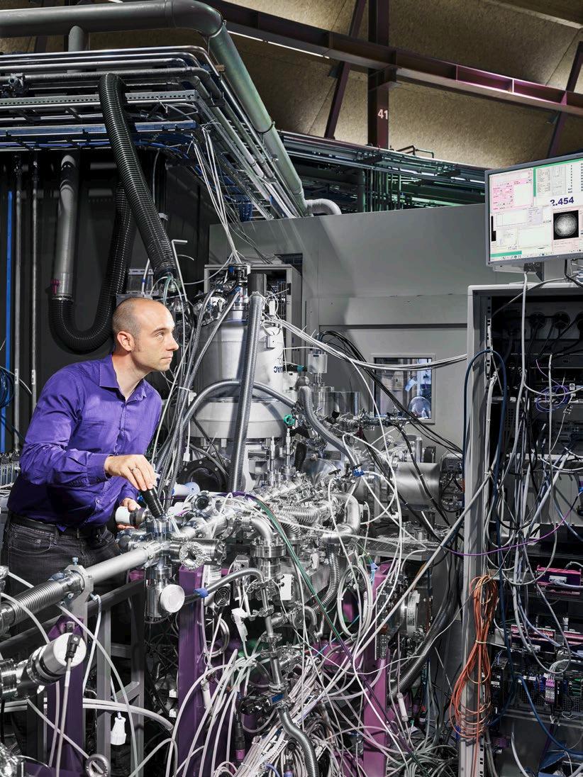
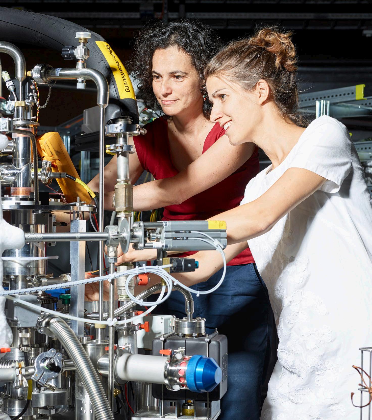 Starting an experiment at the Swiss Light Source SLS.
3 Contents 4 Science with light in 90 seconds 6 Structures of life 6 Repair signals 7 Losing sight at night 7 Nobel prize 8 Human health 8 Brain science 8 Tip-top teeth 10 Materials science and engineering 10 Carbon fibres 10 Fire safety 11 World-beating silicon chips 12 Manipulating magnetism 12 A single atom data store 13 Switching magnets with light 13 Magnetic hexagons 14 Quantum materials 14 Observing orbitons 1 5 Secrets of superconductors 16 Energy and environment 16 Natural gas from wood 1 7 Better batteries 1 7 Painting ships 18 Industry and innovation 18 Investing in innovation 18 Solving industry problems 18 Developing new technologies 19 Spin-off companies 20 Inside the Swiss Light Source SLS 22 Matter and light 22 Atoms are everywhere 23 Light is everywhere 23 Experiments with light 24 The extraordinary light of the Swiss Light Source SLS 25 Super-fast electrons 25 Electrons and light 25 Brighter and brighter 26 SwissFEL, the Swiss X-ray free-electron laser at PSI 26 What is an X-ray free-electron laser? 26 At the frontiers of science 26 Worldwide collaboration 27 Diamond technology 28 We make it work 31 PSI in brief 31 Imprint 31 Contacts Cover photo Extremely bright X-ray and ultraviolet light beams enable researchers at the Swiss Light Source SLS to understand structures of materials at a scale one million times smaller than a grain of sand. Scientists can discern how atoms and molecules are connected and how these connections change in real time.
Science with light in 90 seconds
The Swiss Light Source SLS at the Paul Scherrer Institute PSI is a super-microscope that can reveal features a million times smaller than a grain of sand. The SLS makes super-bright, pin-point sharp beams of X-rays and ultraviolet light. They are used to learn how the outer appearance and behaviour of objects is linked to what is inside. They can reveal how atoms and molecules inside an object are joined together and what they are doing.
X-rays have been used for over 100 years in medicine to see inside the human body. They are routinely used by doctors and dentists to identify broken bones, image tumours and check-up on the health of teeth.
But for research at the forefront of science and engineering, a hospital X-ray machine is not powerful enough — a much more advanced machine is needed. This is why the Swiss Light Source SLS was built.
The beams of X-rays made by the Swiss Light Source SLS are a billion times brighter than those produced by a hospital X-ray machine and have other unique qualities. This means that thousands of very precise and highly detailed measurements can be made in just a few seconds.
A new X-ray light source started operating at the Paul Scherrer Institute PSI in 2016. The SwissFEL X-ray free-electron laser is complementary to the Swiss Light Source SLS and will allow investigations into extremely fast processes to become routine.

PSI is Switzerland’s largest natural and engineering sciences research insti-
tute, and every year over 2500 scientists travel to PSI from Switzerland and around the world to use its world-leading large scientific research facilities. The institute is also home to two other large research facilities: the Swiss Spallation Neutron Source SINQ and the Swiss Muon Source SμS.
Innovation and discovery
Experiments at the Swiss Light Source SLS are essential for advancing science and solving problems in industry. They are helping to address contemporary issues in medicine, energy supply, the environment, and materials for new technology.
At SwissFEL, ultrashort pulses of laser-like X-ray light will allow completely new types of experiments in biology, chemistry, physics and materials science to be performed.
Working together, the Swiss Light Source SLS and SwissFEL will ensure Swiss science and engineering continues to be at the leading edge of innovation and discovery for many years to come.
You can read more about PSI research using super-bright beams of X-ray light and ultraviolet light in the following pages.
4

5
The comforts and convenience of modern life would be impossible without many decades of research into new materials.
Structures of life
Modern medicine has discovered that an incredible world of molecular activity lies at the centre of life – making cells, destroying infections and repairing damage. Despite enormous advances, there is still much more to be learnt in the fight against disease.
Repair signals
X-ray studies at the Swiss Light Source SLS are enabling drugs to be designed to repair defective blood vessels or to limit the growth of tumours.
A cut on the finger damages blood vessels and reduces the oxygen supply to surrounding tissue. Cells around the cut make a call for help by releasing a signalling molecule called a growth factor. The growth factor spreads through the tissue away from the cut causing other cells to spring into action and make new cells and blood vessels to repair the damage.
There are many types of growth factors, each highly specialised for growing cells of a particular type, for example, for skin, for nerve cells, or for blood or lymph vessels.
Each growth factor molecule has to align with a particular receptor molecule present on a target cell surface in order to activate it and start making new cells. This requires the molecular structures of the growth factor and the receptor, each made up from thousands of atoms, to be correctly positioned and oriented.
Mapping and analysing these incredibly complex molecular structures can

only be made with the state-of-the-art X-ray instruments available at the Swiss Light Source SLS.
Recent experiments have determined the exact structure of a growth factor and receptor responsible for the growth and repair of blood and lymph vessels.
With this knowledge, a new generation of drugs can be developed to limit tumour growth in the body by blocking the activity of specific growth factors involved in vessel building to cut off the blood supply.
6
The structure of a ribosome, one of the most important molecules in an organism. The scientist Venkatraman Ramakrishnan and his research group have determined the position of several hundred thousand atoms in this gigantic molecule. The experiments conducted within this endeavour were performed at the Swiss Light Source SLS and other light sources. They finally earned Ramakrishnan – along with two other researchers – the Nobel Prize in Chemistry in 2009.

Losing sight at night
Night vision relies on a light-sensitive molecule in the retina of the eye called rhodopsin. Research at PSI is learning how genetic defects affect rhodopsin and cause conditions such as congenital night blindness.
Rhodopsin is a protein molecule that is extremely sensitive to light and is used for seeing in the dark. Rhodopsin belongs to a large family of proteins known as G protein-coupled receptors. These molecules are found in the membrane wall surrounding a cell and their purpose is to sense changes outside the cell and pass a signal to the inside of the cell. When light hits rhodopsin, it changes shape and triggers a cascade of signals that pass to the brain.
Using the Swiss Light Source SLS, PSI researchers have been able to capture pictures of rhodopsin in its extremely short-lived light-activated state. They now have a precise picture of the first step in the process of seeing.
From the experiments, it has been found that night blindness is directly linked to a defect in the structure of rhodopsin. The defect causes rhodopsin to be constantly activated, even when it is not receiving light. The visual system becomes desensitised, as it tries to ignore the sense of seeing a constant low light.
Understanding the molecular cause of the condition opens the way for precisely targeted medicines. Although this approach would not cure night blindness, it could prevent progression of a related condition, retinitis pigmentosa, towards severe loss of daytime sight.
Nobel prize
Venkatraman Ramakrishnan from the MRC Laboratory of Molecular Biology in Cambridge, UK was one of the winners of the 2009 Nobel Prize in Chemistry. The prize was awarded for determining the structure of the ribosome, one of the largest and most important molecules in the cell. Some of the data used to achieve this was collected at the state-of-the-art X-ray instruments available at the Swiss Light Source SLS.
Ribosomes are complex, bead-like structures, which exist in multiple copies in each cell. The ribosome translates genetic information, producing tens of thousands of different proteins that in turn control the activity of living organisms.
To solve the structure, the position of each and every one of the hundreds of thousands of atoms that make up a ribosome was identified. It is one of the most complex atomic-resolution maps that has been made.
The experiments have been crucial for understanding how the ribosome works and the differences between bacterial and human ribosomes.
There is also a practical use: many modern antibiotics work by blocking the activity of bacterial ribosomes without affecting human ones. Infections are cured by selectively killing or inhibiting bacteria.
7
Human health
Novel imaging techniques developed at the Swiss Light Source SLS reveal extraordinary detail and are helping scientists understand a range of medical conditions.
Brain science
Damage to myelin, the insulating coat around nerve fibres in the body’s nervous system, can lead to medical conditions like multiple sclerosis. A new imaging method developed at the Swiss Light Source SLS can make pictures of the myelin coating of neurones in animal brain tissue with extremely fine detail.
The nervous system contains millions of neurones (nerve cells) that are highly specialised to carry messages from one part of the body to another. All neurones have a cell body and nerve fibres that can vary in length from a few millimetres to over a metre. The sciatic nerve is the longest nerve fibre in the human body. It connects the base of the spinal cord to the big toe. Most neurones are coated with myelin, a good insulator that allows electrical signals to travel along nerve fibres as fast as 400 km/hour. In multiple sclerosis patients, the myelin coating is damaged causing messages from the brain to be passed on poorly or not at all. This is like a power cable short circuit if the insulation is damaged. No
one knows what causes multiple sclerosis. Usually, the disease is mild, but some people lose the ability to write, speak or walk.
A new 3D imaging technique at the Swiss Light Source SLS is being used to study the role of myelin at the molecular level in rat brains. The 3D images are made without needing to cut into the brain, a significant advantage over typical study methods.
To create the 3D image, the rat brain is slowly rotated and an X-ray picture taken after each small step. The 800,000 pictures are combined by specially written software to give a complete overview of the concentration and thickness of myelin around the brain. The results show that the highest myelin concentration is around the bundle of nerve fibres connecting the left and right hemispheres of the brain. This new imaging approach opens up the possibility of studying myelin changes in relation to different patterns of illness.
Tooth enamel is the hardest substance in the human body. A human tooth has a hard outer layer of enamel covering a slightly softer layer of dentine. Unlike other hard tissues in the body such as bone, enamel can’t repair itself after damage, and teeth need the help of a dentist to bring them back to good health.
X-rays are used routinely by dentists to look for hidden problems with teeth, but these can only see details with the smallest size around one hundredth of a millimetre.
A study of healthy and decayed teeth with the powerful X-rays at the Swiss Light Source SLS has been able to record details ten thousand times smaller.
The molecular design of the enamel and dentine in teeth is extremely intricate when viewed at such small scales, but it is responsible for making teeth tough and hard-wearing.
Tip-top teeth
Sugary foods and poor toothbrushing can lead to painful cavities, a trip to the dentist, and fillings. X-ray studies of the molecular structure of healthy and damaged teeth suggest that new types of fillings could be developed that would last much longer than current treatments.
The study was able to map the arrangement of very fine collagen fibres in the dentine and study the region where enamel and dentine meet. The fine structure was found to be extremely similar in many different teeth and, surprisingly, was unaffected in teeth suffering from tooth decay.
Currently dentists repair teeth with materials that ignore the molecular design of teeth. The results of this study suggest that longer lasting treatments could be developed by making fillings that have a better match with the natural structure of teeth.
8
The molecular design of the enamel and dentine in teeth is extremely intricate when viewed with X-rays, but it is responsible for making teeth tough and hard-wearing.

9
Materials science and engineering
Technology can take big leaps forward in form and function by using new materials. They can be quickly tested using the bright beams of X-rays and ultraviolet light at the Swiss Light Source SLS.

Carbon fibres
Carbon fibres are very stiff and are used to reinforce other materials to make them tougher. Manufacturing carbon fibre is expensive, so engineers are investigating ways to reduce the cost.
Carbon fibre composite materials are strong and lightweight and ideal for high performance engineering in aircraft, yacht masts and artificial limbs. Carbon fibres are most often made from a carbon-rich starting material called polyacrylonitrile. Long strands of the starting material are heated to a very high temperature in an oxygen-free enclosure so that they do not burn. This process, called carbonisation, leaves the fibre composed of long, tightly interlocked chains of carbon atoms with only a few non-carbon atoms remaining.
Making carbon fibre is expensive, so engineers at Honda R&D Europe (Deutschland) are investigating new ways to manufacture carbon fibres with improved performance and reduced cost. Lignin, which is found in wood, is one new starting material for carbon fibre that is being explored.
Working with PSI scientists, Honda engineers have used advanced X-ray tomography imaging to build highly detailed 3D maps of the internal structure
of the lignin-based fibres to compare them with standard commercial fibres. In the commercial fibre, the internal structure was quite simple — a dense core wrapped in a less dense outer layer. In the lignin-based fibre, a sponge-like structure developed with tiny pores around 1000 times smaller than a grain of sand. With this unique view of carbon fibres, engineers are developing a deeper understanding of the links between carbon fibre structure and performance.
Fire safety
Intense heat from fires in buildings causes wooden beams to crack even when they are far from the flames of the fire. High-speed X-ray imaging can show how the inner structure of wood affects its mechanical strength.
Trees grow fastest in spring and early summer, laying down a ring of lightcoloured wood. A ring of darker, denser wood follows in the autumn. Across the rings, lines of ‘ray cells’, that stretch from the centre of the tree out to the bark, are used to store water.
At the Swiss Light Source SLS, wood can be rapidly heated to hundreds of degrees in a specialised laser furnace, with the cellular structure and cracking patterns being measured at the same time by X-ray tomographic microscopy. In beech wood, for example, a durable hardwood construction material widely used across Europe, thermal cracks have been found to mostly start along the ray cells and in the junctions of the seasonal growth layers.
Structural fire engineering research combines data from many different sources to gain a better understanding of materials and structures at fire temperatures. The data can be used to design and improve safety regulations.
Scientists from PSI can create the world’s smallest patterns on reflective silicon wafers which will be needed in future computer chips. The manufacturing technique was developed at the Swiss Light Source SLS.
10
World-beating silicon chips
Extreme ultraviolet light at the Swiss Light Source SLS is used by the semiconductor industry to develop new manufacturing techniques. PSI holds the world record for the smallest feature ever made on a silicon chip.

Lithography – the technology to print tiny circuits on silicon chips – has enabled e-mail, mobile phones, streaming video, and safer cars, trains and planes. The building block of all silicon chip circuits is the transistor, a precise switch that can be turned on and off
millions of times every second. The average silicon chip contains millions of transistors in a square millimetre.
Silicon chips are made by laying down a light-sensitive masking layer on the silicon and then using light to make a pattern with the fine detail of the circuits.
In fine art painting, painting smaller details needs a smaller brush. In lithography, finer details are made using light with shorter wavelength.
Commercial lithography machines have gone from using ultraviolet light with a wavelength of 365 nanometers to using deep ultraviolet light of 193 nano-
metres. The next step is to use extreme ultraviolet light with a wavelength of 13.5 nanometres.
At the Swiss Light Source SLS, PSI scientists have made the smallest structures in the world – rows of wires just 14 nanometres apart. For comparison, a human hair is about 50,000 nanometres wide and grows at 5 nanometres per second.
The PSI lithography capability is some 5–10 years ahead of standard industry methods, and it is widely used by companies and universities to test manufacturing methods for making the next generation of silicon chips.
11
Manipulating magnetism
The ability to manipulate magnetism is vital for modern computer technology. Music, photos and videos can all be stored by recording their information in magnetic patterns at the scale of atoms. Future technology is demanding new ways to store information and quickly access rapidly growing mountains of data.
A single atom data store
Researchers from Swiss universities working with IBM in the United States have prepared single atoms in the lab into a state that could, in the future, see stable single atom magnets being used to store data in computers.
MRAM is a type of computer memory where information can be stored permanently without the need to continually refresh the data. MRAM is used in aircraft and satellite control systems as it is not affected by cosmic radiation. Shrinking the size of MRAM components would allow more data to be stored, but the atomic structure of the material sets an ultimate size limit. A single atom is the smallest structure for storing data that can ever be achieved.
The Swiss/US team have shown, through experiments at PSI, that single atoms of cobalt placed on an ultra-thin magnesium oxide surface can be placed into the high energy state that is the necessary first step towards creating a stable magnet out of a single atom.

12
PSI researchers are at the forefront of studying new materials where the magnetic properties can be changed using a pulse of light from a laser.
Switching magnets with light
In a new class of materials, pulses of laser light can change the magnetic properties. Research into this unusual behaviour is at an early stage, but the new compounds are expected to find wide use in technology.
PSI researchers are at the forefront of studying new materials where the magnetic properties can be changed using a pulse of light from a laser. This novel feature has the potential for many applications including ultrafast data storage. Computer hard drives store data on rapidly spinning disks with a magnetic coating. As the disks fly underneath small read-write heads, data is recorded in tiny patterns of north and south poles. In a conventional hard drive, the time taken to switch a north pole to a south pole is typically a few nanoseconds. Using the new materials and a laser pulse, the switching time can be made 1000 times faster, around 1 picosecond (a picosecond is a million-millionth of one second).

Some of the extremely precise and demanding experiments can only be carried out using a new generation of large experimental facilities called X-ray free-electron lasers (XFELs). For many years, PSI scientists have travelled to do experiments at the LCLS facility in the United States, and the SACLA facility in Japan. Soon the Swiss X-ray freeelectron laser SwissFEL at PSI, and the European XFEL in Hamburg, will allow these studies to take place in Europe as well.
The experiments rely on synchronising a laser pulse, to alter the magnetism of the material, with an ultrashort X-ray pulse that immediately follows, to take a snapshot picture of the magnetic poles.
Using light for magnetic switching clearly works. But what exactly is happening is still the subject of ongoing debate and exploration in the research community.
Magnetic hexagons
Precise geometric patterns of tiny magnets can be made on silicon with different shapes and layouts. When the magnet size is less than a micrometre, new phenomena emerge. These structures could be used in future electronic devices for memory applications or to perform logic operations.
A research group at PSI has developed a method to make regular patterns of tiny magnets and study them with an X-ray microscope built at the Swiss Light Source SLS.
The tiny magnets are shaped like grains of rice. When the magnets are laid out in hexagons, the six magnets making up the hexagon arrange themselves with north and south poles touching to form a stable ring.
Under the X-ray microscope, the arrangements of north and south poles can be followed as more hexagons are joined together. As the network of hexagons becomes larger and larger, the tiny magnets do not have a clear choice on the direction their north and south poles should point. The whole network must rearrange itself into a stable configuration.
The PSI research group has developed precise mathematical models to explain what is seen. Using this well-controlled system, the group can accurately test situations found in real materials.
13
Quantum materials
Research in quantum materials explores the complex and unexpected behaviour that takes place when large numbers of electrons interact with each other in solids. Harnessing these strange effects has the potential to transform the next generation of electronic materials.
Observing orbitons
An unusual synchronised motion of electrons in a solid that was predicted over 30 years ago has finally been observed by physicists at the Swiss Light Source SLS.
Motion in a solid is extremely complicated. Every electron and atomic nucleus gets pushed and pulled by all the other electrons and nuclei in the solid which may themselves be in motion. The strong interactions, and the huge number of electrons and protons involved, make it very difficult to predict and understand the behaviour of solids.

14
Physicists simplify the description of solids using the idea of a ‘quasiparticle’. These are not real objects inside the solid, but a shorthand way of describing how a large number of electrons and protons are moving together in a coordinated way.
The complicated motions of all the electrons in a solid can be imagined as the much simpler motion of quasiparticles, which behave more like isolated particles that ignore each other. An individual electron cannot be split into smaller components — it is described as a ‘fundamental particle’. But in the 1980s, physicists predicted that many electrons travelling up and down a chain of atoms could be described as three quasiparticles: a ‘holon’ carrying the electron’s charge, a ‘spinon’ carrying its magnetism and an ‘orbiton’ carrying its energy and momentum. In an experiment of great technical achievement, physicists at PSI have been able to measure orbiton and spinon quasiparticles in a material. A beam of X-rays was fired at a group of electrons, causing it to absorb energy and allowing a spinon and orbiton to form, moving with different speeds in different directions.
Orbitons could be of use in a quantum computer which would perform calculations much faster than today’s computers.
A major stumbling block for quantum computers is that memory states are typically destroyed before calculations can be performed. Orbiton transitions are extremely fast, taking just femtoseconds. That’s so fast that using spinons and orbitons may offer a good way of storing and manipulating information in a realistic quantum computer.
Secrets of superconductors
Superconductors are one of the great scientific discoveries of the 20th century. Their incredible ability to let electricity flow freely without resistance at low temperatures is steadily being harnessed. Scientists working at PSI are at the forefront of the worldwide effort to explain how they actually work.
Superconductivity was discovered in 1911. In superconducting wires, electric current can flow freely without causing the wires to heat up. They can carry a current more than 100 times that of a copper cable of the same size. Superconductors need to be cooled to low temperatures, around 290 degrees colder than room temperature, using liquid nitrogen or liquid helium. They are used inside hospital MRI scanners, as electronic filters in mobile phone base stations, and in some power grids to transfer large amounts of power between nearby installations. The simplest superconducting materials are well-understood, but more and
more superconductors are being discovered that defy explanation. The newer superconductors can work at warmer temperatures — around 190 degrees colder than room temperature — and are known as ‘high-temperature superconductors’.
The most important building blocks of a typical high-temperature superconductor are layers of copper and oxygen atoms arranged on a square grid. The copper atoms act like tiny magnets and seem to be connected in some way with the high superconducting temperature. This is surprising, as superconductivity and magnetic fields are normally seen as rivals with magnetic fields destroying the superconducting state. One research team from the USA have mastered the art of making extremely thin films of superconductors with just two copper-oxygen layers.
In a unique collaboration, the team travelled to Switzerland to work with scientists at PSI and use the extremely sensitive and advanced X-ray instruments at the Swiss Light Source SLS. PSI is the only place in the world where this collaboration could get the results they needed.
The experiments showed that in these super-thin films of superconductor material, magnetism had not gone away and could be understood using very simple descriptions. The experiments show that magnetism is very important for producing the high superconducting temperature.
This result is one step closer to the ultimate dream of making a room-temperature superconductor.
15
The complex behaviour of electrons in superconductors can be measured in great detail using the state-of-the-art X-ray instruments at the Swiss Light Source SLS.
Energy and environment
Making the best use of Planet Earth’s limited fuel resources is important for society. Science at PSI is playing its part to reduce the impact of human activity on the environment.
PSI researchers are working hard to get the best performance out of new materials for prototype sodium-ion rechargeable batteries. Using X-rays at the Swiss Light Source SLS, changes to battery materials can be followed in real-time on an atomic scale while the battery is charging and discharging.

Natural gas from wood
Wood is a versatile, renewable energy resource that can be sustainably harvested from Swiss forests. PSI has developed technology that allows wood to be efficiently converted into natural gas.
Biomass waste from homes, farms and sewage treatment plants is fermented into biogas in many locations in Switzerland. However, wood is not easily fermented, and is usually burnt to provide heat.
The Paul Scherrer Institute PSI has developed an alternative way to process wood. First, it is heated to high temper-
16
ature and converted into hot gases. Then the hot gases are recombined to form synthetic natural gas. Gas from wood can be supplied into the gas grid giving greater flexibility on where the fuel can be used.
The hot gas mixture from wood is a mixture of carbon monoxide and carbon dioxide, hydrogen, and steam. There are also unwanted by-products including tars and sulfur-containing compounds.
Sulfur-containing compounds must be removed. They are corrosive to pipe-

lines and damage the materials used to convert the hot gas mixture into methane, the main component of natural gas.
Scientists at PSI have used results from experiments at the Swiss Light Source SLS to develop a material that can successfully work in the hot gas stream to remove the sulfur.
The new material is based on the element molybdenum. It was developed by using X-ray beams to study chemical reactions in real time and identify how the material should be adapted to have the best performance.
Better batteries
Lithium-ion batteries are a common source of energy for laptops, tablets and mobile phones. New types of sodium-ion battery could cost less to make and have almost the same performance.
A big challenge for modern society is how to store energy. Lithium-ion batteries are widely used for electronic gadgets and are increasingly being used in electric cars and to store energy produced by wind turbines or solar plants.
Swapping lithium for sodium could be a way to make a new type of rechargeable battery. Sodium is similar to lithium in terms of its chemical properties, and is much more abundant. Sodium is also up to fifty times less expensive to buy.
Like lithium-ion batteries, sodium-ion batteries must be able to provide a constant operating cell voltage and be chemically and structurally stable when charging and discharging.
At PSI, the future potential of sodium-ion batteries is being studied. Using X-rays, it is possible to see inside
prototype sodium-ion batteries as they are charging or discharging and to observe the changes on an atomic scale. Measurements with X-rays at the Swiss Light Source SLS can be made very quickly and give many useful details on the structure of the different materials inside the batteries. Many tens of prototype batteries can be tested at the same time.
The long-term goal is to develop sodium-ion batteries that can be used as easily as lithium-ion batteries. PSI researchers are working hard to get the best performance out of this new technology.
Painting ships
Ships and ocean structures are covered with specialised marine paints to protect them from corrosive seawater. 3D X-ray images of the microscopic structure of marine paint show how it offers such good protection.
Ocean-going ships are usually painted with an epoxy coating mixed with aluminium or glass flakes. The flakes overlap each other, like the tiles on a roof, forcing water to take a much longer path before it can reach the steel below.
A partnership between the London Centre for Nanotechnology, University College London, PSI and AkzoNobel is working to optimise paint coatings for ships and ocean structures. X-ray images show the shapes and arrangement of individual flakes in the paint on the nanometre scale.
Endurance studies of painted metal in salt water can take many years. By using the X-ray information in computer simulations, the performance of new paints can be better predicted and product research and development time is shorter.
17
Industry and innovation
PSI actively encourages industry to make use of its research.
Investing in innovation
Access to state-of-the-art synchrotron light facilities is essential for companies in the life science sector to be able to develop new medicines to tackle conditions like Alzheimer’s, arthritis and cancer.
Proteins are tiny molecular machines that perform all of the jobs needed to keep cells alive. Their activity can be modified by drugs and medicines, that can, in many cases, target a very specific protein. Atomic structures of proteins and drugs measured with synchrotron light can show how drugs and proteins work together at the molecular level, and indicate how to modify them to change their activity.
At the Swiss Light Source SLS, two experimental stations for measuring protein and drug structures are supported by industry: one by the Swiss pharmaceutical companies Novartis and Hoffmann La Roche and the German Max Planck Society; the second partially funded by a partnership between the Paul Scherrer Institute PSI, Novartis, Actelion, Boehringer Ingelheim, Proteros, and Mitsubishi Chemical in Japan.
Solving industry problems
Industry users frequently visit the Swiss Light Source SLS, and companies will often combine their knowledge and expertise with that of university research teams and the expert scientists working at PSI.
The examples below are just a few of the many where industry is using the stateof-the-art instruments at the Swiss Light Source SLS to solve immediate problems, refine procedures for later use in product development and manufacturing, or build a full understanding of new materials.
A partnership between the London Centre for Nanotechnology, University College London, PSI and AkzoNobel is working to optimise paint coatings for ships and ocean structures.
Honda R&D Europe (Deutschland) are studying prototype carbon fibres for improved performance and reduced manufacturing cost.
Conventional oil production leaves approximately 50–70% of the oil behind.
Shell Global Solutions International B.V. are working with PSI and Mainz University in Germany to develop safe methods to extract oil and gas trapped inside small pores in sedimentary rocks.
The feel of food when eaten is of crucial importance for manufacturing commercial food products. The Swiss food company Nestlé uses the Swiss Light Source SLS to understand how the texture of ice cream changes with temperature.
IBM is working at the Swiss Light Source SLS with a number of Swiss universities
to understand how magnetic data storage for computers can be miniaturised beyond current limits.
Intel and ASML work with researchers at PSI to evaluate advanced lithography techniques using extreme ultraviolet light that could be used to manufacture the next generation of silicon chips.
Developing new technologies
An advanced imaging technique developed at the Swiss Light Source SLS is a promising new method for the diagnosis of breast tumours. It is being tested at the Kantonsspital hospital in Baden with industry partner Philips.
Mammography is a medical technique used to diagnose and locate breast tumours. Doctors looking at an X-ray picture of a breast can identify if a tumour is present or could be forming. Seeing the difference between healthy and unhealthy tissue can sometimes be very difficult. A technique pioneered at the Swiss Light Source SLS for materials science can make significantly better X-ray pictures of breast tissue. A conventional X-ray picture relies on X-rays being absorbed by different amounts as they pass through different types of tissue.
The new technique also records how the X-rays change direction. This additional information significantly improves the image in the X-ray picture. It offers the prospect of earlier diagnosis of breast tumours.
18
Spin-off companies
Four spin-off companies based around synchrotron light have been created from technology and processes developed at PSI.
DECTRIS is the leading producer of hybrid photon counting X-ray detectors in the world. Their detectors have transformed research at synchrotron light sources as well as industrial and med-

ical X-ray applications. The technology used in the most successful product, the Pilatus detector, was developed by the DECTRIS company founders at PSI and laid the foundation for a successful international market position. DECTRIS now employs over 70 people. The founders of Eulitha have developed novel lithography techniques using the extreme ultraviolet light produced at the Swiss Light Source SLS. Eulitha provides nano-patterning services and
equipment for applications in photonics, optoelectronics, displays, biotechnology and other areas under the brand name PHABLE. Its products are used for industrial scale manufacturing, research and development.
Expose and Excelsus collect data for pharmaceutical and bio-technology companies at the Swiss Light Source SLS and help to analyze the collected data.
19
The underlying technology of DECTRIS X-ray detectors was originally developed at PSI. It has transformed research at synchrotron light sources as well as industrial and medical X-ray measuring methods.

Inside the Swiss Light Source SLS

Matter and light
Everything is made of atoms, and atoms are tiny. Tens of thousands of them can fit across the width of a human hair. At PSI, researchers use super-bright, pin-point sharp beams of X-ray light to find out where atoms are and what they are doing.

Atoms are everywhere
Atoms are the building blocks of the natural world. At the centre of an atom, protons and neutrons are tightly bound together into a nucleus. A cloud of electrically charged electrons sur-
rounds the nucleus. Atoms join up in countless different ways to make up all the substances and materials in the universe.
Light is everywhere
Visible light is a narrow slice of a much wider spectrum of light ranging from radio waves to gamma rays. Light carries energy that can be absorbed by the atoms that make up matter.
Radio waves and microwaves are the lowest energy light. Medium-energy light is called visible light or ultraviolet light, high-energy light is called X-rays, whilst gamma rays have even higher energy. Microwaves can be used to heat up food. Visible light striking the retina in
the eye is turned into an electrical signal to the brain. Ultraviolet light in sunlight causes melanin to combine with oxygen, darkening the skin to give a suntan. X-rays allow a doctor to look inside the human body.
Experiments with light
At the Swiss Light Source SLS, beams of X-ray and ultraviolet light are used to illuminate materials and objects to understand their properties at the scale of atoms.
When an object is placed in a beam of X-ray light, the beam passes through and is scattered by the atoms inside the object.
The scattered light is captured by detectors (that act like cameras) placed around the object. From the detected signals, the locations and movements of atoms inside the object can be worked out. Rapid sequences of snapshots can be combined into 3D movies of the atomic action.
Experiments can be performed simultaneously at up to 16 experimental stations at the Swiss Light Source SLS. Each station specialises in a different type of experiment with light.
SwissFEL will have six experimental stations when fully operating.
At the Swiss Light Source SLS, beams of ultraviolet and X-ray light are used to illuminate materials and objects to understand their properties at the scale of atoms.

23
The extraordinary light of the Swiss Light Source SLS
The synchrotron light at the SLS is produced in the storage ring which is built inside a circular concrete tunnel. Here, electrons circulate at almost the speed of light, making a million full circles in every second. A team of accelerator operators ensures smooth operations of the accelerator 24 hours a day.

24
To create the extraordinary light needed for ground-breaking scientific experiments, researchers and engineers have come together to build an enormous machine of great technical precision: The Swiss Light Source SLS. The light source is housed inside a striking circular building in the grounds of the Paul Scherrer Institute PSI on the western side of the River Aare.
Under a magnificent curved wooden roof, the intricate and sophisticated equipment of the Swiss Light Source SLS is fascinating to see. At first, the maze of pipes, cables and laboratories can be confusing. But after walking around the building for a while, it becomes easier to see how everything is connected.
Super-fast electrons
At the SLS, so called synchrotron light is emitted by a beam of electrons that have been accelerated to extremely high speed by a sequence of particle accelerators. The electrons travel inside a storage ring – a highly evacuated tube surrounded by a concrete tunnel with a circumference of 288 meters. Hundreds of magnets along the tube keep the electrons at the centre of the constantly curving path.
The electron beam at the Swiss Light Source SLS is as thin as spider silk, and the electrons can circulate for hours. From time to time, electrons in the beam collide with each other and get pushed out of the beam. To compensate for the loss, new electrons are regularly added into the beam to keep

the resulting light beams at a constant brightness.
The Swiss Light Source SLS works continuously through the day and night for more than 220 days a year. After a break in operation, it takes only a few minutes to refill the storage ring with all of the required electrons.
Electrons and light
Whenever super-fast electrons are forced to change their direction, they emit light. At the SLS, devices known as undulators wobble the electrons rapidly from side to side, like a slalom. This causes them to emit light in a high quality beam that can be used in experiments.
Whilst the electrons continue on their curved path inside the storage ring, the emitted light beam speeds straight ahead to the experimental stations. Special mirrors guide and focus the light, concentrating an extremely bright, pin-point sharp beam of ultraviolet or X-ray light onto the materials being studied.
Brighter and brighter
A new development in vacuum technology could allow the Swiss Light Source SLS to have even brighter beams of light. This major upgrade could be ready to operate in 2021. It would extend the lifetime of the light source by 20 years and open up new areas of science.
25
SwissFEL –the X-ray free-electron laser at PSI
The SwissFEL X-ray free-electron laser is the newest large research facility of PSI. Its unique X-ray light opens the way for important experiments in the fields of energy, environment, medicine, materials, and new technologies. Thus SwissFEL reinforces Switzerland’s leading position internationally in science and research. And the economy benefits too.
SwissFEL generates ultrashort pulses of X-ray light that have the characteristics of laser light. They are a billion times as bright as the light produced by the Swiss Light Source SLS. The X-ray pulses are so short and intense that they make it possible to produce films of the movement of atoms and molecules. For this reason, the work of SwissFEL is complementary to that of SLS. Together these two facilities can accommodate the increasing demand for state-of-the-art X-rays and ultraviolet light.
is routed through a long undulator section. Here, with the aid of magnets, the electrons are forced onto a fast slalom course. Through their constant change in direction, the electrons emit ultrashort pulses of X-ray light in close succession.
At the forefront of research
The experiments at SwissFEL make it possible to understand matter and materials on an entirely new level –whether in biology, chemistry, engineering or materials sciences.
What is an X-ray free-electron laser?
An X-ray free-electron laser concentrates X-ray light into an unimaginably bright, ultrashort X-ray laser pulse. Worldwide, there are only a few comparable facilities.

In an X-ray free-electron laser, a beam of electrons is accelerated to nearly the speed of light. Then this electron beam
The Swiss Light Source SLS has great successes to show: The static structures of numerous important proteins have been investigated here. With SwissFEL it is now also possible to track movement within these proteins. This opens up completely new insights into processes in the human body. The chemical composition of a substance, together with the geometric structure, determines how it behaves in a chemical reaction. At SwissFEL, researchers can observe the individual steps in such reactions. SwissFEL broadens our understanding of how magnetic properties of materials arise and how they can be altered. With that SwissFEL is paving the way for the computers of the future, which will be expected to store ever more data in an ever smaller space. Researchers can investigate, for example, how magnetic data can be selectively stored with the help of light, and how to achieve significant increases in the speed of information transfer.
Strengthening Switzerland as a business location
SwissFEL is strongly oriented towards the demands of the Swiss universities and industry and takes their research interests and requirements into account.
In the long term, SwissFEL will strengthen Switzerland as a research location, contributing at the same time to the competitiveness of the Swiss economy. This competitive capability is based mainly on bringing innovative products to market before the competitors can. First-rate research possibilities in a company’s home country allow the timely development of new knowledge as well as novel methods and tools that address global challenges. But Swiss industry also profits immediately from the new research possibilities at SwissFEL, whether through collaboration with PSI and the universities or through studies at the Swiss-
26
The SwissFEL X-ray light originates in the undulators. They were produced in collaboration with the Daetwyler group: Peter Daetwyler (left) with SwissFEL project leader Hans Braun with the undulators, installed and ready, shortly before the start of operations at the newest large research facility of PSI.
FEL facility within the framework of their own development work.
Still more SwissFEL, starting in 2020
In 2020, a second beamline will begin operating at SwissFEL. It allows a still greater variety of experiments.

The experiment stations at SwissFEL were conceived to exactly meet the expected requirements of its users.
That’s because every question to be probed – whether biological, chemical, or physical – sets different requirements for the experimental setup and the method best suited for the study. But the type of X-ray light is also decisive in what can best be investigated: At the moment, the researchers at SwissFEL can conduct their tests with so-called “hard” X-rays. This X-ray light has an extremely short wavelength and is optimally suited, for example, to track how and where atoms move during an ultrafast process.
However, if the researchers want to understand more precisely what happens with atoms or molecules while they are forming a new chemical bond, or how they react to external influences such as electromagnetic fields or light, they need “soft” X-ray light with a longer wavelength. A second beamline at SwissFEL will generate exactly this kind of light. It goes into service in 2020.
27
We make it work
Plumber
Perfecting pipework carrying cooling water and compressed air
Technical coordinator
Supervising projects and coordinating plans of action
Radiation protection technician Ensuring a safe working environment for all
Cleaner
Cleaning carefully around delicate and expensive equipment
Accelerator physicist
Delivering high quality synchrotron light for experiments

Assistant to head of department
Helping the head of department with all administrative and organisational tasks
Software developer Writing code to control high-precision experiments
Electronics engineer
Developing electronic components for precise positioning of scientific instruments
28
Many different people with a wide range of skills keep the PSI facilities working day and night. Meet a few of them:
Electrician
Installing main power and providing complex electric cabling of the beam line
Crane driver
Skillfully moving tonnes of equipment around every day
Vacuum engineer

Making air-free paths for particle beams
Beamline technician
Maintaining the beam line and building new components
Detector physicist
Making ultra-fast detectors to capture experimental results
Scientist
Designing experiments to make new discoveries
Multi-skilled mechanic
Creating precision components for unique scientific equipment
29

30
Aerial view of the Paul Scherrer Institute PSI. The circular building in the background is the Swiss Light Source SLS, on the west bank of the River Aare. The SwissFEL X-ray free-electron laser is located in the forest across the river, on the left of the picture.
PSI in brief
The Paul Scherrer Institute PSI is a research institute for natural and engineering sciences, conducting cutting-edge research in the fields of future technologies, energy and climate, health innovation and fundamentals of nature. By performing fundamental and applied research, we work on sustainable solutions for major challenges facing society, science and economy. PSI develops, constructs and operates complex large research facilities. Every year more than 2500 guest scientists from Switzerland and around the world come to us. Just like PSI’s own researchers, they use our unique facilities to carry out experiments that are not possible anywhere else. PSI is committed to the training of future generations. Therefore about one quarter of our staff are post-docs, post-graduates or apprentices. Altogether PSI employs 2200 people, thus being the largest research institute in Switzerland.
Imprint
Concept/Text/Editing
Dr. Martyn J. Bull
Editing committee
Dr. Martyn J. Bull, Christian Heid, Dr. Laura Hennemann, Dr. Paul Piwnicki
Photography and Graphics
All Photos Scanderbeg Sauer
Photography except:
page 6: Unchanged picture:
The Structural Basis for mRNA Recognition and Cleavage by the Ribosome-Dependent Endonuclease RelE; Cell, 2009 Dec 11; 139(6): 1084–1095 (doi: 10.1016/j. cell.2009.11.015), copyright: creativecommons.org/licenses/by/4.0/
page 27: Frank Reiser
page 30: Markus Fischer
Layout Mahir Dzambegovic
Printing Paul Scherrer Institut
Available from Paul Scherrer Institut
Events and Marketing
Forschungsstrasse 111
5232 Villigen PSI, Switzerland
Tel. +41 56 310 21 11
Villigen PSI, October 2018
Contacts
Head of the Research Division Photon Science
Prof. Dr. Gabriel Aeppli
Tel. +41 56 310 42 32 gabriel.aeppli@psi.ch
Chief of Staff Photon Science
Elizabeth Bianchi a.i.
Tel. +41 56 310 43 77 elizabeth.bianchi@psi.ch
Science Coordinator and CEO SLS Techno Trans AG
Stefan Müller
Tel. +41 56 310 54 27 stefan.mueller@psi.ch
Head of the Macromolecules and Bioimaging Laboratory
Dr. Oliver Bunk
Tel. +41 56 310 30 77 oliver.bunk@psi.ch
Head of the X-ray Nanoscience and Technologies Laboratory
Dr. Yasin Ekinci
Tel. +41 56 310 28 24 yasin.ekinci@psi.ch
Head of the Condensed Matter Laboratory
Prof. Dr. Frithjof Nolting
Tel. +41 56 310 51 11 frithjof.nolting@psi.ch
Head of the Femtochemistry Laboratory
Prof. Dr. Christoph Bostedt
Tel. +41 56 310 35 94 christoph.bostedt@psi.ch
Head of the Advanced Spectroscopy and X-ray Sources Laboratory
Prof. Dr. Luc Patthey
Tel. +41 56 310 45 62 luc.patthey@psi.ch
Head of the Nonlinear Optics Laboratory
Prof. Dr. Adrian Cavalieri
Tel. +41 56 310 30 79 adrian.cavalieri@psi.ch
Head Corporate Communications
Dr. Mirjam van Daalen
Tel. +41 56 310 56 74 mirjam.vandaalen@psi.ch
Paul Scherrer Institut :: 5232 Villigen PSI :: Switzerland :: Tel. +41 56 310 21 11 :: www.psi.ch SLS_Science with light_e, 5/2023
