
Original Article
Clinics of Oncology
ISSN: 2640-1037 Volume 5
Transfection of Cis-element ‘Decoy’ AP-1 DNA Binding Sequences Inhibits expression of uPA, uPAR and VEGF in Salivary Gland Cancer Cells
Ishibashi H1*, Sugimoto N2, Yamada S3, Sumitomo S1 and Muramatsu Y1
1Department of Oral and Maxillofacial Surgery, School of Dentistry, Asahi University, Japan
2Department of Pathology and Laboratory Medicine, Kanazawa Medical University, Japan
3Department of Physiology, Kanazawa University, Graduate School of Medicine, Japan
*Corresponding author:
Hrioaki Ishibashi,
Department of Oral and Maxillofacial Surgery, School of Dentistry, Asahi University, 1891
Hozumi, Mizuho-city, Gifu 501 0296, Japan, E-mail: hiro-i@dent.asahi-u.ac.jp
Keywords:
Salivary gland carcinoma; Urokinase type plasminogen activator; Urokinase type plasminogen activator receptor; Vascular endothelial growth factor; AP-1 decoy
1. Abstract
Received: 19 July 2021
Accepted: 02 Aug 2021
Published: 07 Aug 2021
Copyright:
©2021 Ishibashi H et al. This is an open access article distributed under the terms of the Creative Commons Attribution License, which permits unrestricted use, distribution, and build upon your work non-commercially.
Citation:
Ishibashi H, Transfection of Cis-element ‘Decoy’ AP-1 DNA Binding Sequences Inhibits expression of uPA, uPAR and VEGF in Salivary Gland Cancer Cells. Clin Onco. 2021; 5(3): 1-6
The effects of inhibition of transcription factor AP-1 on synthesis of urokinase type plasminogen activator (uPA), uPA Receptor (uPAR), and Vascular Endothelial Growth Factor (VEGF) were determined using cultured salivary gland carcinoma cells (ACCS and ACCT cells). The expression of uPA, uPAR and VEGF was induced in the presence of EGF. To inhibit AP-1 activation, we transfected double-stranded synthetic oligonucleotides containing the binding sequence of AP-1 (AP-1 decoy ODNs) using a novel, Hemagglutinating virus of Japan-, liposome method. Transfection of AP-1 decoy ODNs into ACCS and ACCT cells inhibited EGF-induced uPA and uPAR mRNA expression. AP-1 decoy ODN transfection reduced the in vitro invasion activity of both cells in the presence of EGF to 27.0 and 39.4%, respectively. EGF-induced mRNA expression of VEGF was simultaneously suppressed by AP-1 decoy ODNs. Inhibition of AP-1 by decoy ODNs has potential as a novel method for the simultaneous inhibition of salivary gland carcinoma cell invasion and angiogenesis.
2. Introduction
Because salivary gland carcinomas are highly invasive and tolerate chemoradiotherapy, resection is often required. However, complete resection is frequently impossible because of the complicated anatomy of the head and neck regions. New strategies are required
to inhibit the invasion of salivary gland cancers and enhance patient survival. Many proteases, such as Matrix Metaloproteases (MMPs) [1, 2] and the uPA-uPAR system [3] are involved in cancer cell invasion. uPA is a critical factor for the fibrinolytic system through activation of plasminogen to plasmin, while MMP-1 activates pro-MMP-1 [3]. uPA regulates not only cleavage of the Extracellular Matrix (ECM), but also cell migration via binding to uPAR [4]. The uPA-uPAR system has important functions in tumor cell progression; several pathways are activated through binding of uPA to uPAR resulting in the stimulation of cell adhesion, induction of chemotaxis and activation of the MAP kinase pathway, which induces cell invasion and migration [5-7]. The inhibition of the uPA-uPAR system via transfection of antisense RNA [8], neutralizing antibody [9], soluble uPAR [10] and dominant-negative uPAR [11] suppressed tumor cell migration and invasion. These reports suggest that inhibition of the uPA-uPAR system may suppress cancer cell invasion and metastasis. Expression of uPA and uPAR is induced by several cytokines [12]. Among them, Epidermal Growth Factor (EGF) is expressed commonly by salivary gland carcinomas [13], and induces either/or uPA and uPAR synthesis in cancer cells [14] through the activation of the transcription factor, AP-1 [15].
Vasculature development is an important aspect of the growth and
clinicsofoncology.com 1
metastasis of solid tumors. Among the angiogenic factors produced by tumor cells, Vascular Endothelial Growth Factor (VEGF) is considered to be the most potent and pathologically important [16]. The synthesis of VEGF is also modulated through AP-1, following stimulation by EGF. The inhibition of AP-1 activation in the presence of EGF may be effective in regulating the invasion of tumor cells and angiogenesis. If interference with AP1 activity results in the simultaneous inhibition of uPA- and uPAR-induced cell migration, and VEGF-related angiogenesis, tumor growth could be dramatically suppressed. In the present study, we applied a new ‘decoy system’ by transfecting double-stranded Oligonucletides (ODNs), containing the binding sequence of AP-1, into cancer cells. The decoy AP1 binding sites in cancer cell nuclei should interfere with the binding of activated AP-1 in the presence of EGF to regulatory regions of target genes and thus act as a sink for AP1. As a result of the inhibition of AP-1, mRNA expression of uPA, uPAR and VEGF may also be affected. The purpose of this study was to examine the effects of inhibiting AP-1 activation by introducing decoy AP-1 into cells. The cell invasive and angiogenic activities of salivary gland cancer cells carrying the AP-1 decoy were also determined. For transfection of AP-1 decoy ODNs, we used the Hemagglutinating Virus of Japan (HVJ)-liposome method [17]. The significance of our findings is discussed.
3. Materials and Methods
3.1. Cells
ACCS and ACCT cells (from Kyushu University Hospital, Fukuoka, Japan), derived from adenoid cystic carcinoma cells of the salivary gland were cultured in DMEM medium supplemented with 10% fetal bovine serum (FBS, Flow Laboratories, Stanmore, Australia), penicillin (100 units/ml) and streptomycin (100 μg/ml) in a humidified 5% CO2 incubator (Multi-Gas incubator BL3200, Astec Co., Fukuoka, Japan) at 37oC.
3.2.
Synthesis of ODNs
The sequences of the decoy ODNs were selected as follows. ODNs were composed of the binding sequences (underlined) for the transcriptional factor, AP-1, at the central site and dummy sequences upstream and downstream of the AP1 binding sequences [18]. Two bases (small letters) of the AP-1 binding sequence of AP-1 decoy ODNs (mt-AP-1 decoy ODNs) were mutated, and a lack of homology to binding sequences for other transcription factors was confirmed using Databases on Transcriptional Regulation [19].
AP-1 decoy ODNs:
5’ -ATTACCGGGCGGGCGGGCTAC- 3’
3’ -GTAGCCCGCCCGCCCGGTAAT- 5’
mt-AP-1 decoy ODNs:
5’ -ATTACCGG t a GG t a GGGCTAC- 3’
3’ –GTAGCCC t a CC t a CCGGTAAT- 5’
To enhance the intranuclear shift of the transferred decoy ODNs, high mobility group proteins (HMG)-1 and -2 were incubated with the ODNs at 20oC for 20 min to facilitate complex formation.
3.3. Transfection of Decoy ODNs using the HVJ-Liposome Method
We transferred ODNs into cancer cells using the HVJ-liposome method [17]. Briefly, phosphatidylserine (sodium salt), phosphatidylcholine and cholesterol (all from Sigma Chemical Company, St. Louis, Missouri) were mixed at a ratio of 1:4.8:2 by weight in 3.9 ml tetrahydrofuran. The lipid mixture (10 mg) was deposited on the sides of a flask by removal of the organic solvent in a rotary evaporator. The dried lipid was hydrated in 200 µl AP-1 decoy ODNs or mt-AP-1 decoy ODNs, and the mixture was vigorously shaken in a Vortex mixer for 30 seconds. After the addition of 300 µl of BSS (140 nmol/l, NaCl, 5.4 nmol/l, KCl, 10 mmol/l, Tris-HCl, pH 7.6), the sample was placed on a mechanical reciprocal shaker (120 strokes/min) at 37oC for 30 min. Purified HVJ (Z strain) was inactivated by fragmentation of genomic RNA by ultraviolet (UV) irradiation (11J/m2/s) for 120 s, and 30,000 hemagglutination units of the HVJ solution were added to the liposome suspension. The total volume was brought to 4 ml by addition of BSS. The mixture was incubated at 4oC for 10 min to allow the HVJ to adhere to the liposomes and then at 37oC for 60 min with gentle shaking to allow the liposome membrane and HVJ to fuse. Free HVJ was removed from the HVJ-liposomes by sucrose density gradient centrifugation at 60,000 × g at 4oC for 3 h. The HVJ-liposomes containing wild-type or mt AP-1 decoy ODNs were stored at 4oC and used for transfections. HVJ-liposomes without ODNs (empty HVJ vehicles) were used as controls. After cultivation of cancer cells to 50% confluency, 10% FBS-supplemented RPMI 1640 was replaced with FBS-free RPMI 1640 and incubated for 12 h. Serum-free medium was exchanged with HVJ-liposome solution containing either decoy ODNs or no ODNs was added in balanced salt solution (10 mM Tris-HCl, 137 mM NaCl, 5.4 mM KCl) at a concentration of 100 HVJ-liposome particles/ cell. After incubation at 37oC for 12 h, cells were washed with Phosphate Buffered Saline (PBS) five times to completely remove the un-transferred HVJ-liposomes. Cells were immediately used for the following experiments.
3.5. Flow Cytometry
To detect intracellular decoy ODNs in the cultured cells after transfection, ACCS and ACCT cells were transfered with FITC-labeled AP-1 decoy ODNs or mt-AP-1 decoy ODNs by HVJ-liposome method and subjected to flow cytometry. Cells were trypsinized, washed three times with PBS, and analyzed on a FACScan II (Becton Diskinson, Mountain View, CA), gated for live cells. Fluorescence was monitored at 488 nm with a 525 nm band pass filter. ACCS and ACCT cells that had been transfected with mt-AP-1 decoy ODNs using a simple liposome method and cells with mt-
clinicsofoncology.com 2 Volume 5 Issue 3 -2021 Original Article
AP-1 decoy ODNs alone in the culture medium were included as controls.
3.6. Electrophoretic Mobility Shift Assays
Nuclear proteins from cancer cells were isolated [20] 30 and 60 min after serum-free media containing EGF (100 ng/ml) replaced conditioned media supplemented with serum. Twenty micrograms of nuclear extracts were subjected to Electrophoretic Mobility Shift Assay (EMSA), as reported previously [21]. AP-1 decoy ODNs or mt-AP-1 decoy ODNs (0.35 pmol/µl) were end-labeled using T4 polynucleotide kinase, [γ-32P] dATP in binding buffer (GIBCO BRL, Life Technologies, Inc., Rockville, MD) for 30 min incubation at 37 oC in a total volume of 10 µl, and the reaction was stopped with EDTA. Ten micrograms of nuclear extract and 2.0 µg of poly (dI-dC) were incubated with radiolabeled ([γ-32P] dATP) wild-type or mt AP1 decoy ODNs in a total volume of 15 µl. ODN-protein complexes were analyzed by electrophoresis through 5% polyacrylamide gels in 22.3 mM Tris, 22.3 mM boric acid, and 0.5 mM EDTA. A BAS 2000 bioimage analyzer (Fuji Photo Film Co., Tokyo, Japan) was used to visualize bands.
3.7. Northern Blot Analysis
ACCS and ACCT cells were treated with EGF (100 ng/ml) for 24 h, and total RNA was collected using guanidine isothiocyanate extraction, as described previously [24]. Twenty micrograms of total RNA were electrophoresed on 1.2% agarose/3% formaldehyde RNA gels and transferred onto N nylon membranes (Amersham Life Sciences, Amersham, UK). Membranes were pre-hybridized in 1 mM EDTA, 0.5 M NaHPO4, pH 7.2, and 7% SDS at 52 oC for 2 h. Hybridization was performed with radiolabeled uPA, uPAR or VEGF cDNAs in the above solution at 52 oC for 16 h. The membranes were initially washed in 0.1× SSC/0.1% SDS at 52oC. Visualization was performed using the BAS 2000, and mRNA expression was evaluated by densitometry.
cDNA probes (uPA, uPAR and VEGF) were radiolabeled with [α-32P] dCTP (Amersham) using a DNA Labelling Kit (α-dCTP; Pharmacia Biotech). Total RNA was routinely standardized using human GAPDH.
3.8. In Vitro Invasion Assay
The Boyden chamber was used to evaluate cell migration activity, as described elsewhere [21], in a 24-microwell chemotaxis chamber (Falcon). The upper and lower wells were separated by a polyvinyl-pyrolidone-free polycarbonate filter (8 µm pore size) coated with Matrigel. EGF (100 ng/ml)-containing medium was placed in the lower wells. ACCS and ACCT cell suspensions, 1.2×10⁴ cells in 50 µl of serum-free medium, were added to the upper wells. The chamber was incubated at 37 oC for 6 h; the filters were then removed and fixed in methanol overnight. Non-migrating cells on the upper surface of the filter were removed with a cotton swab. Cells were stained with Giemsa and 10 random fields per well
were counted under 40× magnification. Migration was assayed by measuring the number of cells that had moved across the filter. Each experiment was performed in triplicate. Migrated cell numbers were expressed as mean ± S.D, and compared with the number of migrating cells without any treatment.
3.9. Statistical Analysis
Results were expressed as the mean ± S.D. Statistical comparison of the multiple means was carried out by an analysis of variance; a comparison of the two means was performed with Student’s t-test. All p values were analyzed.
4. Results
4.1. Effect of EGF on uPA, uPAR and VEGF Synthesis in Salivary Gland
Carcinoma Cells
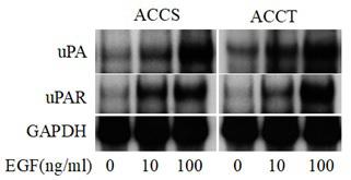
To examine the effects of EGF on uPA and uPAR mRNA expression in ACCS and ACCT cells, after treatment with EGF (0, 10 and 100 ng/ml) total RNA was isolated and subjected to Northern blot analysis. Expression of uPA and uPAR mRNAs was highest in both cells after stimulation with 100 ng/ml EGF (Figure 1), although no morphological changes were apparent (data not shown). Thus, we treated ACCS and ACCT cells with 100 ng/ml EGF in subsequent experiments.
Figure 1: Northern blot analysis of uPA and uPAR mRNA in ACCS and ACCT cells. After treatment for 24 h with the indicated concentration of EGF (0, 10, 100 ng/ml) total RNA was isolated from cells. For each lane, 20 µg of total RNA was electrophoresed on a 1.2% agarose gel, transferred to a nitrocellulose membrane, and hybridized with radiolabeled uPA or uPAR cDNA probes. Human GAPDH was the internal control.
4.2. EGF Activates AP-1
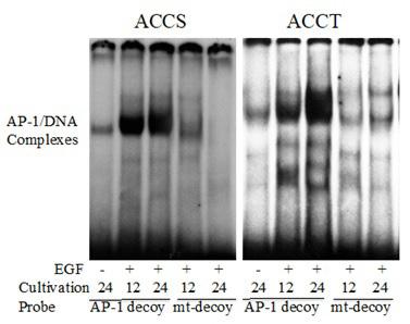
To confirm the transactivation of AP-1 in ACCS and ACCT cells after EGF (100 ng/ml) treatment for 30 and 60 min, EMSA was performed using radiolabeled AP-1 decoy ODNs as probes. Nuclear proteins extracted from ACCS and ACCT cells formed complexes with radiolabeled AP-1 decoy ODNs following stimulation with EGF for 60 min, whereas no complexes were detected with radiolabeled-mt-AP-1 decoy ODNs (Figure 2). These results indicate that EGF treatment induced the transactivation of AP-1 in ACCS and ACCT cells. Also, as expected the AP-1 decoy ODNs and mt-AP-1 decoy ODNs functioned as positive and negative probes, respectively.
Volume 5 Issue 3 -2021 Original Article clinicsofoncology.com 3
Figure 2: The effects of EGF stimulation on AP-1 activity in ACCS and ACCT cells. Nuclear extracts were prepared from ACCS and ACCT cells incubated for the indicated times in the presence of EGF (100 ng/ml). Following incubation with 32P-labeled AP-1 decoy ODNs or mt-AP-1 decoy ODNs, cells were analyzed by EMSA. Solid arrowheads indicate the position of the specific complex of AP-1 and decoy ODNs.
4.3. Gene Transfer of Decoy ODNs by HVJ-liposomes
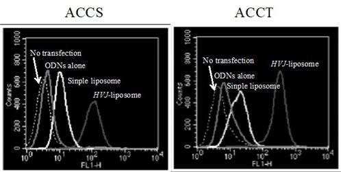
We transferred AP-1 decoy ODNs or mt-AP-1 deocy ODNs to ACCS and ACCT cells using HVJ-liposomes. To determine the transfer efficiency, FITC-labeled AP-1 decoy ODNs or mt-AP-1 decoy ODNs were transferred into cancer cells, and the frequency of cells that had taken up the lipofected DNA was determined
by flow cytometry. FITC-labeled AP-1 decoy ODNs and mt-AP-1 decoy ODNs were both transferred to 100% of both cancer cell types (Figure 3, and data not shown), respectively. Relative to the HVJ-liposome method, both the simple liposome method and ODNs alone had extremely low efficiencies (data not shown).
transfection of FITC-labeled
to determine
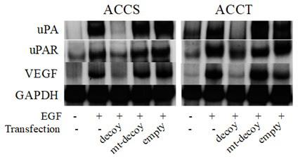
4.4. Effect of AP-1 Decoy ODNs on EGF-induced uPA, uPAR and VEGF mRNA Expression in ACCS and ACCT cells
Next, the effect of the transfected AP-1 decoy ODNs on EGF-induced uPA, uPAR and VEGF mRNAs expression were determined in ACCS and ACCT cells. Transfections of mt-AP-1 decoy ODNs and empty HVJ-liposomes were also performed as controls. Although transfection of AP-1 decoy ODNs dramatically reduced EGF (100 ng/ml)-induced uPA, uPAR and VEGF mRNA synthesis in both cell types, neither mt-AP-1 decoy transfection nor the empty HVJ-liposome influenced uPA, uPAR and VEGF mRNA expression (Figure 4).
4.5. In vitro invasion activities of ACCS and ACCT cells transfected with AP-1 decoy ODNs
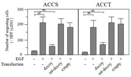
To examine the invasion activity of ACCS and ACCT cells in the presence or absence of EGF (100 ng/ml), in vitro invasion assays were performed. The number of invading cells was dramatically increased in the presence of EGF (100 ng/ml) as compared to that in medium alone (Figure 5). We also examined the effects of AP-1 decoy ODNs on EGF-mediated in vitro invasion activities. AP-1 decoy ODNs inhibited EGF-induced invasion by ACCS and ACCT cells to 27 and 39.4%, respectively. In addition, neither transfection of mt-AP-1 decoy ODNs nor treatment with empty HVJ-liposome influenced EGF-induced invasion activity of either cell type.
Volume 5 Issue 3 -2021 Original Article clinicsofoncology.com 4
Figure 3: In vitro expression of FITC-labeled AP-1 decoy ODNs in ACCS and ACCT cells introduced using the HVJ-liposome method. Three hours after
AP-1 decoy ODNs into ACCS (a) and ACCT (b) cells using the HVJ-liposome method, both cell types were subjected to flow cytometry
the transfection efficiency. Cells transfected with simple liposomes or ODNs alone were included as controls.
Figure 4: Effects of decoy ODNs on EGF-induced uPA, uPAR and VEGF mRNA expression. Total RNA was isolated from ACCS and ACCT cells cultured for 24 h in the presence of EGF (100 ng/ml) after transfection of AP-1 decoy ODNs (AP-1 decoy), mt-AP-1 decoy ODNs (mt-decoy) and treatment with empty HVJ liposomes (Empty). Both cell lines were cultured for 24 h in the presence or absence of EGF (100 ng/ml). Twenty micrograms of total RNA were electrophoresed on a 1.2% agarose gel, transferred to a nitrocellulose membrane, and hybridized with radiolabeled cDNA probes. The number on the lanes indicates the relative intensity identified by densitometry. Human GAPDH mRNA was included as an internal standard.
[22]. Binding of uPA to its receptor, uPAR, accelerates uPA activation from the enzymatically inactive pro-uPA [23]. uPA is also key factor in activation of MMP-1 [24]. In addition, uPA regulates cancer cell invasion by not only degrading the ECM, but also inducting cell migrating activity, independent of its proteolytic activity [22]. There have been several reports that inhibition of the uPA-uPAR system suppressed tumor cell migaration and invasion, via transfection of antisense RNA [8], neutralizing antibodies [9], soluble uPAR [10] and dominant-negative uPAR [11]. These reports suggest that inhibition of the uPA-uPAR system may suppress cancer cell invasion and metastasis. However, these methods have clinical barriers, such as side effects and transfer efficiency. In the present study, we propose a safe method for use as a therapeutic tool, a decoy strategy using the HVJ-liposome method that was developed in Japan.
Figure 5: Effects of AP-1 decoy ODNs on the invasion ability of ACCS and ACCT cells. ACCS and ACCT cells were transfected with AP-1 decoy ODNs (AP-1 decoy) or mt-AP-1 decoy ODNs (mt-decoy). ACCS and ACCT cells treated with empty HVJ-liposomes (empty) were also prepared. Cells were suspended at a density of 1.2 × 10⁴ cells in 50 ml of serum-free medium and seeded on the upper surface of the filter of the Matrigel chemotaxis chamber. Medium with EGF (100 ng/ml) was added to the lower wells and incubated at 37°C for 6 h. The number of cells on the lower surface of the filter was then counted. The data represent the number of cells/well ± S.D. Each experiment was performed in triplicate. The means were compared statistically by variance analysis, and pairs of means were compared using Student’s unpaired t-test. P values were analyzed on two sides.
5. Discussion
The most important characteristic of salivary gland cancer cells is their capacity to invade surrounding tissue, which depends on many proteases such as MMPs and uPA. uPA plays an important role in pericellular fibrinolyis during cell migration and tissue remodeling by physiological activation of plasminogen to plasmin
EGF-induced transactivation of AP-1 could be identified in ACCS and ACCT cells using EMSA and labeled AP-1 decoy ODNs, but not using labeled mutant type AP-1 decoy ODNs. Thus, the AP-1 decoy ODN used in this study was functional, and the mutant ODN could be used as a negative control for the decoy strategy. We demonstrated that transfection of AP-1 decoy ODNs using the HVJ-liposome method led to the inhibition of EGF-induced uPA and uPAR synthesis in ACCS and ACCT cells. The decoy strategy was originally reported by Morishita et al. [25], who used nuclear factor-kappa beta decoy ODNs to suppress acute inflammatory processes in myocardial ischemia and reperfusion injury. We selected the decoy system to inhibit AP-1’s role in regulating EGF-induced uPA and uPAR synthesis, since uPA and uPAR expression are transactivated by AP-1 [15]. While uPA and uPAR overexpression in the presence of EGF was inhibited by transfection of AP-1 decoy ODNs, neither mt-AP-1 decoy ODNs nor treatment with empty HVJ-liposome influenced uPA or uPAR expression. In addition, the number of invading cells detected in the in vitro invasion assay was reduced by transfection of AP-1 decoy ODNs. These in vitro results suggested that the AP-1 decoy strategy is a novel and efficient therapeutic tool for the inhibition of uPA and uPAR expression and suppression of cell invasion.
Another important characteristic of cancer is angiogenic activity, and VEGF is an important factor in the development of stromal neovascularization. In the present study, transfection of wild-type AP-1 decoy ODNs also reduced EGF-induced VEGF mRNA synthesis in ACCS and ACCT cells. These results indicate that AP-1 decoy transfection by the HVJ-liposome method could simultaneously suppress the angiogenic and invasive activities of ACCS and ACCT cells by inhibiting VEGF synthesis and the uPA-uPAR system.
The HVJ-liposome method has several advantages, including a high efficiency of transferring ODNs and proteins, a possible use in gene transfer, because this vector is non-immunogenic, and fi-
Volume 5 Issue 3 -2021 Original Article clinicsofoncology.com 5
nally HVJ virus when inactivated by UV-irradiation is non-virulent in humans. In fact, when we followed the transfer efficiency and time-sequential localization of FITC-labeled ODNs in cancer cells by flow cytometry and fluorescent microscopy, we found that the transfer efficiency was 100% at 3 h after transfer, without apparent cytotoxic effects on cancer cells. In fact, FITC-labeled ODNs were mainly localized within the cytoplasm at 3 h and then in cancer cell nuclei 6 h after transfection. These results indicate that the HVJ-liposome method is both useful and effective for ODN transfer. However, animal studies are required to further study the inhibitory effects of decoy ODNs on AP-1.
References
1. Babykutty S, Suboi P, Srinivas P, Nair AS, Chandramohan K, Gopala S. Insidious role of nitric oxide in migration/invasion of colon cancer cells by upregulating MMP-2/9 via activation of cGMP-PKG-ERK signaling pathways. Clin Exp Metastasis. 2012; 29: 471-92.
2. Kim S, Han J, Lee SK, Choi MY, Kin J, Lee J, et al. Berberine Suppresses the TPA-Induced MMP-1 and MMP-9 Expressions Through the Inhibition of PKC- α in Breast Cancer Cells. J Surg Res. 2011; 176: 21-9.
3. Ryu J, Ku BM, Lee YK, Jeong JY, Kang S, Choi J, et al. Resveratrol reduces TNF-alpha-induced U373MG human glioma cell invasion through regulating NF-kappaB activation and uPA/uPAR expression. Anticancer Res. 2011; 31: 4223-30.
4. Mekkawy AH, Morris DL, Pourgholami MH. HAX1 augments cell proliferation, migration, adhesion, and invasion induced by urokinase-type plasminogen activator receptor. J Oncol. 2012; 2012: 950749.
5. Yang J, Duh EJ, Caldwell RB, Behzadian MA. Antipermeability function of PEDF involves blockade of the MAP kinase/GSK/beta-catenin signaling pathway and uPAR expression. Invest Ophthalmol Vis Sci. 2010; 51: 3273-80.
6. Li C, Cao S, Liu Z, Ye X, Chen L, Meng S. RNAi-mediated down-regulation of uPAR synergizes with targeting of HER2 through the ERK pathway in breast cancer cells. Int J Cancer. 2010; 127: 1507-16.
7. Cheng X, Shen Z, Yin L, Lu SH, Cui Y. ECRG2 regulates cell migration/invasion through urokinase-type plasmin activator receptor (uPAR)/beta1 integrin pathway. J Biol Chem. 2009; 284: 30897-906.
8. Nalabothula N, Lakka SS, Dinh DH, Gujrati M, Olivero WC, Rao JS. Sense p16 and antisense uPAR bicistronic construct inhibits angiogenesis and induces glioma cell death. Int J Oncol. 2007; 30: 669-78.
9. Luo W, Pla-Roca M, Juncker D. uPAR as anti-cancer target: evaluation of biomarker potential, histological localization, and antibody-based therapy. Anal Chem. 2011; 83: 5767-74.
10. Koga T, Okada A, Kawashiri S, Kita J, Suzuki T, Nakashima Y, et al. Soluble urokinase plasminogen activator receptor as a useful biomarker to predict the response to adalimumab in patients with rheumatoid arthritis in a Japanese population. Clin Exp Rheumatol. 2011; 29: 811-5.
11. Jo M, Thomas KS, Marozkina N, Amin TJ, Silva CM, Parsons SJ,
et al. Dynamic assembly of the urokinase-type plasminogen activator signaling receptor complex determines the mitogenic activity of urokinase-type plasminogen activator. J Biol Chem. 2005; 280: 17449-57.
12. Kim MJ, Kim DH, Na HK, Surh YJ. TNF-α induces expression of urokinase-type plasminogen activator and β -catenin activation through generation of ROS in human breast epithelial cells. Biochem Pharmacol. 2010; 80: 2092-100.
13. Sheikh Ali MA, Gunduz M, Nagatsuka H, Gunduz E, Cengiz B, Fukushima K, et al. Expression and mutation analysis of epidermal growth factor receptor in head and neck squamous cell carcinoma. Cancer Sci. 2008; 99: 1589-94.
14. Ueda M, Fujii H, Yoshizawa K, Terai Y, Kumagai K, Ueki K, et al. Effects of EGF and TGF-alpha on invasion and proteinase expression of uterine cervical adenocarcinoma OMC-4 cells. Invasion Metastasis. 1998; 18: 176-83.
15. Amos S, Redpath GT, Dipierro CG, Carpenter JE, Hussaini IM. Epidermal growth factor receptor-mediated regulation of urokinase plasminogen activator expression and glioblastoma invasion via C-SRC/ MAPK/AP-1 signaling pathways. J Neuropathol Exp Neurol. 2010; 69: 582-92.
16. Ng YS, Krilleke D, Shima DT. VEGF function in vascular pathogenesis. Exp Cell Res. 2006; 312: 527-37.
17. Ishibashi H, Nakagawa K, Onimaru M, Castellanous EJ, Kaneda Y, Nakashima Y, et al. Sp1 decoy transfected to carcinoma cells suppresses the expression of vascular endothelial growth factor, transforming growth factor beta1, and tissue factor and also cell growth and invasion activities. Cancer Res. 2000; 60: 6531-6.
18. Rauscher FJ 3rd, Sambucetti LC, Curran T, Distel RJ, Spiegelman BM. Common DNA binding site for Fos protein complexes and transcription factor AP-1. Cell. 1988; 52: 471-80.
19. Heinemeyer T, Wingender E, Reuter I, Hermjakob H, Kel, AE, Ignatieva EV, et al. Databases on transcription regulation: TRANSFAC, TRRD and COMPEL. 1998; 26(1): 362-7.
20. Andrews NC, Faller DP. A rapid micro-preparation technique for extraction of DNA-binding proteins from limiting numbers of mammalian cells. Nucleic Acid Res. 1991; 19: 2499-505.
21. Dignam JD, Lebovitz RM, Roeder RG. Accurate transcription initiation by RNA polymerase II in soluble extracts from isolated mammalian nuclei. Nucleic Acid Res. 1983; 11: 1475-89.
22. Brekhman V, Neufeld G. A novel asymmetric 3D in vitro assay for the study of tumor cell invasion. BMC Cancer. 2009; 9:415.
23. Kwaan HC, McMahon B. The role of plasminogen-plasmin system in cancer. Cancer Treat Res. 2009; 148: 43-66.
24. Wun TC. Plasminogen activation: biochemistry, physiology, and therapeutics. Crit Rev Biotechnol. 1988; 8: 131-48.
25. Kranenburg O, Gebbink MF, Voest EE. Stimulation of angiogenesis by Ras proteins. Biochim Biophys Acta. 2004; 1654: 23-37.
26. Morishita R, Sugimoto T, Aoki M, et al. In vivo transfection of cis element ‘decoy’ against nuclear factor-kB binding site prevents myocardial infarction. Nature Med. 1997; 3: 894–9.
Volume 5 Issue 3 -2021 Original Article clinicsofoncology.com 6