ODA FEATURE
Differential Diagnosis: Radiographic Lesion By: Glen D. Houston, DDS, MSD
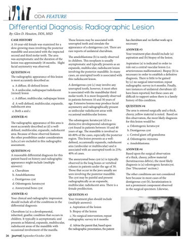
CASE HISTORY A 10-year-old female was examined for a slow-growing mass involving the posterior mandible and associated with the impacted second and third molar teeth. The area was asymptomatic and the duration of the lesion was approximately 18 months. Slight facial asymmetry was observed. QUESTION #1 The radiographic appearance of this lesion is most accurately described as: a. A diffuse, ill-defined lesion b. A unilocular, radiopaque/radiolucent (mixed) lesion c. A diffuse, multilocular, radiopaque lesion d. A well-defined, multilocular, expansile, radiolucent area e. Both a and c ANSWER #1 The radiographic appearance of this area is most accurately described as (d) a welldefined, multilocular, expansile, radiolucent area. Because of these observed features the other possibilities under consideration (a,b,c,e) are excluded in this radiographic assessment. QUESTION #2 A reasonable differential diagnosis for this patient based on history and radiographic appearance might include (multiple answers): a. Cherubism b. Ameloblastoma c. Dentigerous cyst d. Odontogenic keratocyst e. Aneurysmal bone cyst ANSWER #2 Your clinical and radiographic impression should include all of the conditions in the differential diagnosis: Cherubism (a) is a developmental, inherited, gnathic condition that occurs in children. It typically is asymptomatic and presents as bilateral, expansile, multilocular, radiolucent areas of the mandible with occasional involvement of the maxilla. 26 journal| September/October 2020
These lesions may be associated with unerupted teeth and simulate the appearance of a dentigerous cyst. There are rare reports of unilateral cherubism. The ameloblastoma (b) is rarely observed in children. This neoplasm is usually asymptomatic and typically presents as an expansile, multilocular, radiolucent lesion involving the posterior mandible. In many cases, an unerupted tooth is associated with this radiolucent lesion. A dentigerous cyst (c) may involve any unerupted tooth; however, it most often is associated with the mandibular third molar tooth. It is most frequently observed in patients between 10 and 30 years of age. Extensive lesions may produce facial asymmetry and radiographically present as unilocular radiolucent areas with occasional multilocular lesions. The odontogenic keratocyst (d) is a distinctive developmental odontogenic cyst found in patients between 10 and 40 years of age. The mandible is involved in 60-80% of the cases, especially the posterior region. This lesion presents as a welldefined, occasionally expansile, radiolucent area (unilocular or multilocular) and is associated with an unerupted tooth in 25%40% of cases. The aneurysmal bone cyst (e) is typically observed in the long bones or vertebral column in patients under the age of 30. Those that occur in the jaws usually are seen involving the posterior mandible. The cyst may be painful and present radiographically as an expansile, multilocular, radiolucent area. There is a female predilection. QUESTION #3 Your treatment plan should include (multiple answers): a. Aspiration of the lesion b. Biopsy of the lesion c. No surgical intervention; repeat radiographic survey in 6 months d. Advise the parent that, based upon the radiographic presentation, the patient
has cherubism and no further work-up is necessary ANSWER #3 Your treatment plan should include (a) aspiration and (b) biopsy of the lesion. Aspiration (a) is indicated in order to rule out a central vascular lesion (i.e. arteriovenous malformation). Biopsy (b) is necessary in order to establish a definitive diagnosis. There is little to be gained by (c) no surgical intervention; repeat radiographic survey in 6 months. Finally, rare instances of unilateral cherubism (d) have been reported, but these cases are difficult to support unless there is a family history of this condition. QUESTION #4 The area is entered surgically and a thick, cheesy, yellow material is noted. Based on this observation, the most likely diagnosis for this lesion would be: a. Odontogenic keratocyst b. Dentigerous cyst c. Central giant cell granuloma d. Odontogenic myxoma e. Ameloblastoma ANSWER #4 Based upon the surgical observation of a thick, cheesy, yellow material (keratinaceous debris), the most likely diagnosis is (a) odontogenic keratocyst. (See "Discussion" section.) The other conditions are not considered here because in most cases of the dentigerous cyst (b), keratinization is not a prominent component observed in the surgical specimen. Likewise,


