HOW TO: DETERMINE JOINT INVOLVEMENT IN LIMB WOUNDS# Brendon Bell brendonb@vetsouth.co.nz
The most common cause of limb synovial infections in adult horses is from wounds that penetrate or breach synovial structures, most often joints. The early and aggressive treatment of any wound that also involves a joint minimises the deleterious consequences of joint infection. It thus follows that if presented with a wound over or close to a joint, the earlier any joint involvement is recognised and treated, the more successful the outcome likely will be. In the early stages of a wound involving a joint, the joint may be contaminated and not yet actively infected, so that if joint involvement is recognised early the area can be decontaminated, greatly reducing the chances of bacterial colonisation and subsequent infection. When presented with a wound on a limb an appreciation of the precise location and extent of synovial structures in relation to the wound is necessary. The excellent reference by Moyer et al, (2011) has colour coded diagrams showing the extent of joint limits in relation to other limb structures. Downloading a copy to your phone allows horse-side reference to help gauge possible joint involvement. When first presented with the horse and its wound, taking a full history is important. Often owners have no idea how the wound occurred but there are occasions when they witnessed the injury. Factors like impaling on objects such as nails or spikes will help to determine the possible depth of wounds and whether a joint has been breached. Initially, perform a quick clinical examination to determine if the horse has any systemic involvement such as pyrexia. Next, assess the degree of lameness - horses with established joint infections are normally moderately to severely lame. Inspect the wound carefully for evidence of a clear/yellow sticky discharge coming out and running down the limb? This may be joint fluid, but there often will be a serous inflammatory discharge from the wound surfaces. The differentiation between inflammatory discharge and synovial fluid can be difficult but synovial fluid tends to be stickier and more globular compared to the waterier inflammatory discharge. If you gently flex or extend the joints near the wound this may pump out
#
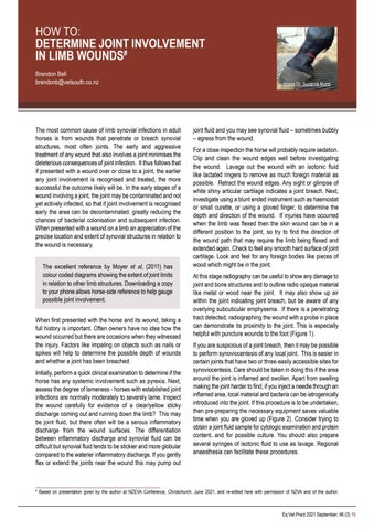
Image Dr. Suzanne Mund
joint fluid and you may see synovial fluid – sometimes bubbly – egress from the wound. For a close inspection the horse will probably require sedation. Clip and clean the wound edges well before investigating the wound. Lavage out the wound with an isotonic fluid like lactated ringers to remove as much foreign material as possible. Retract the wound edges. Any sight or glimpse of white shiny articular cartilage indicates a joint breach. Next, investigate using a blunt ended instrument such as haemostat or small curette, or using a gloved finger, to determine the depth and direction of the wound. If injuries have occurred when the limb was flexed then the skin wound can be in a different position to the joint, so try to find the direction of the wound path that may require the limb being flexed and extended again. Check to feel any smooth hard surface of joint cartilage. Look and feel for any foreign bodies like pieces of wood which might be in the joint. At this stage radiography can be useful to show any damage to joint and bone structures and to outline radio opaque material like metal or wood near the joint. It may also show up air within the joint indicating joint breach, but be aware of any overlying subcuticular emphysema. If there is a penetrating tract detected, radiographing the wound with a probe in place can demonstrate its proximity to the joint. This is especially helpful with puncture wounds to the foot (Figure 1). If you are suspicious of a joint breach, then it may be possible to perform synoviocentesis of any local joint. This is easier in certain joints that have two or three easily accessible sites for synoviocentesis. Care should be taken in doing this if the area around the joint is inflamed and swollen. Apart from swelling making the joint harder to find, if you inject a needle through an inflamed area, local material and bacteria can be iatrogenically introduced into the joint. If this procedure is to be undertaken, then pre-preparing the necessary equipment saves valuable time when you are gloved up (Figure 2). Consider trying to obtain a joint fluid sample for cytologic examination and protein content, and for possible culture. You should also prepare several syringes of isotonic fluid to use as lavage. Regional anaesthesia can facilitate these procedures.
Based on presentation given by the author at NZEVA Conference, Christchurch, June 2021, and re-edited here with permission of NZVA and of the author.
Eq Vet Pract 2021 September; 46 (3) 15