32
SF | ALLERGOLOGY
March 2021 | Vol. 21 No. 3 www.medicalacademic.co.za
CD4+ cells collaborate with neighbouring B cells to produce IgE through the process of immunoglobulin class switching. This process requires not only physical contact between T and B cells (cognate interaction) but also simulation of B cells by cytokines, especially interleukin (IL)-4 but also IL-5, IL-6 and IL-13. Recently it has been shown that CD4+ T cells in fact comprise two groups of cells, the so-called TH1 and TH2 populations. These two sets of T helper cells secrete different cytokines. The TH2 population produces the cytokines which augment IgE production, while TH1 cells suppress IgE production by B lymphocytes. Clearly, atopic individuals have a TH2 subset, and exciting possibilities may exist in this regard for prevention of atopy.
Cells in allergy The mast cell is the primary cell active in allergy induced inflammation. Mast cell granules contain and subsequently release mediators such as histamine, prostaglandin D2, leukotrienes C 4 and D 4, thromboxane A 2, and platelet activating factor. Mast cells also appear to be an important source of the cytokines which stimulate IgE synthesis viz IL‑4, IL-5 and IL-6. Eosinophils also play a pivotal role in allergic inflammation, probably controlling the late phase of these conditions. Their presence in the airway epithelium is a result of a complex interaction of adhesion molecules, chemokines, cytokines and other pro-inflammatory mediators. The nett result of eosinophil activation is the release of a number of products which produce disease. Of all the products of the eosinophil, the granule proteins are most well-known. The T lymphocyte (especially TH2) also plays an important role in allergy, from the induction of IgE production to the elaboration of controlling cytokines.
Cytokines and chemokines The various cells, which orchestrate allergy, communicate with one another through a group of molecules that allow this cell-tocell communication and thereby facilitate immune and inflammatory responses. Initially thought to be produced only by the cells of the specific immune and haematological systems, they are now known to be produced by many cells. Chemokines are cytokines with potent chemotactic function, attracting cells to the site of inflammation. Cytokines include the lymphokines, monokines, interleukins, interferons and haemopoietic growth factors, and are low molecular weight proteins with a short half-life. IL-5, together with IL-3 and granulocyte
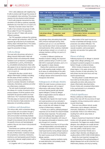
Table 1: AR: Major symptoms and responsible mediators Pathological event Symptoms elicited
Mediator(s) responsible
Pruritus
Tickling, palatal ‘clicking’
Histamine (H1), prostaglandins
Mucosal oedema
Nasal obstruction
Histamine (H1), eicosanoids, kinins, prostaglandins
Sneezing
Sneezing or feeling of the need to sneeze
Histamine (H1), eicosanoids
Mucus secretion
Runny nose, postnasal drip
Histamine (H1 ± H2), eicosanoids, muscarinic discharge
Late-phase allergic reactions
Congestion, nasal hyperirritability
Inflammatory factors, eicosanoids, chemotactic factors
macrophage colony stimulating factor (GMGSF), enhance eosinophil activity. Other cytokines such as IL-2, IL-8, and RANTES have recently been shown to be eosinophil chemoattractants. Other cytokines, implicated in IgE synthesis, include IL-4, IL-5 and IL-6, whereas interferon γ (IFNγ), IL-8, and IL-12 are inhibitory. A number of different cytokines are therefore involved in allergy, the important specific cytokines being IL-5 (with IL-3 and GM-CSF) in eosinophil activation, and IL-4 in IgE regulation in atopic disease. Clearly other cytokines are implicated and, while targeting single mediators may be of value, an understanding of the cell/s of origin, and control of cytokine synthesis in allergic disease will be essential for future therapeutic intervention.
Pathogenesis of allergic rhinitis Within the nasal mucosa a process occurs with allergen exposure, with a range of inflammatory cells present. Mast cells are found, bearing specific IgE directed against allergens. Contact by allergen leads to degranulation and the release of inflammatory mediators. Basophils also migrate into nasal mucosa and nasal secretions and are activated in similar fashion to mast cells. Eosinophils also play a crucial role, with release of a large number of inflammatory mediators. In rhinitis, as in asthma, pathophysiological responses can be divided into early/immediate and late-phase responses. The early-phase occurs immediately after degranulation of both mast cells and basophils, with many of the responses attributable to histamine, which acts through its H1 receptor. The late-phase occurs four to 24 hours after mast cell degranulation, and eosinophils are probably most important. Chronic, ongoing nasal hyperreactivity during allergen exposure is the result of accumulation of neutrophils in the nasal mucosa as part of the late-phase allergic reaction. The late-phase response manifests mainly as nasal obstruction, and probably results from cellular infiltration.
Inflammation of the nasal mucosa is a function of epithelial cell damage, mucous production and cellular infiltration. The sources of nasal secretions (mucous) are vascular exudation, submucosal gland secretion and secretion from goblet cells (Table 1).
Phases of allergy Following exposure to an allergen or physical trigger factor, allergic pathology, and consequent symptoms progress in an orderly fashion through a number of phases. Within minutes of exposure, the earlyphase begins. In the upper airway this is mainly a function of mast cell activity. The early-phase may last some hours and may resolve spontaneously. The early-phase is usually followed some hours later by the late-phase, in which eosinophils and other cells produce the classic inflammation described before. Chronic rhinitis with unresolved inflammation is the usual scenario in an untreated or partially-treated patient.
Clinical significance Rhinitis is defined as inflammation of the lining of the nose with the occurrence of nasal congestion, itching, sneezing and/or watery or mucoid rhinorrhoea. There are many different stimuli, usually inflammatory, due to allergens, infections, chemicals, medications, or irritants. However, occasionally rhinitis is non-inflammatory, resulting from cold air, spicy foods, or strong odours. Atopy is of paramount importance in allergic rhinitis, with both genetic and acquired components. The triggers are usually inhalant allergens. Aero-allergens are usually derived from natural organic sources such as house dust mite, pollens, mould spores, insect emanations and animals. Particle size is usually 2-60μm diameter, and allergens are usually proteins of MW 10 000-40 000 daltons. Clearly the most important message from our understanding of the pathophysiology of allergic diseases such as AR is that this is an inflammatory disease, and only by treating