SF | ALLERGOLOGY
March 2021 | Vol. 21 No. 3 Photo credit: Shutterstock.com
www.medicalacademic.co.za
This article was independently sourced by Specialist Forum.
There's something new in the AIR AIR Before considering the role of a new agent to treat and improve care of allergic rhinitis (AR), it seems wise to follow the immunological journey from exposure to an antigen to the final overwhelming inflammatory cascade it creates. But first, let’s consider why some of us become allergic. New thoughts on the origin of allergy: Foetal origins A TH2-like cytokine profile maintains the pregnant state. In fact, atopic compared to nonatopic mothers are more likely to have several children. The newborn destined to become atopic has low levels of interferon gamma and this continues to occur over the first two years of life. The cause of this defect remains to be discovered. In addition, the foetus is capable of mounting allergen–specific responses from 23 weeks of gestation. Why we all do not become atopic is probably due to deletion of allergen–reactive T-cells from the host by anergy or apoptosis as a consequence of high-dose antigen exposure in the gut and the development of oral tolerance.
The gastrointestinal flora at birth An important part of postnatal regulation of immunoglobulin E (IgE) reactivity seen at birth, may be the acquisition of the appropriate commensal gut flora (also known as the microbiome). There is evidence for this in both mouse and man. Studies comparing the gut commensal flora of neonates from Sweden (with its high prevalence of allergic disease) to those from Estonia (low prevalence of disease) found less intensive colonisation with lactobacilli in Swedish children and more frequent clostridia. In addition or alternatively, there may be impaired recognition and response to bacterial products derived from the microbiome in children who develop atopic disease. The gut flora is required for successful oral tolerance as gramnegative products such as lipopolysaccharides stimulate tissue macrophages to produce anti-TH2 cytokines, IL-1 and TNF-alpha.
The ‘hygiene hypothesis’ Since atopic children have lower circulating levels of interferon gamma, greater exposure to bacteria or their products during early life may increase interferon gamma. Factors that have damped down this mechanism include: » Improvements in public health and hygiene » Changes in infant diets » Early use of antibiotics » Elective caesarean section » Reduced exposure to bacteria with smaller family sizes (less siblings). Several studies have demonstrated an inverse relationship between atopy, seasonal AR (and asthma) and size of family. A possible explanation for this phenomenon may be the TH1/TH2 paradigm. In children living in small families where infections are less common, TH2 cells may develop instead of TH1 cells. As a consequence, IgE responses are produced. In children of large families, where infections are common, the immune system may be TH1
By Prof Robin J Green, Department of Paediatrics and Child Health, University of Pretoria
cell orientated. Atopy is also less common in individuals who grow up in a farming environment and here exposure to a less sterile environment is the postulated explanation.
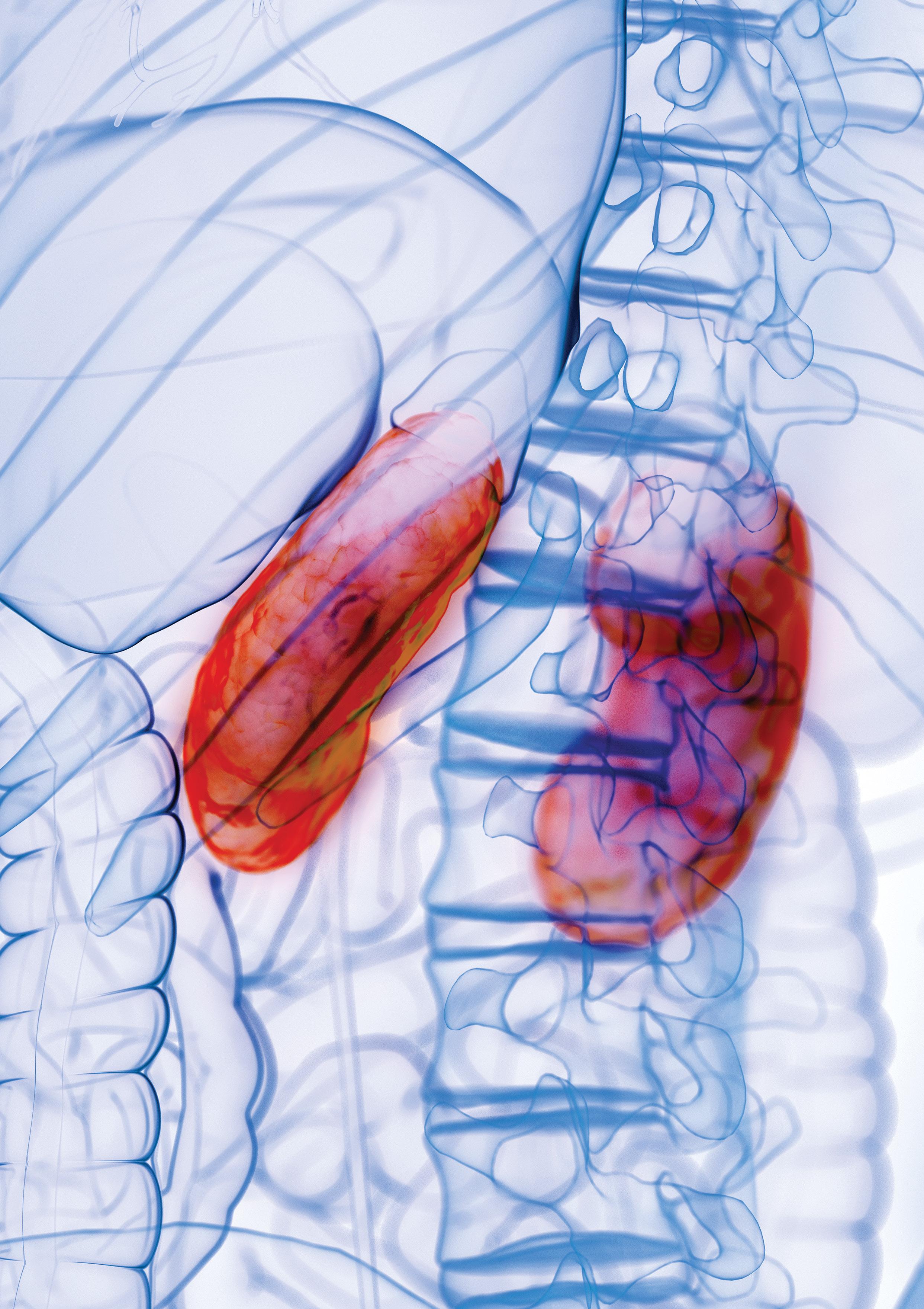
Allergens and IgE For antigens/allergens to trigger an immunological event they are required to be presented to T lymphocytes. In the airway such antigen-presenting cells (APC) are known as dendritic cells. Dendritic cells are formed from blood monocytes and move to the epithelium of the airway under the influence of chemokines. The antigen-containing dendritic cells then move into regional lymph nodes where interaction with T cells occurs. Within the APC, peptide components of the antigen combine with HLA Class II molecules and are expressed on the surface of the cell. It is this complex which T cells recognise and bind to via the T cell receptor. This bonding is further augmented by cellular adhesion molecules of the immunoglobulin super-family class. (Figure 1).
Figure 1: Antigen presentation to T-cells and resultant activation of mast cells
31