8 minute read
Sun poisoning – it’s real
Sun poisoning – it’s real
Researchers from the University of the Witwatersrand recently found that our summers last six months. Our love of spending time outdoors may put us at high risk of photodermatoses, also known as sun poisoning.
Advertisement
Although it is not generally lifethreatening, photodermatoses cause considerable suffering. Some people can go into anaphylactic shock following full body exposure.
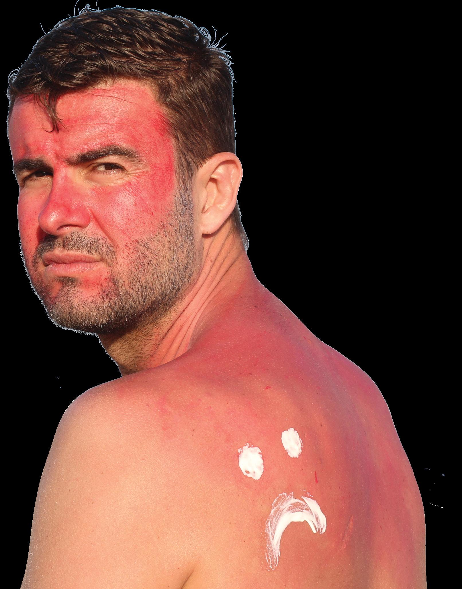
Photodermatoses are ‘abnormal’ cutaneous reactions to sunlight induced by excessive UV radiation – even for short periods of time (<30 minutes) – that can take several weeks to resolve. Sunburn on the other hand, is considered a ‘normal’ reaction to the sun, causing redness that go away after a few days.
While not all sunburns require medical attention, all cases of photodermatoses require immediate medical attention. Apart from anaphylactic shock, photodermatoses can cause dehydration, skin lesions, scarring, blisters, scaling, pruritus and hyperpigmentation.
Apart from the physiological impact of photodermatoses, a recent British study showed that 31%-39% of patients experienced a very large impact on their quality of life. Patients had around double the rates of anxiety and depression found in the general population.
Risk factors
Polymorphic light eruption (PMLE), which is often incorrectly labelled as sun allergy, is the most common form of the condition (prevalence 10% to 20%). Other, rarer types such as hydroa vacciniforme (characterised by recurrent fluid-filled blisters [hydroa] that heal with pox-like [vacciniform] scars) affects an estimated 0.34 people per 100 000.
People at greatest risk of photodermatoses are those who:
» Have fair skin
» Have a family history of skin cancer
» Are taking antibiotics or oral contraceptives
» Use certain herbal supplements, such as St. John’s wort
» Apply citrus oils to the skin prior to sun exposure
» Live in a region near the equator
» Reside in high altitudes (such as mountainous regions)
» Spend long periods in the sun – especially at the beach (sunlight reflects more intensely off sand and water)
» Engage in regular snow activities during the winter (sunlight reflects off snow)
» Use alpha hydroxy acids (AHAs), such as chemical peels.
Types of photodermatoses
Two types of photodermatoses can be distinguished, namely primary and secondary. Primary photodermatoses are induced by photosensitising substances such as industrial chemicals, some medications (eg antibiotics), some plants and other agents that increase the sensitivity to UV damage.
Secondary photodermatoses are associated with systemic diseases such as lupus erythematosus, metabolic disorders such as porphyrias, or disorders of DNA repair such as xeroderma pigmentosum.
Presentation, treatment and prevention
Photodermatoses are divided into:
1. Phototoxic and photoallergic reactions (photosensitisers are known)
2. Idiopathic photodermatoses (photosensitiser are unknown).
Examples of photodermatoses with known photosensitiser include:
Photoallergic dermatitis
Reactions occur after exposure to a specific allergen. Important topical photoallergens include halogenated salicylanilide, fenticlor, hexachlorophene, bithionol, and in rare cases also sunscreens. The skin is slightly inflamed, red, lichenified and scaly. Other signs include erythemas, papular vesicles, severe pruritus, and sometimes blistering. The affected skin areas differ from those parts of the body protected from light by clothing.
Treatment
Sun protection using both dense clothing and sunscreen (UV-A filters) is essential.
Phototoxic dermatitis
Phototoxic skin reactions are more common than photoallergic reactions. They usually present as dermatitis and symptoms are similar to sunburn. Phytophotodermatites (grass dermatitis) and phototoxic reactions induced by medications such as tetracyclines are clinically significant. Symptoms are similar to sunburn and include acute dermatitis with reddening, oedema, vesicles or blisters, and often severe pigmentation.
Treatment and prevention
The use of all medications and cosmetics with phototoxic actions must be discontinued. Systematic use of fragrancefree sunscreen is essential. Pronounced depigmentation can be achieved using a combination of 0.1% retinoic acid, 5.0% hydroquinone and 1% hydrocortisone. Occasionally, however, persistent hyperpigmentation develops. In these cases, laser therapy (Rubin laser) can help.
Examples of idiopathic photodermatoses include:
Polymorphous light eruption
The cardinal symptom is severely pruritic skin lesions. Skin lesions develop a few hours to several days after sun exposure. Initially, patchy erythema develops, accompanied by pruritus. Distinct lesions then develop. The upper chest, upper arms, backs of the hands, thighs, and the sides of the face are the primary localisations. The skin lesions resolve spontaneously within several days of ceasing sun exposure and do not leave behind any traces. Many patients develop tolerance over the course of the sunny period of the year, meaning that ultimately even prolonged sunbathing can be tolerated later in the season.
Treatment and prevention
Differentiation between symptomatic treatment of manifested PMLE and prevention is extremely important. Avoiding further sun exposure should be emphasised as this can lead to the rapid and spontaneous remission of symptoms. Remission can be accelerated by topical glucocorticoids. Antihistamines may alleviate pruritus, but their value should not be overestimated. The same applies to topical antihistamines. Prevention is extremely important. Light tolerance or hardening can be accelerated using phototherapy before the sunny period of the year. This should only be administered under specialist medical supervision and not in a solarium in order to ensure minimal UV exposure. Topical application of broad-spectrum sunscreen is useful. This predominantly benefits patients with UV-B-induced PMLE. With an extremely low UV-A threshold, PMLE episodes cannot be prevented even with very potent UV-A filters. General sun protection measures such as covering with clothes and appropriate, sensible behaviour are also useful. An interesting new approach to preventative external therapy involves topical application of suitable antioxidants, because pathophysiologically, inflammatory reactions are most likely mediated by free radicals generated in the skin. Photochemotherapy (PUVA) is exceptionally effective but should, however, be reserved for extremely light-sensitive patients.
Solar urticaria
Although rare, solar urticaria is a severe condition that can result in anaphylactic shock. Urticarial skin lesions appear a few minutes after exposure – even if the skin is covered with thin clothing. Patients may report pruritus, erythema, and wheal formation of varying degrees after a short period (<30 min) of sun exposure. Anaphylactic shock may occur after wholebody exposure.
Treatment
PUVA has become established as the method of choice for severe forms of solar urticaria because this procedure can achieve longer remissions (two to three weeks) compared to radiation without psoralene (a few days). Prior to starting it is recommended to develop tolerance using repeated provocative radiation over the entire integument. PUVA treatment can then be initiated overlapping with this light hardening. After verification of the presence of a plasma factor, which is hypothetically formed by UV absorption and mediates type I reactions, treatment using plasmapheresis can achieve an improvement.
Hydroa vacciniforme
Has an acute onset in early childhood and is characterised by necrohaemorrhagic lesions that appear on uncovered areas of skin, which became crusted then gradually heal leaving varioliform scars, hence the denomination. The course is chronic, characterised by periods of activity and remission. Recently, an association has been reported in some cases between latent Epstein-Barr virus (EBv) infections.
Treatment
Treatment is based on solar protection, and in severe cases, systemic corticoids can be used. The use of PUVA proved to be beneficial when used before sun exposure. Recent studies described the treatment of hydroa vacciniforme associated with EBv infection with acyclovir/valacyclovir therapy, with a good clinical response. After treatment, the patients reported having less fatigue, rashes, and scars. They were able to spend more time outdoors without causing new eruptions. The risk of lymphoproliferative malignancy in children with hydroa vacciniforme and chronic infection by EBv should be observed carefully and follow-up is recommended.
Chronic actinic dermatitis
Chronic actinic dermatitis (CAD) is an umbrella term that includes conditions such as persistent light reaction, actinic reticuloid, and photosensitive eczema. CAD is characterised by chronic, usually lichenified dermatitis on sun-exposed skin areas also spreading to other areas which, although covered by clothing, are not adequately protected from the sun. The skin appears inflamed, red and hard. The distressing pruritus leads to excoriation. The most commonly affected sites are the forehead, cheeks, ears, nape, throat, and the backs of the hands and in severe cases the entire integument.
Treatment and prevention
PUVA treatment has become the method of choice in addition to systemic glucocorticosteroids, azathioprine and cyclosporine A. Initiating the treatment can be very difficult due to the extreme light sensitivity. Initial doses below the eczema threshold must be selected. A combination of immunosuppressants is useful in this initial phase. A combination of cyclosporine A and PUVA should be avoided due to the risk of photocarcinogenesis. Avoiding radiation that triggers the dermatitis has the highest priority. Due to the large action spectrum, the use of particularly intensive sun protection is important. In extreme cases artificial lighting at work can also contribute to continued persistence of the eczema. Shifting leisure activities to the evening and night, wearing light-blocking clothing, and full-coverage, tinted make-up preparations can also help.
Actinic prurigo
Onset is in childhood and persists into adulthood. The condition presents as pruriginous skin lesions that develop on chronically sun-exposed areas of the skin such as the face, often centrofacial, the nape, the ears, the backs of the hands and the lower arms. Lichenified erythematous plaques, cushion-like infiltrations, and nodular prurigo develop.
Treatment
Treatment is very difficult. The treatment of choice is thalidomide, the use of which must be examined in depth considering the serious adverse reactions (teratogenicity, irreversible neuropathies). Other than that, no local or systemic medication has yet been able to achieve a substantial improvement. Even light hardening using phototherapy has often had little effect on the clinical symptoms.
References available on request. SF


