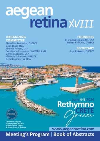
aegeanretinaXVIII
ORGANIZING COMMITTEE
Efstathios Detorakis, GREECE
Dean Eliott, USA
Thomas Friberg, USA
Constantin Pournaras, SWITZERLAND
Dimitra Skondra, USA
Miltiadis Tsilimbaris, GREECE
Demetrios Vavvas, USA
FOUNDERS
Evangelos Gragoudas, USA
Ioannis Pallikaris, GREECE
SECRETARY
Irini Kokolaki, GREECE
CRETE Rethymno June 2024 6-9 G r e e c e
Meeting’s Program Book of Abstracts www.aegeanretina.com Under the auspices of the Hellenic Society of Ophthalmic Plastic & Reconstructive Surgery
aegean retina xviii
Rethymno Crete 6th-9th of June 2024
Welcome to the 18th Aegean Retina Meeting
Rethymno Crete 6th - 9th of June 2024
Aegean Retina, since its inception in 1989 by Evangelos Gragoudas of the Massachusetts Eye and Ear Hospital and Ioannis Pallikaris of the University of Crete, has been a unique forum for the scientific interaction of innumerable international retinal specialists. The spectacular scenery of the Aegean region is a special allure, and over three decades devoted retina specialists have formed the “Aegean Retina Family” This family is continuously enriched by new and repeat participants, so welcome to you all.
We expect this year's meeting, the 18th of a successful series, to be unforgettable!
The Organizing Committee.
Ef Detorakis D Eliott, T Friberg, C Pournaras, D Skondra, M Tsilimbaris, D Vavvas
With the approval of:
The Hellenic Drug Organization (EOF)
The Hellenic Association of Pharmaceuticals Companies (SFEE)
1
2
OVERVIEW OF EACH DAY
Thursday June 6th, 2024
16:00 - 18:00
Registration and distribution of conference material
16:00 - 18:00 Plenary Session I - Posters
20:00 Welcome Cocktail at Kritamo Restaurant - Amira Luxury Resort
Friday June 7th, 2024
07:45
Registration and distribution of conference material.
08:15 - 08:30 Welcome and opening comments
08:30 - 11:00 Plenary Session ΙΙ
11:00 - 11:30 Coffee Break
11:30 - 14:00 Plenary Session ΙΙΙ
Saturday June 8th, 2024
08:30 - 11:00 Plenary Session ΙV
11:00 - 11:30 Coffee Break
11:30 - 14:00 Plenary Session V
21:00 Dinner at Almyra Restaurant - Amira Luxury Resort
Sunday June 9th, 2024
09:00 - 11:00 Plenary Session VI
11:00 - 11:20 Closing remarks, end of the meeting, conclusions
layout and printing by: www.visionadv.gr 2810 314919
SCIENTIFIC PROGRAM
Thursday June 6th, 2024
16.00 - 18.00 Session I - Posters
Friday June 7th, 2024
07:45 08:15
08:30 - 11:00 08:30 08:45 09:00 09:15 09:30 09:45 10:00 10:15 10:30 10:45
3
11:00 - 11:30
Registration
Welcome and opening comments
Session II (Moderators: Miltiadis Tsilimbaris, Thanos Papakostas)
George Pappas PVR management.
George Papadakis
Successful closure of large macular holes with a novel surgical technique using intraoperative perfluorocarbon (PFCL).
George Pappas
Optic disk pit management
Evangelia Gkaragkani
Sunrising after the storm of a trabeculectom
Thanos Papakostas
Vitreoretinal Surgery with an Augmented Reality Digital Exoscope
Rafaela Datseri
Macuar contour evolution after ERM removal Case report
Romy Bejjani
Long-term visual outcomes of photodynamic therapy in patients with central serous chorioretinopathy in a tertiary care center and in the IRISΒ® Registry.
Ioannis Roumeliotis
Central serous chorioretinopathy treated with Subthreshold Micro-pulse Laser Photocoagulation assisted by Navilas laser 577s.
Saghar Bagheri
A mystery case of sudden vision loss
Sotiris Plainis
Longitudinal changes in objective accommodative response, pupil size and spherical aberration: a case study
Coffee break
aegean retina xviii
Rethymno Crete 6th-9th of June 2024
Friday June 7th, 2024
End of 1st day 11:30 - 14:00
Session III (Moderators: Demetrios Vavvas, Asimina Mataftsi)
Asimina Mataftsi
Retinopathy of prematurity in Greece today- management preferences and implications.
Nimesh A. Patel,
Validation of the TWO-ROP Algorithm at a Tertiary Referral Multi-site.
Aikaterini Seliniotaki
Optimizing retinopathy of prematurity screening: the MyMiROPS and stressROP Trials
Stefan Sacu Retinal detachments in children.
Efstathios Detorakis
The role of Ru-106 brachytherapy in the therapeutic paradigm of ocular surface neoplasia.
Kostas Perisinakis
Radioactive plaque episcleral brachytherapy for ocular melanoma: how to reduce uncertainties of delivered treatment dose
Kalogeropoulos Chris Intraocular lymphomas
Michael Tatarakis
High Intensity Laser-Based Secondary Sources for Biomedical Applications using the Zeus 45TW laser and pulsed power high current plasma X-ray sources at IPPL.
Sotiris Plainis
Μyopia: a failure of emmetropisation in children, a disease in adulthood.
Aggeliki Gleni
Implementing silent reading speed and oculomotor indices as a clinical measure of functional vision
4
11
12
12
12
12
45 13:00 13:15 13:30 13:45 14:00
11:30
:45
:00
:15
:30
:
SCIENTIFIC PROGRAM
Saturday June 8th, 2024
08:30 - 11:00 08:30 08:45 09:00 09:15 09:30 09:45 10:00 10:15 10:30 10:45
Session IV (Moderators: Sofia Androudi , Paris Tranos)
Alexandros Charonis
Long-term intravitreal pharmacotherapy for choroidal neovascularization secondary to angioid streaks: Pearls and pitfalls.
Demitrios Vavvas
Proteomics in Age-related Macular Degeneration (AMD).
Sofia Androudi
Laser flare photometry in eyes receiving brolucizumab intravitreal injections for age related macular degeneration
Stella Blazaki
Progression of macular atrophy in eyes with nAMD compared to fellow dry-AMD eyes.
Nikolaos Gkouliopoulos
The efficacy of subthreshold micropulse laser on the treatment of polypoidal choroidal vasculopathy.
Irini Chatziralli
Cardiovascular disorders and other risk factors for diabetic retinopathy in patients with type 2 diabetes
Ioanna Ploumi
Impact of Predominant Peripheral Lesions on Retinal Microvasculature and Risk of Vitreous Hemorrhage in Proliferative Diabetic Retinopathy.
Paris Tranos
New trends in monitoring inflammation.
Aleksandra Poluianova
Assessing the Tolerability of Silica Microparticle Gels for Drug Delivery
Achilleas Gravanis
Eye Regenerative Pharmacology: from neurogenic compounds to neuroimplants
5
11:00 - 11:30
Coffee break
aegean retina xviii
Rethymno Crete 6th-9th of June 2024
Saturday June 8th, 2024
Session V (Moderators: Ioannis Pallikaris, Chris Kalogeropoulos)
Robert McLaren
Surgical techniques in retinal gene therapy.
Robert McLaren
Gene therapy for X-linked retinitis pigmentosa.
Petros Petrou
Gene therapy for RPE65:The Greek Experience
Tryfon Rotsos
Genetic Treatment in Greece Current condition and trends of the future
Atsushi Kuwahara
Self-organizing stem cell culture and preclinical studies of allogeneic iPSC-derived retinal sheets for retinitis pigmentosa.
Maram Abdalla
Hyperautofluorescent Ring Progression in Ciliopathies follows an Exponential Decay Function.
Miltiadis Tsilimbaris
Scleral IOL fixation Our approach
Ioannis Pallikaris
fixOflex an artificial capsule, a tool that may improve retinal disorders.
Sandra R Montezuma
Surgical techniques for optimal lens management during metallic intraocular foreign body removal: a case series.
Thanasis Nikolakopoulos
3D vitrectomy in macular hole surgery
6
End of 2nd day 11:30 - 14:00 11:30 11:45 12:00 12:15 12:30 12:45 13:00 13:15 13:30 13:45 14:00
SCIENTIFIC PROGRAM
Sunday June 9th, 2024
09:00 - 11:15 09:00 09:15 09:30 09:45 10:00 10:15 10:30 10:45 11:00 11:15 11:20
Session VI (Moderators: Efstathios Detorakis, Saghar Bagheri)
Nikolaos Gkouliopoulos
The intake of resveratrol oral supplements is associated with fewer intravitreal injections in cases of wet AMD.
Plaka Argyro
Cytomegalovirus retinitis in an immunocompetent patient: A case report.
Spyridon Charisis
Bilateral Combined Cataract and Macular Hole Surgery
George Bontzos
Vascular parameters after vitrectomy for rhegmatogenous retinal detachment, an optical coherence tomography angiography study.
Evangelia Bagkaki
RNFL changes after successful rhegmatogenous retinal detachment operations.
Chris Kalogeropoulos
“Mass like” lesions of the fundus
Anastasios Stavrakakis
Reaching the T-junction Presentation of a case
George Makrygiannis
Grade IV Hypertensive Retinopathy and increased Intracranial Pressure in an adult with an extra-adrenal paraganglioma.
George Kontadakis
Treatment of Dry Age Related Macular Degeneration using Photobiomodulation 9.30Plaka ArgyroCytomegalovirus retinitis in an immunocompetent patient: A case report.
Closing remarks
End of meeting
7
aegean retina xviii
Rethymno Crete 6th-9th of June 2024
Friday, 7 JUNE
Session ΙI (08.30)
PVR management.
George Pappas
Venizeleion General Hospital of Heraklion, Crete, Greece
PVR i an existing complication of retinal detachment. So far there is no standardised treatment for this condition. We will present proposed pharmaceutical and surgical approaches in order to minimise the possibility of developing PVR as well as affecting the final retinal integrity
8
ABSTRACTS
Friday, 7 JUNE
Session II (08:45)
Successful closure of large macular holes with a novel surgical technique using intraoperative perfluorocarbon (PFCL).
Papadakis Georgios, Banderas-Garcia Sandra, Sabatino Francesco, Patton Nial, Dhawahir-Scala Felipe
- Manchester Royal Eye Hospital, Manchester, UK
- Norfolk and Norwich University Hospitals NHS Foundation Trust, Norwich, Norfolk, UK
Macular hole (MH) is a full thickness defect of the neurosensory retina at the fovea level. Surgery for MH repair has evolved significantly in recent years, with pars plana vitrectomy (PPV) combined with internal limiting membrane (ILM) peel and gas tamponade as standard with over 90% closure rates. Chronic, large, persistent and secondary macular holes are at higher risk of failure to close. Novel techniques have been described to reduce the closure failure on the use of autologous platelet concentrate , various ILM flap techniques , various tissue flaps and macular detachment. Chronic macular holes have shown excellent closure rates if passive suction is applied to the edges intraoperatively We present a novel surgical technique, initially developed by Marzio Chizzolini (FLOREtina 2017), which combines in vivo observation of the macular hole closure with intraoperative OCT using perfluorocarbon (PFCL). We demonstrated successful repair of 5 MH cases with an average size of 761µm performed at the Manchester Royal Eye Hospital, by two experienced surgeons. This technique essentially involves the aspiration of remaining fluid at the hole base intraoperatively, facilitating apposition of the edges, reducing the tangential traction forces and enhancing inner retina contraction. Later on, during the postoperative time, gas tamponade provides further barrier for fluid to accumulate at the hole while cell proliferation consolidates.
9
aegean retina xviii
Rethymno Crete 6th-9th of June 2024
Friday, 7 JUNE
Session II (09:00)
Optic disk pit management.
George Pappas
Venizeleion General Hospital of Heraklion, Crete, Greece
Various theories have proposed for the optic disc pit maculopathy mechanism as well as the ways of treating this condition. A systematic review of the literature and the concussions extracted from the management of 14 cases in our department is described. Videos of various techniques as well as tips and purples how to achieve the best results will be presented.
10
ABSTRACTS
Friday, 7 JUNE
Session II (09:15)
Sunrising after the storm of a trabeculectomy.
Evangelia Gkaragkani, Theodor Stappler
Jules Gonin Eye Hospital, Lausanne, Switzerland
Suprachoroidal hemorrhage, although rare, presents a significant challenge following trabeculectomy, often leading to devastating consequences for visual acuity and intraocular pressure (IOP). Here, we report a case of a 73-year-old male patient with uncontrolled primary open-angle glaucoma who underwent trabeculectomy in his right eye. He has had previous vitrectomy and internal limiting membrane peeling in the same eye and had to undergo two surgical revisions within the first week post-trabeculectomy due to elevated IOP Subsequently, he presented with sudden vision loss in the right eye, attributed to a suprachoroidal hemorrhage.
Upon examination, the patient exhibited perception of light visual acuity, remarkably low IOP (4mm Hg), and a shallow anterior chamber, with b-scan revealing hemorrhagic kissing choroidals. Prompt intervention was crucial, involving revision of trabeculectomy, closure of the scleral flap, and coverage with donor pericardium, alongside drainage of the suprachoroidal hemorrhage.
One month post-operation, the patient's visual acuity improved to 6/24, with IOP stabilizing at 16mm Hg without the need for glaucoma medication. We attribute the choroidal hemorrhage to rapid decompression and resultant hypotony, particularly in the context of a previously vitrectomized eye and the patient's cardiovascular risk factors.
This case underscores the importance of swift recognition and management of hypotony following trabeculectomy, especially in patients with predisposing factors. Early intervention, including timely management of hypotony and drainage of suprachoroidal hemorrhage, can significantly impact visual outcomes, highlighting the critical role of vigilant post-operative care in glaucoma management.
11
aegean retina xviii
Rethymno Crete 6th-9th of June 2024
Friday, 7 JUNE
Session II (09:30)
Vitreoretinal Surgery with an Augmented Reality Digital Exoscope.
Thanos Papakostas
The Retina Institute, St. Louis, USA
Purpose: To describe the experience in VR surgery with the first augmented reality digital exoscope available in the market.
Methods: 100 cases were performed with the Beyeonics system. The principles behind the AR visualization are discussed and setup of the device is presented. Representative videos of common VR pathologies such as epiretinal membranes, macular holes, vitreous hemorrhage and retinal detachment are presented. Color enhancement techniques are presented that aid the VR surgeon in critical steps of surgery are discussed and additional emphasis is placed on ergonomics.
Results: Visualization with the first AR digital exoscope results in superior ergonomics during surgery and safe and efficient surgery The integration of the various foot pedal settings in the AR headset allows the surgeon to move the scope, focus and zoom with simple head gestures. Color enhancement can aid certain key steps such as peeling maneuvers.
Conclusions: The Beyeonics system can provide superb visualization for VR surgery and superior ergonomics when compared to analog microscopes and other heads up digital display systems.
12
ABSTRACTS
Friday, 7 JUNE
Session II (09:45)
Macuar contour evolution after ERM removal. Case report.
Rafaela Datseri, Anastasis Stavrakakis, Miltiadis Tsilimbaris
University Eye Hospital of Heraklion, Crete, Greece
We present two cases with epiretinal membranes and partial thickness macular defects in which the surgical removal of the ERM resulted in normalization of macular contour We discuss the role of foveal cellular microanatomy in the presurgical morphology and the possible reversibility after successful removal of the ERM.
13
aegean retina xviii
Rethymno Crete 6th-9th of June 2024
Friday, 7 JUNE
Session II (10:00)
Long-term visual outcomes of photodynamic therapy in patients with central serous chorioretinopathy in a tertiary care center and in the IRIS® Registry.
Romy Bejjani, Eric Goldberg, Connor Ross, Elizabeth J. Rossin, Demetrios G. Vavvas, Tobias Elze, Alice C. Lorch, and Joan W. Miller
Mass Eye and Ear Infirmary, Harvard Medical School, Boston, USA
Purpose: To compare long-term (≥5 years) visual outcomes in central serous chorioretinopathy (CSCR) eyes treated with photodynamic therapy (PDT) to untreated eyes in a tertiary care center and the IRIS® Registry (Intelligent Research in Sight).
Methods: We reviewed CSCR patients followed for ≥5 years at Massachusetts Eye and Ear (MEE) from 2007-2024 and in the IRIS® registry (2000-2017), excluding eyes with choroidal neovascular membrane, focal laser, severe retinopathy, or anti-VEGF injections before PDT We estimated and plotted best visual acuity (VA) trends in PDT-treated and untreated eyes using mixed linear and linear splines regression models.
Results: There were 26 CSCR eyes treated with PDT and 37 untreated eyes from the MEE database. Median (IQR) baseline VA was 0.3 (0.16-0.47) and 0.1 (0-0.18) in eyes with PDT and untreated eyes, respectively Corresponding final VAs were 0 (0, 0.3) and 0 (0, 0.1). Mixed linear and linear splines models showed a flat VA trend overall, with no significant difference among subgroups. In the IRIS® Registry, there were 167 eyes treated with PDT and 4445 without treatment. Baseline VA was 0.25 (0.1, 0.4) and 0.1 (0, 0.22) respectively, which improved after 5-6 years to 0.1 (0, 0.29) and 0.08 (0, 0.18), respectively Regression models of a matched subset (163 patients) showed a small significant trend of long-term VA improvement but no intergroup difference.
Conclusion: Findings from both the MEE sample and IRIS® Registry show that despite worse vision at baseline, PDT is associated with a long-term trend of VA stabilization or improvement similarly seen in untreated eyes.
14
ABSTRACTS
Friday, 7 JUNE
Session II (10:15)
Central serous chorioretinopathy treated with Subthreshold Micro-pulse Laser Photocoagulation assisted by Navilas laser 577s.
Roumeliotis Ioannis, Gouliopoulos Nikolaos, Ligerou Meropi, Tzanidaki Malvina, Karageorgiou Georgia, Minakakis P.
- Tzaneio General Hospital of Piraeus, Pireaus, Greece
- Attikon University Hospital, Athens, Greece
- Omma Institution, Athens, Greece
Background: Central serous chorioretinopathy (CSCR) constitutes an idiopathic chorioretinal disorder characterized by serous detachment of the retina in the macula region related to leakage at the level of the retinal pigment epithelium (RPE). Subthreshold micro-pulse laser (SML) may help with management by selectively targeting RPE cells without inducing chorioretinal damage or scarring. Targeted RPE cells have been shown to down-regulate cytokine production and inflammation resulting in faster resolution of the retinal detachment.
Method and Results: We had 20 cases of CSCR which were diagnosed with fluoresceine angiography, that were successfully treated with SML using the Navilas laser 577s. We will be presenting 5 of these cases which had the most significant improvement.
Conclusion: SML using the Navilas laser appears to be safe and effective for patients with CSCR and leakage near the fovea. This technique should be considered by fellow ophthalmologists as an alternative treatment option, especially when photodynamic therapy is unavailable or contraindicated.
15
aegean retina xviii
Rethymno Crete 6th-9th of June 2024
Friday, 7 JUNE
Session II (10:30)
A mystery case of sudden vision loss.
Saghar Bagheri MD, PHD
Mass Eye and Ear, Harvard Medical School, Boston, USA
16
ABSTRACTS
Friday, 7 JUNE
Session II (10:45)
Longitudinal changes in objective accommodative response, pupil size and spherical aberration: a case study.
1 1 2
S Plainis , S Panagopoulou and W N Charman
1 Laboratory of Optics & Vision (LOV), School of Medicine, University of Crete, GR
2 Division of Pharmacy and Optometry, Faculty of Biology, Medicine and Health, University of Manchester, UK
Purpose: Previous transverse studies have shown that the slope of the static accommodation response/stimulus curve declines as complete presbyopia is approached. Changes in pupillary miosis and ocular spherical aberration are also evident. This study further investigates longitudinal changes in the relationships between the static accommodative response, pupil diameter and spherical aberration of a single adult.
Methods: A wavefront analyzing system (COAS), was used in conjunction with a Badal optometer to allow continuous recording of the aberration structure of the dominant eye of a low myope for a range of accommodative demands over a period of 17 years, until the age of 50. Monocular accommodative response was calculated as the equivalent refraction minimizing wavefront error The associated longitudinal changes in pupil size and spherical aberration with accommodation were also recorded. Results: A decrease in accommodation response with age was found at almost all target vergences, with the changes being greatest for the higher vergences. In addition, although absolute pupil diameter decreased with age, the rate of change in pupil diameter with accommodation stimulus was approximately constant with age. Pupil constriction occurred for near stimuli even in full presbyopia. Spherical aberration changed linearly with accommodation response at all ages.
Conclusions: Objective amplitude of accommodation declines linearly with age as complete presbyopia is approached, while the slope of the response/stimulus curve also falls. retinal image blur associated Τhe with the larger accommodation lags, found with higher accommodation stimuli, is reduced by pupil constriction and the resultant lower levels of spherical aberration.
17
aegean retina xviii
Rethymno Crete 6th-9th of June 2024
Friday, 7 JUNE
Session III (11:30)
Retinopathy of prematurity in Greece today- management preferences and implications.
A.
Mataftsi, S. Moutzouri, S. Basiakos, N. Ziakas
2nd Department of Ophthalmology, School of Medicine, Aristotle University of Thessaloniki, Thessaloniki, Greece
A prospective national 12-month incidence study of treatment-requiring retinopathy of prematurity (ROP) was completed in Greece for the period June 2020-May 2021 and showed that 3.8% of at-risk infants were treated. Centers providing treatment are mostly public, and do not have all treatment modalities available. Availability of fundus imaging and fluorescein angiography is also limited across centres, hindering documentation which can help in establishing criteria for treatment, as well as diagnosis of persistent peripheral retinal ischemia in high risk patients.
Preference for anti-VEGF intravitreal injection as first line treatment is on the rise, mainly due to reduced access to laser treatment and ease of application. Treatment with VEGF inhibitors is known to have a high risk of recurrence and the lack of recommendations for follow-up can lead to missed diagnoses of relapses in the first years of life, when fundus examination without anesthesia is a significant challenge.
18
ABSTRACTS
Friday, 7 JUNE
Session III (11:45)
Validation of the TWO-ROP Algorithm at a Tertiary Referral Multi-site.
Nimesh A. Patel MD, Francisco Altamirano , Sandra Hoyek , Hanna De Bruyn , Ryan Gise , Iason S. 5 6 Mantagos , Deborah K. VanderVeen Md1
Harvard Medical School / Mass Eye and Ear / Boston Children's Hospital
Purpose: We aim to validate the previously published TWO-ROP algorithm on an external dataset. Methods: Retrospective consecutive study with infants screened for retinopathy of prematurity (ROP) between January 2013 and August 2023 at a tertiary referral multi-site. Infants with birth weight (BW) and longer gestational age (GA) were included and stratified into three groups as follows: group 1 (BW<1500 g, GA≥30 weeks), group 2 (BW≥1500g, GA< 30 weeks), and group 3 (BW≥1500g, GA≥30 weeks) Outcomes were the rate of ROP, treatment-warranted ROP (TW-ROP), and number of inpatient examinations were evaluated in the three groups.
Results: 1,095 (33.8%) patients met the inclusion criteria. The number of patients in groups 1, 2, and 3 were 837 (76.4%), 72 (6.6%), and 186 (17.0%), respectively ROP was detected in 120 (11.0%) patients; the rate was 9.8% in group 1, 20.8% in group 2, and 12.4% in group 3 (p=0.013). The overall mean number of inpatient examinations for patients undergoing traditional, TWO-ROP 36-week, and TWO-ROP 40-week screening systems were 1.95, 1.43, and 0.99, respectively (p<0.001). Stage 3 was found in 9 eyes of 5 patients (0.5%, all zone II). Three eyes of 2 patients (0.2%) had plus disease. Two patients had bilateral laser treatment at 44 and 39.4 weeks corrected GA; 3 out of 4 of these eyes met Type 1 treatment criteria. Overall, the ROP screening burden saved was 9.0% and 16.7% for the TWO-ROP 36-week and 40-week systems, respectively. The sensitivity for TW-ROP was 100% for TWO-ROP 36-week system and 99.4% for TWO-ROP 40-week system.
Conclusion: The TWO-ROP algorithm can reduce the number of inpatient examinations while maintaining safety. To ensure timely management, we recommend that the single first ROP examination occurs at 38-39 weeks post-conception age. .
1 2 3 4
19
aegean retina xviii
Rethymno Crete 6th-9th of June 2024
Friday, 7 JUNE
Session III (12:00)
Optimizing retinopathy of prematurity screening: the MyMiROPS and stressROP Trials.
Seliniotaki A.K., Moutzouri S., Lithoxopoulou M., Gika H., Haidich A.-B., Ziakas N., Mataftsi A.
1. 2nd Department of Ophthalmology, School of Medicine, Faculty of Health Sciences, Aristotle University of Thessaloniki, Papageorgiou General Hospital, Thessaloniki, Greece
2. Department of Hygiene, Social-Preventive Medicine & Medical Statistics, School of Medicine, Faculty of Health Sciences, Aristotle University of Thessaloniki, Thessaloniki, Greece
3. Department of Ophthalmology, School of Medicine, University of Crete, University General Hospital of Heraklion, Heraklion, Crete, Greece
4. 2nd Department of Neonatology & NICU, School of Medicine, Faculty of Health Sciences, Aristotle University of Thessaloniki, Papageorgiou General Hospital, Thessaloniki, Greece
5. Laboratory of Forensic Medicine & Toxicology, School of Medicine, Aristotle University of Thessaloniki, Thessaloniki, Greece
Retinopathy of prematurity screening exposes infants to repetitive eye examinations that are known to be stressful and can result in systemic adverse events (AE). The induced stress has been attributed to both mydriasis and fundoscopy. Optimizing procedures of ROP screening remains the most significant strategy for minimizing induced pain, and targeting efficiently the population truly at risk helps avoiding unnecessary examinations. The MyMiROPS Trial, a non-inferiority crossover randomized controlled trial (RCT) of 83 infants, assessed the efficacy and safety of mydriatic microdrops (6.5µL) of combined phenylephrine 1.67% and tropicamide 0.33% compared with standard drops (28-34µL) of the same regimen. It is the first study establishing non-inferiority of microdrops compared with standard drops of a diluted mixture regarding mydriatic efficacy at T45, T90, and T120, showing reduced cardiorespiratory AE after microdrops during a 48h follow-up period, and determining the pharmacokinetic profile of phenylephrine eyedrops in preterm infants. The stressROP Trial, a non-inferiority crossover RCT of 41 infants, assessed the safety and efficacy of fundoscopy with (Sp) and without (speculum free, SpF) the use of lid speculum and scleral indentation, using a pain scoring system. The induced pain and stress were shown to be significantly higher when a lid speculum and scleral indentor were used, while the speculumfree exam proved efficient in identifying the presence or absence of treatment-requiring disease in all cases. Future studies may contribute to confirm our findings and extrapolate the results in different regimens, different settings, different populations.
20
ABSTRACTS
Friday, 7 JUNE
Session III (12:15)
Retinal detachments in children.
Prof Stefan Sacu MD
Department of Ophthalmoloy and Optometry, Medical University of Vienna, Vienna, Austria
Pediatric retinal detachments account for 3.2% to 6.6% of all retinal detachments and include both congenital and acquired diseases. As there is usually no detailed medical history, the indication for surgery is often difficult to establish. The surgical procedure and results differ considerably from treatment in adults.
The incidence is around 0.5 per 100,000 children. The most common causes of retinal detachment in children are trauma, myopia or prior ocular surgeries (60%), other causes are aphakia, retinopathy of prematurity, persistent hyperplastic primary vitreous, morbus coats, FEVR, Norrie disease, inflamatory and tumor associated retinal detachments, etc.
Outcomes of own clinical study, as well as clinical cases with different causes of retinal detachments and treatment strategies will be presented.
21
aegean retina xviii
Rethymno Crete 6th-9th of June 2024
Friday, 7 JUNE
Session III (12:30)
The role of Ru-106 brachytherapy in the therapeutic paradigm of ocular surface neoplasia.
Efstathios T. Detorakis, Gogo Solomou, Kostas Perisinakis
1 Department of Ophthalmology, University Hospital of Heraklion, Crete, Greece
2 Department of Medical Physics, University Hospital of Heraklion, Crete, Greece
Ocular surface neoplasia includes a variety of different potentially sight and life threatening conditions such as conjunctival melanoma and conjunctival squamous cell carcinoma. Surgical removal with clear margins is the mainstay of treatment but often lesions recur aggressively either on the ocular surface or posteriorly into the orbit, sometimes necessitating orbital exenteration. Auxiliary topical treatment, such as cryotherapy on the wound margins or the use of topical chemotherapeutic agents, such as mitomycin C, has been widely employed, whereas systemic immunotherapy with various agents, such as checkpoint inhibitors or PD-1 blockers, has also offered significant improvement in prognosis but still some tumors recur, stressing the need for complete tumor destruction on site. Ru-106 transepithelial brachytherapy for the destruction of residual neoplasia may be another useful and minimally invasive option for the achievement of local tumor control in high risk cases, with low morbidity and high success rates.
22
ABSTRACTS
Friday, 7 JUNE
Session III (12:45)
Radioactive plaque episcleral brachytherapy for ocular melanoma: how to reduce uncertainties of delivered treatment dose.
Gogo Solomou, Kostas Perisinakis, Dimitrios A. Liakopoulos, Efstathios T. Detorakis
1 University Hospital of Heraklion, Medical Physics Department, Crete Greece
2 University of Crete, Medical School, Medical Physics Department, 71003 Heraklion, Crete, Greece
3 General Hospital of Rhodes 'Andreas Papandreou', Rhodes, Greece
4 University of Crete, Medical School, Department of Ophthalmology, 71003 Heraklion, Crete, Greece
Ru-106 applicators are increasingly used for treatment of ocular choroidal tumors with apical height up to 7 mm and base diameter up to 20 mm. Dosimetry planning is crucial for an effective therapeutic result. A dedicated treatment planning software package is commonly used for the determination of time required to achieve a prescribed dose to tumor apex and tumor margins. The conventional episcleral plaque brachytherapy treatment planning employs a) tumor metrics, such as the apex height and basal diameter, usually derived from ultrasound imaging and b) the tumor location and the size of the eye, derived from pretreatment tomographic imaging. Commercially available Ru-106 plaques have standard dimensions and curvature. However, the curvature of the treated eye globe at the site of tumor may be different than the curvature of the Ru-106 plaque used. Also, the delivered treatment dose during brachytherapy with Ru106 plaques depends on the accuracy of surgical plaque placement. The uncertainties in delivered treatment dose due to a) the plaque and eye globe curvature mismatch and b) the difference of plaque position from the position assumed in treatment planning will be discussed and methods to suppress these uncertainties will be proposed.
23
aegean retina xviii
Rethymno Crete 6th-9th of June 2024
Friday, 7 JUNE
Session III (13:00)
Intraocular lymphomas.
Chris Kalogeropoulos
Department of Ophthalmology, University Of Ioannina, Ioannina, Greece
There are different types of lymphomas involving the intraocular tissues. Primary vitreoretinal lymphoma (PVRL) is a rare ocular lymphoid malignancy, which consists a subset of primary central nervous system lymphoma (PCNSL) and the most common type of intraocular lymphoma. The involvement of eyes is estimated to be approximately 20% of PCNL, but the brain involvement may up to 80% of PVRL. Typically, PVRL is a high grade B-cell malignancy of the retina. Although PVRL tends to have a good response to the initial treatment the prognosis is poor and the survival restricted due to the high relapse rates of CNS involvement. Other intraocular lymphomas arise in the uveal tract. Uveal lymphomas are subdivided to those that present as a primary disease and those that represent a secondary localization of systemic nonHodgkin lymphoma (NHL). Additionally, in Hodgkin disease anterior uveitis, vitritis and chorioretinitis occur and represent manifestations of a masquerade syndrome. The main of neoplastic origin masquerade syndrome in elderly is the PVRL.
In addition, T-cell angioimmunoblastic lymphoma, a T-cell lymphoma with potentially intraocular expansion may be confused as uveitis or more specifically as opportunistic infection of the eye, a marker of poor prognosis.
In cases with intraocular lymphomas an accurate diagnosis includes cytology/pathology, immunohistochemistry, flow cytometry, molecular pathology and cytokine profile analysis.
24
ABSTRACTS
Friday, 7 JUNE
Session III (13:15)
High Intensity Laser-Based Secondary Sources for Biomedical Applications using the Zeus 45TW laser and pulsed power high current plasma X-ray sources at IPPL.
Georgia Andrianaki , Ioannis Tazes , Alexandros Skoulakis , Stelios Petrakis , Christos Karvounis , Ioannis
Fitilis , Evangelos Kaselouris , John Chatzakis , Efthimios Bakarezos , Emmanouil P. Benis , Vasilios
Dimitriou , Nektarios A. Papadogiannis and Michael Tatarakis ,* Emmanouel Xylouris , Helen A. 8 5 8 9 9 5 Papadaki , Aggeliki Doulis , Aristea K Batsali , Ioannis Charalampopoulos , Ioanna Zota , Thomas G. Maris 6 and Efstathios Detorakis
1. Institute of Plasma Physics & Lasers, University Research & Innovation Centre, Hellenic Mediterranean University, 74100 Rethymno, Crete, Greece
2. Department of Electronic Engineering, Hellenic Mediterranean University, 73133 Chania, Greece
3. Physical Acoustics and Optoacoustics Laboratory, Department of Music Technology & Acoustics, Hellenic Mediterranean University, 74100 Rethymnon, Greece
4. Department of Physics, University of Ioannina, 45110 Ioannina, Greece,
5. Department of Medical Physics, School of Medicine, University of Crete, Heraklion, Greece
6. Department of Ophthalmology, School of Medicine, University of Crete Heraklion, Greece
7. Institute of Vision and Optics, School of Medicine, University of Crete, Heraklion, Greece
8. Department of Haematology, School of Medicine, University of Crete, Heraklion, Greece
9. Department of Pharmacology, School of Medicine, University of Crete, Heraklion, Greece
High-intensity laser-generated electron, ion as well as photon secondary sources have attracted widespread interest during the last two decades because of the important applications, among others in biomedicine. In the field of biomedical applications in particular, laser-driven particle beams as well as laserdriven X-ray sources are a promising field of study The ZEUS 45 TW laser system as well as other high current pulsed power plasma devices developed at IPPL (https://ippl.hmu.gr/) offer unique opportunities for research in laser-driven particle and X-ray sources. Information about the facility and the experiments performed for establishing the baseline platforms for secondary plasma sources will be presented. In addition, results from recent experiments on melanoma cells using energetic pulsed electron beams produced by the Zeus laser system will be discussed.
1 1 1 1 1
1,2 1,3 1,2 1,3 1,4
1,3 1,3 1,2 7
25
aegean retina xviii
Rethymno Crete 6th-9th of June 2024
Friday, 7 JUNE
Session III (13:30)
Μyopia: a failure of emmetropisation in children, a disease in adulthood.
S Plainis and W N Charman
1Laboratory of Optics & Vision (LOV), School of Medicine, University of Crete, GR
2Division of Pharmacy and Optometry, Faculty of Biology, Medicine and Health, University of Manchester, UK
Myopia has long been considered as a common refractive error of the eye, the outcome of the optical imbalance between ocular axial length and its total refractive power It is now well established that the earlier myopia appears, the higher the risk of developing pathological myopia in adulthood, since excessive axial elongation leads to structural changes in the posterior segment of the eye. Genetics alone cannot explain the dramatic change over the last fifty years in the prevalence of myopia and environmental risk factors have a key role in myopia development and progression. The observation that myopic eyes show a tendency to relative hypermetropia in the periphery of the retina compared to axial refraction leads to the hypothesis that conventional correction of myopia with glasses or contact lenses may lead to its "overcorrection" in the peripheral retina. This may lead to further axial elongation, especially when children are deprived of outdoor activities, where the visual environment is dioptrically homogeneous. This presentation offers a summary of the current evidence-based knowledge on the optical factors influencing the emmetropisation mechanism, leading to the development of myopia in children. In addition, it re-examines the underlying patterns of individual refraction at different ages, leading to the increased prevalence of myopia in Asian children populations.
Myopia is a major public health problem that requires immediate planning for comprehensive vision care services. It is important for all eye clinicians to start applying evidence-based techniques for controlling myopia and to act for the benefit of myopic children, preventing the development of a pathological condition.
26
ABSTRACTS
Friday, 7 JUNE
Session III (13:45)
Implementing silent reading speed and oculomotor indices as a clinical measure of functional vision.
Angeliki Gleni, Emmanouil Ktistakis, Miltiadis K Tsilimbaris, Panagiotis Simos Sotiris Plainis
- Laboratory of Optics and Vision (LOV), School of Medicine, University of Crete, Greece
- Institute of Computer Science, Foundation of Research and Technology-Hellas, Greece
Purpose: High contrast visual acuity, the preferred test among clinicians, offers limited value as an endpoint for evaluating functional vision deficits. Alternative visual function measures, such as oral reading speed, have been utilised, failing though to naturally simulate everyday reading activities, for which silent reading is preferred. In this study eye movements during reading were recorded, to provide surrogate indicators of silent reading performance.
Methods: Silent reading performance was evaluated binocularly in 53 participants (52±4 years old), using the Greek IReST reading passages, displayed at 40cm distance on a screen. Correction for near was achieved with reading spectacles worn over the distance single vision contact lenses. In a second experiment, reading performance (N=26/53) was evaluated (i) without best-correction for near (“blur”) and (ii) following instructions for “comprehension”. Eye movements were recorded simultaneously during passage reading, using video oculography (Eye-Link II, SR Research Ltd) Data analysis included computation of reading speed, fixation duration, forward fixations and regressions Frequency distributions of fixation durations were analysed with ex-Gaussian fittings.
Results: The strongest correlate of silent reading speed was the number of forward fixations (r=-0.86), accounting for 74% of its variance, while modest correlations were found with the percentage of regressions (r=-0.57) and the ex-Gaussian parameter τ (r=-0.59). Reading speed was found statistically significantly slower in both “blur” and “comprehension” conditions (p<0.001). Comprehensive reading showed an increased number of forward fixations and regressions. Blur had a significant impact on fixation duration, ex-Gaussian parameter µ and the number of fixations.
Conclusions: Silent passage reading strongly predicts visuo-motor vs. cognitive processing, when implementing oculomotor indices, which account for systematic within and between subject variability in reading speed. This approach could provide reliable clinical measures of functional vision.
27
aegean retina xviii
Rethymno Crete 6th-9th of June 2024
Saturday, 8 JUNE
Session IV (08:30)
Long-term intravitreal pharmacotherapy for choroidal neovascularization secondary to angioid streaks: Pearls and pitfalls.
Alexander Charonis MD, Maria Emfietzoglou MD
Athens Vision Eye Institute, Athens, Greece
Purpose: To present and critically analyze the long-term results of anti-VEGF monotherapy in angioid streaks (AS)-related choroidal neovascularization (CNV).
Methods: Retrospective case series and review of the pertinent literature.
Results: We studied 11 eyes of 7 patients (2 males, 5 females; mean age=45 years; range 34-53 years) with a mean follow up of 107 months (36-168 months) and a mean number of 46 injections per patient (7-138), or 29 injections per eye (7-69). Multimodal imaging (FA, OCT, OCTA, BAF, IRAF) was performed upon initiation, as well as during the course of the treatment. At baseline, 7(64%) CNVs were subfoveal, 1(9%) was juxtafoveal and 3(27%) were extrafoveal. While BCVA (Snellen) improved or remained stable in all 11 eyes at 12 months after the initiation of treatment, only 3 (27%) eyes had stable vision at the last follow-up, all of which had initially been diagnosed with extrafoveal CNV This long-term vision loss appeared to be related both to subretinal fibrosis and atrophy of the RPE. Fibrosis was observed in the context of continuous exudation (attention will be given to potential OCT biomarkers of such low grade exudation), whereas atrophy was observed both dependent, as well as independent to the CNV complex, especially in the context of underlying pseudoxanthoma elasticum.
Conclusions: AS-related CNV remains a therapeutic challenge even in the current era of intravitreal antiVEGF pharmacotherapy The underlying degeneration and weakening of Bruch's membrane may be the reason why the CNV has such an aggressive course with high recurrence rates requiring long-term “maintenance” anti-VEGF therapy. While the initial anatomic and functional response to the therapy is often deemed satisfactory, long-term vigilance and avoidance of undertreatment is strongly recommended in order one to maintain such satisfactory outcomes.
28
ABSTRACTS
Saturday, 8 JUNE
Session IV (08:45)
Proteomics in Age-related Macular Degeneration (AMD).
Demetrios G Vavvas, Olympia Sideri, Victor Correa, Nikolaos Ziakas, Ioannis Tsinopoulos and Joan W. Miller
Mass Eye and Ear Infirmary, Harvar Medical School, Boston, USA
Purpose: To systematically review the information on proteomics in non-neovascular age-related Macular Degeneration (AMD).
Methods: Relevant electronic databases were searched with search terms “proteomics” and “age-related macular degeneration” from 2012 onwards. Pathway analysis was performed for proteins that exhibited significant changes more than 1.5 fold.
Results: Twenty-two studies (n= 6932 participants) and diverse tissue samples were identified. Most studies investigated neovascular AMD with few studies focusing on non neovascular AMD and geographic atrophy. There is paucity of data for early AMD. There was high discordance of identified proteins among different studies and body fluid types, with functional and pathway analysis identifying multiple nonintersecting pathways as involved in the pathogenesis of AMD.
Conclusions: Proteomics studies in non-neovascular AMD though limited by sample size suggest that AMD is a heterogeneous group of diseases. Combining proteomic data from different studies could uncover new protein networks and associated functional pathways that might indicate novel potential therapeutic targets for AMD. Future studies will benefit from more sensitive proteomics methodology, increased sample size and improved phenotyping/genotyping of the patients.
29
aegean retina xviii
Rethymno Crete 6th-9th of June 2024
Saturday, 8 JUNE
Session IV (09:00)
Laser flare photometry in eyes receiving brolucizumab intravitreal injections for age related macular degeneration.
Sofia Androudi MD, PHD, Nikolaos Chadoulos, Anna Dastiridou, Andreas Mitsios, Ioannis Tsinopoulos, Christos Kalogeropoulos
- Ophthalmology Department, University of Thessaly, Greece
- Ophthalmology Department, Papageorgiou Hospital, Aristotle University, Thessaloniki, Greece
- Ophthalmology Department, University of Ioannina, Ioannina, Greece
Purpose: To measure aqueous flare levels in treatment naïve eyes suffering from wet age- related macular degeneration (wAMD) and treated with intravitreal brolucizumab.
Patients and methods: Patients with treatment naïve wAMD in one eye were prospectively enrolled. Flare levels were measured with laser flare photometry at baseline, 1 day and 1 month after each of the 3 monthly injections during the loading phase.
Results: Twenty-two eyes from 22 patients aged 6.7+/-6 years were enrolled. Flare values were 10.6±3.7 photons/msec at baseline and 12.6+/-5.8 photons/msec at the last follow up visit, 1 month after the third injection (p=0.289, repeated measures ANOVA). The mean change in flare after the first injection was 4.2+/-3.6. photons/msec, 6.6+/-8.9 photons/msec after the 2nd and 8.6±20.8 photons/msec after the 3rd injection (p=0.640, repeated measures ANOVA). No patient had IOI.
Conclusions: Eyes receiving brolucizumab injections for wAMD showed similar flare at baseline, during and 1 month after completion of three-monthly intravitreal injections. There was no evidence of subclinical inflammation during the loading phase of brolucizumab based on laser flare photometry measurements.
30
ABSTRACTS
Saturday, 8 JUNE
Session IV (09:15)
Progression of macular atrophy in eyes with nAMD compared to fellow dry-AMD eyes.
Styliani Blazaki, Emmanouil Blavakis, Georgios Bontzos, Irini Chatziralli, Demetrios G. Vavvas, Georgios Smoustopoulos, Eleni Dimitriou, Anastasios Stavrakakis, Stamatina Kabanarou, Tina Xirou, Gregory Chlouverakis, Miltiadis K. Tsilimbaris
1. University of Crete Medical School, Heraklion, Crete, Greece
2. Korgialenio-Benakio General Hospital, Athens, Greece
3. National and Kapodistrian University of Athens, Athens, Greece
4. University of Harvard Medical School, Boston, MA, USA
Purpose: Τo evaluate the evolution of macular atrophy (MA) in patients with neovascular AMD (nAMD) under anti-VEGF treatment, compared with their fellow dry AMD eyes (dAMD).
Methods: This is a retrospective study including 124 patients treated with anti-VEGF in their nAMD eye and having dAMD in the fellow eye. Three centers participated in the study Patients without MA at baseline were analyzed to calculate the time to first MA development. Synchronous and unsynchronous time course of MA for nAMD vs. dAMD eyes was also studied. MA was evaluated using near-infrared images, while all available optical coherence tomography (OCT) images were used to confirm the CAM group criteria for complete MA.
Results: MA first detection in nAMD eyes increased significantly from year 2 to 6 compared to dAMD eyes. Over the study's follow-up, 45.1% of nAMD-E developed MA, compared to 16.5% of fellow eyes (p<0.001). When MA in the two eyes was compared in a synchronous paired manner over 4 years, nAMD eyes had an average MA progression rate of 0.275 mm/year versus 0.110 mm/year in their fellow dAMD eyes. Multivariate ANOVA revealed significant time (p<0.001), eye (p=0.003), and time-eye interaction (p<0.001) effects. However, when MA did develop in dAMD eyes and was compared in an asynchronous manner to MA of nAMD eyes, it was found to progress faster in dAMD eyes (dAMD: 0.295 mm/year vs. nAMD: 0.176mm/year) with a significant time-eye interaction (p=0.015).
Conclusions: In this study, a significant difference in MA incidence and progression was documented in eyes with nAMD under treatment, compared to fellow eye exhibiting dAMD. Eyes with nAMD tended to develop more MA compared to fellow dAMD eyes. However, when atrophy did develop in the fellow dAMD eyes, it progressed faster over time compared to MA in nAMD eyes.
31
aegean retina xviii
Rethymno Crete 6th-9th of June 2024
Saturday, 8 JUNE
Session IV (09:30)
The efficacy of subthreshold micropulse laser on the treatment of polypoidal choroidal vasculopathy.
Nikolaos Gouliopoulos, Tzanidaki Malvina-Efthimia, Lygerou Meropi, Roumeliotis Ioannis, Rouvas Alexandros, Datseris Ioannis
- 2nd Department of Ophthalmology, University of Athens Medical School, 'Attikon' General Hospital of Athens, Haidari, Athens, Greece
- 'OMMA' Eye Institute, Athens, Greece
- Department of Ophthalmology,' Tzaneio', General Hospital, Athens, Greece
Purpose: We compared the 1-year outcomes of subthreshold micropulse laser combined with intravitreal injections of anti-VEGF agents versus intravitreal injections of anti-VEGF agents as monotherapy, for the treatment of polypoidal choroidal vasculopathy (PCV).
Materials and Methods: In our prospective study, 35 naive and previously untreated PCV patients were enrolled. They were divided in two groups according to the applied treatment regimen. In the first group 17 patients were included, who were treated with a session of subthreshold micropulse laser and afterwards intravitreal injections of anti-VEGF agents were applied according to need. In the second group, 18 patients were included and they were treated with intravitreal injections of anti-VEGF agents according to need. Results: Between the studied groups, no significant differences were present regarding the participants' baseline demographic data (p>0.05 for all). Over the 12-month period, the changes of best corrected visual acuity were comparable between the two treatment modalities (p=0.162). However, the application of subthreshold micropulse laser was accompanied by significantly fewer intravitreal injections of anti-VEGF agents during the aforementioned period (p=0.008).
Conclusions: We demonstrated that subthreshold micropulse laser is an effective complementary treatment in cases of PCV, highlighting the advantage of requiring fewer intravitreal injections of antiVEGF agents.
32
ABSTRACTS
Saturday, 8 JUNE
Session IV (09:45)
Cardiovascular disorders and other risk factors for diabetic retinopathy in patients with type 2 diabetes.
Irini Chatziralli, Chrysa Agapitou, Stamatios Lampsas, Alexia Risi-Koziona, Fotios Kyratzidis, Eleni Dimitriou, Panagiotis Theodossiadis
2nd Department of Ophthalmology, National and Kapodistrian University of Athens, Greece
Purpose: The purpose of this study is to examine the association between diabetic retinopathy (DR) and the atherosclerotic cardiovascular disease (ASCVD) risk in patients with type 2 diabetes mellitus (DM) and to assess risk factors potentially associated with DR.
Methods: Participants in the study were 215 patients with type 2 DM, who underwent a thorough ophthalmic examination, including best-corrected visual acuity (BCVA) measurement, dilated fundoscopy, fundus photography, optical coherence tomography (OCT) and OCT angiography (OCTA). DR was graded as no apparent retinopathy (NDR), mild non-proliferative (NPDR), moderate NPDR, severe NPDR and proliferative DR (PDR). In addition, a detailed medical history of patients was recorded, while the ASCVD risk estimator plus by the American College of Cardiology was used to calculate the ASCVD risk.
Results: Multivariate logistic regression analysis showed that factors associated with DR independently were: DM duration (multivariate OR=3.16, 95%CI: 1.55-6.44, p=0.002), HbA1c levels (multivariate OR=2.94, 95%CI: 1.37-6.32, p=0.006) and presence of neuropathy (multivariate OR=3.59, 95%CI:1.439.05, p=0.007). At the multivariate multinomial logistic regression analysis, NPDR development was associated with duration of DM (multivariate RR=3.31, 95%CI: 1.57-6.97, p=0.002), HbA1c levels (multivariate RR=2.24, 95%CI: 1.00-5.02, p=0.050) and neuropathy (multivariate RR: 3.94, 95%CI: 1.5410.11, p=0.004), while PDR development was only associated with HbA1c levels (multivariate RR=6.88, 95%CI: 2.19-21.63, p=0.001). The ASCVD score was not found to be significantly correlated with DR (p=0.191).
Conclusions: Factors associated with DR were DM duration, HbA1c levels and presence of neuropathy, while the ASCVD score was not found to be significantly associated with DR.
33
aegean retina xviii
Rethymno Crete 6th-9th of June 2024
Saturday, 8 JUNE
Session IV (10:00)
Imaging Impact of Predominant Peripheral Lesions on Retinal Microvasculature and Risk of Vitreous Hemorrhage in Proliferative Diabetic Retinopathy.
Ioanna Ploumi, Jenny Gan, Xinyi Ding, Francesco Romano, Demetrios Vavvas, John Miller
- Harvard Retinal Imaging Lab, Boston, MA, USA
- Retina Service, Massachusetts Eye and Ear, Department of Ophthalmology, Harvard Medical School, Boston, MA, USA
Purpose: To investigate the association of predominant peripheral lesions (PPLs) with retinal microvasculature on WF SS-OCTA and the risk of VH in eyes with PDR.
Methods: The cross-sectional part of this study included 111 PDR eyes of 84 patients with same-day UWFFP (California; Optos plc., Dunfermline, UK) and WF SS-OCTA (PLEX® Elite 9000; Carl Zeiss Meditec, Dublin, CA). A subset of 101 eyes from 75 patients followed for at least 3 months were included in the prospective part of this study, where the history of complications and treatments was reviewed. UWF-FP were graded by 2 independent graders for the presence of and the number of fields with PPLs - defined as more than 50% of diabetic lesions subjectively residing in the extended field compared to the respective ETDRS field. Only 1 field with PPLs was required for the eye to be graded as having PPLs. Ischemic index (ISI) and common SS-OCTA vascular metrics were calculated on FIJI and ARI network, respectively 6X6- and 12X12mm retinal microvasculature alterations were compared between eyes with and without PPLs utilizing the Mann-Whitney U test. The association between the presence and extent of PPLs and the development of VH was evaluated using generalized estimating equations and multilevel Cox mixed-effects regressions. Results: PPLs were present in 36 out of 111 eyes (32.4%). OCTA parameters, including ISI, vessel density and vessel skeletonized density did not show significant differences in groups with or without PPLs. Over a period of 18.1±10.2 months, 34 VH events occurred in 23 eyes – 14 having a history of VH (recurrent VH) and 9 having no VH history (new VH). There was an association between the presence of PPLs and the development of new VH (p=0.04), but not for recurrent VH. The number of peripheral fields with PPLs was a significant predictor for the new development of VH, after adjusting for age, DM duration and prior antiVEGF as well as PRP (HR= 1.92, p=0.03).
Conclusion: While 6X6- and 12X12-mm OCTA metrics in eyes with or without PPLs are comparable, the presence and extent of PPLs are associated with the new development of VH. Given the advancement of UWF imaging technologies, the development of a new DR grading scheme to account for PPLs may enhance our ability to predict the risk of VH in patients with PDR.
34
ABSTRACTS
35
Saturday, 8 JUNE
Session IV (10:15)
New trends in monitoring inflammation.
Paris Tranos
Ophthalmica Eye Center, Thessaloniki, Greece
Ocular inflammatory diseases account for a significant number of blind registrations worldwide, involving predominantly young patients. Differential diagnosis of uveitis cases maybe challenging as they are infrequent and share common features. With the evolution of multimodal imaging several new signs of inflammation have been described facilitating prompt and effective diagnosis. Non-invasive modalities including OCT, OCT-A and Autofluorescence along with traditional diagnostic techniques such as fluoresceine and indocyanine green angiography provide plausible evidence of activity or remission of inflammation which can be used to determine further management and follow up. This lecture will highlight imaging features essential in efficacious monitoring of uveitis.
aegean retina xviii
Rethymno Crete 6th-9th of June 2024
Saturday, 8 JUNE
Session IV (10:30)
Assessing the Tolerability of Silica Microparticle Gels for Drug Delivery.
Aleksandra Poluianova, Alexandra Robciuc, Kai Kaarniranta, Lasse Leino, Jukka Moilanen
- Doctoral Programme of Clinical Research, Faculty of Health Sciences, University of Eastern Finland, Kuopio, Finland
- Helsinki Eye Lab, Helsinki University Eye Hospital, Helsinki, Finland
- Ophthalmology Department, Kuopio University Hospital and University of Eastern Finland, Kuopio, Finland
- DelSiTech Ltd., Turku, Finland
Polymeric particles, particularly silica microparticles (SiMPs), are well-known for their biodegradability and sustained drug release, yielding silicic acid (SiA) upon degradation. Tetraethyl orthosilicate (TEOS), an inorganic material, acts as a liquid precursor for SiMPs and occurs naturally in trace amounts where silica interacts with water. Our study aimed to evaluate the effects of soluble SiA and TEOS on human epithelial cell models (HCE-2 and ARPE-19). Cells were exposed to serial dilutions, and viability was assessed over 24, 48, and 72 hours using the AlamarBlue assay. LDH release evaluated membrane integrity, while specific biochemical markers provided insight into cellular stress through ELISA assays.
Results showed no significant differences in survival rates between the control group and 30 µg/ml SiA for both cell lines. ARPE-19 viability decreased at lower SiA concentrations, but LDH release, cytokine, or apoptosis markers did not confirm this. HCE-2 cells exhibited increased LDH release and Caspase-3 levels over time, but no difference from the control group was observed. Inflammatory marker levels indicated no cellular stress response activation in either cell line. TEOS affected both cell viability at high concentrations (1.2 - 2.4 mg/ml) and cytokine response.
Our study demonstrates that ocular cell lines tolerate 30 µg/ml SiA without cellular stress. In contrast, TEOS shows significant toxicity at concentrations above 0.6 mg/ml, well beyond expected levels in the final SiMP formulation. These findings support the potential of our SiMPs as safe drug-delivery vehicles for ophthalmic disease treatment.
36
ABSTRACTS
Saturday, 8 JUNE
Session IV (10:45)
Eye Regenerative Pharmacology: from neurogenic compounds to neuroimplants.
Achilleas Gravanis
Dept. of Pharmacology, School of Medicine University of Crete, IMBB-FORTH Heraklion Greece
Neurotrophins play a central role in controlling neuronal survival, microglia activation and neuroinflammation while boosting neurogenesis through neural stem cell proliferation and differentiation towards neurons and astrocytes Our group has designed and synthesised small molecules (Microneurotrophins, MNTs) which activate TrkA and p75NTR neurotrophin receptors inducting postreceptor signaling and controlling expression of gene networks involved in neurodegeneration and neurogenesis (Calogeropoulou et al, J Med Chem 2009, Pediaditakis et al, Neuropharmacology 2016). MNTs have shown to decrease neuronal inflammation in diabetic retina of SZT rats while affecting neuronal loss (Ibán-Arias et al, Diabetes 2018). MNTs have also shown to induce neural stem cell survival and differentiation. BNN27, a synthetic MNT is now under clinical development against diabetic retinopathy. Additionally, our group has developed 3D biomaterial-based scaffolds hosting neural stem cells (NSCseeded PCS neuroimplant) which are successfully tested in reversing paralysis after experiential spinal cord injury (SCI) in mice (Kourgiantaki et al, NPJ Reg Med 2020). Microneurotrophin BNN27 when combined with the NSC-seeded PCS neuroimplant, increased the density of survived implanted NSC-derived cells in SCI mice, while diminishing astrogliosis (Georgelou et al, Biomedicines 2023). This technology is now translated to regenerate optic nerve (ON) in ON-crash animal models. In conclusion, we combine smallmolecule mimetics of endogenous neurotrophins with supporting grafted cell therapies for effective combinatorial treatments and reversal of retinal and optic nerve lesions.
37
aegean retina xviii
Rethymno Crete 6th-9th of June 2024
Saturday, 8 JUNE
Session V (11:30)
Surgical techniques in retinal gene therapy.
Robert E MacLaren
University of Oxford
Retinal gene therapy requires a vitreoretinal procedure in order to deliver the viral vector to the subretinal space. Whilst intravitreal injections have been tried previously, the dose required to obtain an equivalent vector concentration as the subretinal space would be approximately three log units higher and the vitreous cavity does not have the same degree of immune privilege. Hence the subretinal route has the advantage of a lower dose requirement and less tendency to inflammation. The drawback however is that the technique is slightly complicated. Protecting the fovea from stretch and avoiding reflux of vector into the vitreous cavity are key goals of the subretinal technique. Here I will provide an overview of surgical approaches that surgeons may find helpful.
38
ABSTRACTS
Saturday, 8 JUNE
Session V (11:45)
Gene therapy for X-linked retinitis pigmentosa.
Robert E MacLaren
University of Oxford
X-linked retinitis pigmentosa (XLRP) is arguably the most common of the severe forms of inherited retinal degeneration and is caused by mutations in the RPGR gene. This gene has a very repetitive coding sequence in the DNA that makes it unstable and difficult to clone into viral vectors. Here I will give an overview of the background to the world's first clinical trial for XLRP which started in Oxford in the UK in 2017 and which is now in global phase III clinical trials. When applied at the optimal dose, the XLRP gene therapy reverses a significant amount of visual field loss and improves visual acuity in low light. It is also essential to use the correct full length sequence of RPGR in order to get the full benefits on cone function. To do this requires optimization of the DNA code.
39
aegean retina xviii
Rethymno Crete 6th-9th of June 2024
Saturday, 8 JUNE
Session V (12:00)
Gene therapy for RPE65: The Greek Experience.
Petros Petrou, Ilias Georgalas
Department of Ophthalmology, National and Kapodistrian University of Athens, Athens, Greece
We present the surgical experience of the first 10 cases of RPE65 Retinitis Pigmentosa treated with Vitrectomy and Subretinal Voretigene Neparvovec (Luxturna) in Greece.
40
ABSTRACTS
Saturday, 8 JUNE
Session V (12:15)
Genetic Treatment in Greece. Current condition and trends of the future.
Tryfon Rotsos
Ophthalmology Clinic, Medical School of the University of Thrace, Alexandroupolis, Greece
This presentation demonstrates the structure, function and operation of the first Ocular Inherited Diseases Clinic in Greece, in the A Department of Ophthalmology, University of Athens. This clinic co existed with the first Genetic Treatment Service organized and provided in the same Department by Prof. Georgalas and his team.
In this presentation we provide information about the Clinic's work with DNA testing, examining patients and ORPHANET linking as well as organizing AOSLO imaging for certain patients. We provide information about the current situation in our country concerning Genetic diagnosis and treatment and give a glimpse of what the future holds for this exciting sector in Ophthalmology.
41
aegean retina xviii
Rethymno Crete 6th-9th of June 2024
Saturday, 8 JUNE
Session V (12:30)
Self-organizing stem cell culture and preclinical studies of allogeneic iPSC-derived retinal sheets for retinitis pigmentosa.
Atsushi Kuwahara
Sumitomo Pharma, Osaka, Japan
Retinitis pigmentosa, a group of hereditary diseases characterized by photoreceptor loss, is a leading cause of irreversible blindness in developed countries. Three-dimensional retinal organoids show great potential as a source for retinal cell and tissue transplantation therapy In this study, we present an overview of our self-organizing stem cell culture and preclinical studies on retinal sheet transplantation. We collaborated with Dr Sasai at RIKEN to develop a three-dimensional retinal differentiation culture from human pluripotent stem cells (Kuwahara et al. Nat Commun 2015; Kuwahara and Yamasaki et al. Sci Rep 2019). Additionally, we refined a manufacturing process and established a quality control strategy (Watari and Yamasaki et al. Commun Biol 2023). In retinal degeneration model rats, transplanted retinal sheets differentiated into mature photoreceptors and exhibited light responses in ex vivo electrophysiology assays. These findings provide compelling evidence supporting the efficacy and potential of self-organizing retinal sheet transplantation therapy. Moreover, we successfully generated clinical-grade human allogeneic iPSC-derived retinal sheets and supplied them to Kobe City Eye Hospital in October 2020 for the first-in-human clinical research on retinitis pigmentosa (jRCTa050200027). Leveraging the self-organizing stem cell culture technology, the field of tissue and organoid therapy in medicine is rapidly advancing.
42
ABSTRACTS
Saturday, 8 JUNE
Session V (12:45)
Hyperautofluorescent Ring Progression in Ciliopathies follows an Exponential Decay Function.
Maram Abdalla, Salwah Rehman, Amandeep Josan, Robert E MacLare
Oxford Eye Hospital, Oxford, UK
Introduction: Distinct changes in fundus autofluorescence (FAF) are seen in rod-cone dystrophies. The aim of the study was to investigate the relationship between optical coherence tomography (OCT) anatomical measures of photoreceptor loss with FAF measures and investigate FAF changes over time in individuals with ciliopathies.
Methods: 58 consecutive patients with rod-cone dystrophy and hyperautofluorescent rings on FAF were identified. Patients with a clinical diagnosis of a ciliopathy confirmed on molecular genetic testing and who had images with a distinct macular hyperautofluorescent ring were included. Correlation between different FAF and OCT parameters in all 29 patients with early-mid stage disease were calculated. We subsequently performed a retrospective, longitudinal analysis of FAF images.
Results: The internal ring radius on FAF correlates with EZ width on OCT (r=0.94; p<0.001). The external ring radius on FAF correlates with ELM width on OCT (r=0 97; p<0 001) The width of the hyperautofluorescent annulus on FAF correlates with loss of the EZ layer on OCT (r=0.92; p < 0.001). Disease progression follows an exponential decay (r2= 0.995) for each of the three FAF measures.
Conclusion: There is a correlation between: the internal ring radius and ellipsoid zone width; the external ring radius and external limiting membrane width; and ring thickness with the loss of the overlying ellipsoid zone. FAF changes decline very predictably in a logarithmic fashion for each of the three autofluorescence measures. Exponential decay appears to be consistent amongst individuals with ciliopathies, independent of genetic mutation.
43
aegean retina xviii
Rethymno Crete 6th-9th of June 2024
Saturday, 8 JUNE
Session V (13:00)
Scleral IOL fixation. Our approach.
Tsilimbaris Miltiadis, Stavgiannoudakis Ioannis, Stavrakakis, Anastasis Papachristou Anastasia, Seliniotaki Aikaterini K
1. Laboratory of Optics and Vision, Panepistemio Kretes Iatrike Schole, Heraklion, Crete, Greece.
2. Department of Ophthalmology, University General Hospital of Heraklion, Heraklion, Crete, Greece.
Purpose: Failure of the symmetry of the suture passing points in the IOL' s optic, represents one of the pitfalls of the double-flanged scleral fixation technique that may lead to IOL tilting and induced postoperative aberrations. The purpose of our work is to describe and present a novel instrument that permits an easy handling of any IOL during the precise pass of the suspending sutures for double flanged scleral fixation respecting the optic's diameter and the symmetry of the lens' suspending points.
Methods: The device was designed using CAD software and printed using a 3-D printer The design includes 3 parts: 1) a handle that permits single-hand holding of the device and carries the sitting base of an IOL insert and a fixation lever, 2) an engraved, interchangeable IOL insert that permits the positioning of the desired IOL in the appropriate alignment assuring that the passing points of the suspending sutures will be symmetrically positioned at the IOL's optic; different IOL inserts accommodate for various IOL designs while two antidiametrically positioned slots determine the points of suture passage in order to maintain axial symmetry, and 3) a lever that fixes the IOL in the desired position permitting the use of one hand for holding the instrument-IOL complex while the other is used to precisely pass the sutures through the prespecified points of the IOL's optic.
Results: The design of the engraved insert permits an accurate alignment of the IOL so that the two slots expose two areas ideally positioned to offer axial symmetry of the suture passing points. Fixation of the IOL in position using the lever offers freedom to handle the device with one hand. The design of the lever and the insert offer adequate support while the suture needle is advanced through the lens' optic. The combination of the accurate symmetry of the sutures' position in the lens' optic with a corresponding axial symmetry in the desired eye meridian during scleral fixation is expected to minimize IOL tilting and subsequent induced aberrations.
Conclusions: We describe a novel device that facilitates the precise symmetrical positioning of suspending sutures for double-flanged IOL scleral fixation. Axial symmetry of passing points and respect of the lens' optic diameter can result in minimal IOL tilting. The possibility of IOL insert engravement customization make the device suitable for any lens design.
44
ABSTRACTS
Saturday, 8 JUNE
Session V (13:15)
fixOflex an artificial capsule , a tool that may improve retinal disorders.
Ioannis Pallikaris
Laboratory of Optics & Vision (LOV), School of Medicine, University of Crete, Heraklion, Greece
FixOflex artificial capsule will present as a devise used for cataract surgery, that may be beneficial for VR surgery and PVD.
45
aegean retina xviii
Rethymno Crete 6th-9th of June 2024
Saturday, 8 JUNE
Session V (13:30)
Surgical techniques for optimal lens management during metallic intraocular foreign body removal: a case series.
Sandra
R Montezuma, Jade Moon, Pedro Monsalve, Michael Simmons, Tahsin Khundkar, Richard Sather III
University of Minnesota, Minnesota, United States
Purpose: We describe three different approaches for lens management in cases of metallic IOFBs involving the posterior segment.
Methods: All consecutive posterior segment metallic IOFB cases from 2012 to 2023 at a single institution were included in this retrospective case series. Surgical outcomes and surgical videos are presented.
Results: A total of 25 metallic IOFB cases were included. In 9 cases, there was no significant lens involvement, therefore not requiring lens extraction at the time of IOFB removal. In 8 cases with traumatic cataract, lens extraction sparing the anterior lens capsule was performed with IOFB removal. In 8 cases, both lens extraction and lens capsule removal were required at the time of IOFB removal.
Conclusion: We demonstrate our algorithm to capsular management in patients who present with IOFB with good visual outcomes in short-term follow-up.
46
ABSTRACTS
Saturday, 8 JUNE
Session V (13:45)
3D vitrectomy in macular hole surgery.
Athanasios Nikolakopoulos
Drama Ophthalmic, Drama, Greece
The use of heads-up 3D vitrectomy in the managent of macular hole will be presented, emphasizing the advantages offered by this new surgical modality
47
aegean retina xviii
Rethymno Crete 6th-9th of June 2024
Sunday, 9 JUNE
Session VI (09:00)
The intake of resveratrol oral supplements is associated with fewer intravitreal injections in cases of wet AMD.
Datseris Ioannis, Tzanidaki Malvina-Efthimia, Rouvas Alexandros, Geros Vasileios, Nikolaos Gouliopoulos
- 'OMMA' Eye Institute, Athens, Greece
- 2nd Department of Ophthalmology, University of Athens Medical School, 'Attikon' General Hospital of Athens, Haidari, Athens, Greece
Purpose: To determine the impact of resveratrol food supplements (Resvega), given as a complementary treatment, in the number of required intravitreal injections in cases of wet AMD.
Materials and Methods: In our prospective study, 50 naïve and previously untreated patients suffering from wet-AMD were included. They were randomly assigned in two subgroups, each consisting of 25 patients, according to the applied treatment regimen. Every participant was treated with 3 monthly intravitreal injections of 2.0 mg aflibercept followed by injections according to need (PRN protocol). The patients in the second group also consumed daily two tablets of resveratrol enriched oral supplement. The patients were assessed monthly for 2 years. The primary end point was the number of applied anti-VEGF injections throughout the studied period.
Results: No significant changes were present regarding the baseline demographic and clinical data between the studied groups, (p>0.05 for all). Over the 24-month study period, the number of IAIs was significantly lower in the group of patients that were treated with the resveratrol supplement (9.32±1.37 vs. 7.40±1.88, p<0.001), while the rest of the clinical data also did not differ significantly after the completion of the study (p>0.05 for all).
Conclusions: Our findings suggest that resveratrol oral supplements could be considered as an adjunctive therapy in cases of wet AMD, having the significant advantage of the fewer applied intravitreal injections of anti-VEGF agents.
48
ABSTRACTS
Sunday, 9 JUNE
Session VI (09:15)
Cytomegalovirus (CMV) retinitis in an immunocompetent patient: A case report.
Plaka Argyro, Karagiannidou Z, Antoniou E, Kontadakis S, Makrygiannis G
General Hospital Of Chania, Chania, Greece
A 52-year-old female patient was referred to our clinic with a 1-week history of decreased vision in her left eye. She had no history of systemic or ocular disease and no drug consumption. Her BCVA was 8/10 in the right eye and HM in the left eye. Right eye examination was within normal limits except some atrophic retinal regions in the superior quadrant. Examination of her left eye revealed panuveitis: anterior uveitis with fine keratic precipitates, mild vitritis, signs of retinal vasculitis, patches of hemorrhagic retinitis with vessel segmentation, perivascular sheathing, optic disc swelling and exudative retinal detachment. The diagnosis of CMV was made by the ELISA for cytomegalovirus antibody in plasma. An extensive workup showed no evidence of an HIV infection nor any other immunodeficiency syndrome. Intravenous acyclovir 750mg three times per day and topical dexamethasone 0,1% eye drops six times per day for one week were administered. Oral valganciclovir 450mg twice per day and oral methylprednisolone 16mg once per day for one week were also administered. The patient had substantial subjective improvement in her VA. Three months after treatment her BCV improved to 2/10 and the retinitis patches started to fade from the macula. Most cases of CMV retinitis are reported in acquired immunodeficiency syndrome (AIDS) patients but it can also affect other immunocompromised patients. Only few cases are reported in healthy individuals. Ophthalmologists should take into account other infectious causes of retinal vasculitis such as Tuberculosis, syphilis and Herpes viral infections.
49
aegean retina xviii
Rethymno Crete 6th-9th of June 2024
Sunday, 9 JUNE
Session VI (09:30)
Bilateral Combined Cataract and Macular Hole Surgery.
Spyridon Charisis
HPPA, Lisieux, France
This is a rare case of a 67 years old caucasian female patient, without previous maculopathy or retinopathy, who presented a simultaneous rapidly aggravating vision drop due to spontaneous bilateral similar macular holes. Standard macular hole surgery, vitrectomy, ILM peel and gas, was performed, combined with cataract surgery, in both eyes simultaneously, under general anesthesia. Anatomic and visual recovery was very satisfactory, from 4/10 preoperative to 9/10 in both eyes.
50
ABSTRACTS
Sunday, 9 JUNE
Session VI (09:45)
Vascular parameters after vitrectomy for rhegmatogenous retinal detachment, an optical coherence tomography angiography study.
George Bontzos
Korgialenio-Benakio General Hospital, Athens, Greece
Purpose: To analyze vascular changes such as the postoperative foveal avascular zone (FAZ) area, the superficial vessel density (sVD) and deep vessel density (dVD) and their correlation with functional (bestcorrected visual acuity, metamorphopsia) and anatomical outcomes (foveal macular thickness, FMT) after surgery for rhegmatogenous retinal detachment (RRD) repair
Methods: Patients with RRD eyes, successfully treated with a single pars plana vitrectomy (PPV) with gas or silicone oil tamponade and a minimum 6 months follow-up, were re-examined. For patients where silicone oil was applied, measures were taken after its removal. Foveal avascular zone (FAZ) area, sVD, dVD and FAZ area were evaluated by using optical coherence tomography angiography (OCTA) and compared to fellow eye. Moreover, anatomical association was tested between areas of reported metamorphopsias and vascular changes in the corresponding area.
Results: Twenty-one patients with macula-on and 24 with macula-off RRD were included in the study In both groups, no significant difference in macular thickness and FAZ area was found compared to fellow eyes. In macula-on RRD eyes, a lower total dVD (p <0.05) was detected; FAZ area was not related to FMT, and the postoperative BCVA was correlated with total DVD (p <0.05) or FAZ area. In macula-off RRD eyes, sDV (p = 0.012), and dVD (p = 0.05 and p < 0.05, respectively) were observed when compared to fellow eyes. The prevalence of metamorphopsia in macula-on group was 42.86% (9/21), whereas in macula -off this number increased to 62.5% (15/24). For both of these groups we found significant association between sDV and presence of metamorphopsia but only in the inferior paramacular area (p=0.01).
Conclusions: Rhegmatogenous retinal detachment eyes successfully treated with PPV had lower vessel density in the superficial and deep retinal plexus compared to fellow healthy eyes; BCVA was related to vessel density but not FAZ area. Metamorphopsias were also related to lower vessel density inferiorly of the macula.
51
aegean retina xviii
Rethymno Crete 6th-9th of June 2024
Sunday, 9 JUNE
Session VI (10:00)
RNFL changes after successful rhegmatogenous retinal detachment operations.
Tsilimbaris Miltiadis, Bagaki Evaggelia, Vlachou Anastasia, Tsoka Pavlina, Stavrakakis Anastasios
1. University of Crete Medical School, Crete, Greece
2. Laboratory of Vision and Optics, University of Crete Medical School, Heraklion, Crete, Greece.
Purpose: To evaluate the peripapillary nerve fiber layer (RNFL) thickness over time after successful repair of rhegmatogenous retinal detachment (RRD).
Methods: Forty nine eyes, which underwent surgery for primary RRD, were included in the study Successful retinal repair surgery by pars plana vitrectomy, retinopexy and SF6 gas tamponade or pneumatic retinopexy was done in all eyes. Eyes with known conditions that have impact to the RNFL (e.g. history of glaucoma) or eyes that demanded extensive manipulations during surgery were excluded from the study Spectral-domain optical coherence tomography (SD-OCT) was used for the evaluation of the peripapillary RNFL. Values calculated by the device in 6 eripapillary sectors were used. Pre-operative measurements and at 1, 6 and 12 months time post-operative were compared. As study sector of peripapillary RNFL was used the sector that corresponds best to the most severely affected part of the detached retina. Control RNFL sector was the same sector of the fellow eye at each time point as well as the most unaffected sector of the detached eye. Demographic and clinical characteristics of patients were also recorded. Twenty seven patients have completed the 12 month follow up .
Results: 27 patients who completed the twelve month follow up were analysed. Peripapillary RNFL values of the 27 affected (detached) eyes were measured in each follow up (at 1, 6 and 12 month post operatively).
A statistically significant reduction of RNFL thickness over time was found after comparing values of the most affected sector of the detached eye at 1st, 6th and 12th month post-op. Comparison of the same RNFL sector thickness values of the fellow eye at the same time points did not reveal any change over time. Finally, RNFL thickness values of the most unaffected sector of the detached eye were compared at 1st, 6th and 12th month.
A less severe but still statistically significant reduction of thickness was observed in this sector too. Analysis was performed with one-way Anova.
Conclusions: RNFL values in the sectors related to the detached retina seem to be affected over time despite successful retinal detachment repair Even after an anatomically successful repair of RRD, changes can be triggered in the retinal tissue including the retinal nerve fibre layer and these changes could be reflected in the peripapillary area.
52
ABSTRACTS
Sunday, 9 JUNE
Session VI (10:15)
“Mass like” lesions of the fundus.
Chris Kalogeropoulos
Department of Ophthalmology, University Of Ioannina, Ioannina, Greeve
Mass like” lesions of the fundus are a general term of chorioretinal lesions including neoplastic or non neoplastic disease. In some cases the differential diagnostic is not easy and from this point of view the meticulous exploration of those lesions has a specific interest. Concerning neoplastic disease primary intraocular tumors (i.e. vascular tumors, melanomas and retinoblastomas) or metastatic tumors (i.e. choroidal metastasis from lung or breast carcinomas) need differential diagnosis between them or masquerade as non-neoplastic entities. On the other hand there are non-neoplastic diseases presenting a “mass like” type of fundus lesion in cases of posterior uveitis or posterior scleritis. Such an entity can be of infections (i.e. ocular tuberculosis, Toxoplasma, Toxocara retinochoroiditis) or non-infectious origin (i.e. sarcoidosis, polyarteritis nodosa, subretinal hemorrhage). The successful diagnostic approach can be very crucial for the patient either in case of a life-threatening condition or a very dangerous for the vision one. Imaging, laboratory investigation and cytology/pathology may give a diagnostic orientation without delay.
53
aegean retina xviii
Rethymno Crete 6th-9th of June 2024
Sunday, 9 JUNE
Session VI (10:30)
Reaching the T-junction. Presentation of a case.
Anastasios Stavrakakis, Aikaterini Chalkia, Anastasia Papachristou, Miltiadis Tsilimbaris
University Eye Clinic of Heraklion, Crete, Greece
We present a case with initially mild ophthalmic involvement that evolved to a rare and serious systemic condition.
54
ABSTRACTS
Sunday, 9 JUNE
Session VI (10:45)
Grade IV Hypertensive Retinopathy and increased Intracranial Pressure in an adult with an extraadrenal paraganglioma.
Makrygiannis G, Plaka A, Karagiannidou Z, Zoidakis D, Antoniou E
Ophthalmology Clinic, General Hospital of Chania, Chania, Greece
A 28-year-old male attended our emergency department complaining of bilateral painless progressive decrease of vision over the last month. He also had a history of headaches and dizziness over the same period of time. He reported no health problems or systemic diseases, no previous surgery and no use of any drugs.
On examination, his Best-Corrected Visual Acuity (BCVA) was Counting Fingers at four metres (CF at 4m) in the right eye and No Light Perception(NLP) in the left eye. Relative Afferent Pupillary Defect (RAPD) was positive in the left eye. Intraocular Pressure (IOP) was 22mmHg bilaterally Fundal examination revealed features of Grade IV hypertensive retinopathy with bilateral macular star and papilloedema. Systemic examination revealed high Blood Pressure of 200/125�mm Hg. An increased opening Pressure of 35mmHg on lumbar puncture revealed a raised intracranial pressure but the Brain CT and MRI scans were normal. After an extensive workup the diagnosis of extra-adrenal paraganglioma was suspected on the basis of increased urinary norepinephrine and VMA levels as well as increased serum rennin and aldosterone levels. Therefore, a contrast-enhanced CT scan of the abdomen was performed which showed a mass next to the left adrenal gland (measuring 25X18mm), very suggestive of an extra-adrenal paraganglioma. The patient was started on medical treatment to control the hypertension and subsequently surgical removal of the tumour was performed.
55
aegean retina xviii
Rethymno Crete 6th-9th of June 2024
Sunday, 9 JUNE
Session VI (11:00)
Treatment of Dry Age Related Macular Degeneration using Photobiomodulation.
Kontadakis GA, Gouliopoulos N, Karageorgou G, Roumeliotis I, Tzanidaki M, Datseris I.
OMMA Eye Institute of Athens, Greece
Pammakaristos Hospital, Athens, Greece
Purpose: To evaluate prospectively the anatomical and functional results after Photobiomodulation (PMD) therapy in patients with Dry Age Related Macular Degeneration (AMD), using spectral domain-optical coherence tomography (SD-OCT).
Methods: 98 patients with dry AMD were treated with 2-3 circles of PBM therapy within 1 year and were followed for 12 months to 3 years. Corrected distance visual acuity, contrast sensitivity and SD-OCT were assessed, and adverse events were recorded.
Results: Visual acuity demonstrated improvement of various degree in comparison to baseline in all patients. Contrast sensitivity also improved in the course of follow up in 82% of the patients, while in the rest remained stable. Anatomical findings were improved in 56% of the patients and in the rest no deterioration was observed. No adverse events were observed.
Conclusions: The current study demonstrated overall favorable outcomes for the treatment of dry AMD with PBM. None of the patients deteriorated in the course of follow up, and both functional and anatomical findings were improved during follow up in the majority of the patients of this study
56
57 NOTES
aegean retina xviii
Rethymno Crete 6th-9th of June 2024
58
59 NOTES
The Aegean Retina XVIII Meeting thanks for their support
 VIANEX