Case report Instruments and methodology Extraction technique when removing an impacted or semi-impacted mandibular third molar is extremely important in order to prevent damage to the surrounding anatomical structures such as the lingual nerve, the inferior alveolar nerve, and the periodontium of the second molar. The surgical instruments used are of paramount importance. In the case that
follows, an innovative instrument, the mechanical periotome Luxator LXÂŽ (Directa) was used to perform a mandibular third molar extraction. The instrument allows to cut the Sharpey fibers surrounding the tooth between cement and alveolar bone (Feneiss, et al., 1952) by luxating the periodontal ligament as seen in Figures 4 to 6. Presentation The patient was a 22-year-old female in good health. She visited our clinic in Monza (Italy), reporting pain coming from the LL8 and spreading through the whole lower arch. The first panoramic picture shows
compression of the mandibular nerve that touches the lower roots of the LL8 — physical inclusion of the mucosa and partial bone inclusion in close correlation with the inferior alveolar nerve. Physical examination showed edematous and erythematous mucosa distal to the LL7. No sensibility alteration in the emiarch concerned. A second X-ray showed the position of the inferior alveolar nerve at the distolingual apex as confirmed by CT. Treatment The patient was given anesthesia and plexus nerve block with a 2% vasoconstrictor. The LL8 was exposed, and an
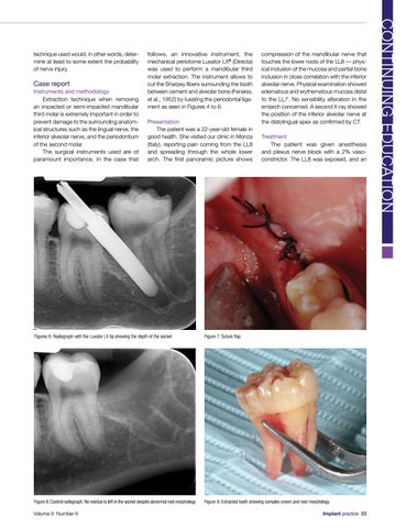
Figures 6: Radiograph with the Luxator LX tip showing the depth of the socket
Figure 7: Suture flap
Figure 8: Control radiograph. No residue is left in the socket despite abnormal root morphology
Figure 9: Extracted tooth showing complex crown and root morphology
Volume 9 Number 6
Implant practice 33
CONTINUING EDUCATION
technique used would, in other words, determine at least to some extent the probability of nerve injury.