CLINICAL
Figure 18: Workflow of integrating intraoral scanning and in-office 3D printing
to the previous example of in-office CAD/ CAM, the high cost of this equipment has been a critical factor limiting the integration into private clinical practices. Recently, 3D printing has become more accessible to practitioners due to the rapid development of consumer-grade stereolithography (SLA) printers such as the Form 1+ (Formlabs, Somerville, Massachusetts) (Figure 17). These consumer-grade printers, often costing just a few thousand dollars, are a significant departure from the currently available dental laboratory printers, which can cost almost 10-20 times more than a consumer-grade printer. The workflow for 3D printing begins with an intraoral optical scan of the dentition, and generation of a STL file and results in a 3D printed model (Figure 18). While some intraoral scanners do not allow for exporting of open STL file, this author uses a scanner that easily converts an intraoral optical scan into an open STL (3M™ True Definition Scanner, 3M ESPE). The STL file is opened in a digital-modeling software (netfabb basic, netfabb GmbH, Lupburg, Germany) where missing parts of the scan can be filled in, and a base can be added. Alternatively, software that comes with a 3D printer will allow for automatic processing of an STL file to make it ready for printing (Figure 19) (Proform, Formlabs). A variety of colors and physical properties are available with some offering printing accuracy of between 25-200µm. Greater accuracy prints require more time, often up to 2-4 hours per dental model, because they are printing in thinner layers that require more layers to fabricate a dental model. The STL file is imported into the printer software, and the resin tank is filled with liquid resin. The 32 Implant practice
Figure 19: 3D printing software allows for orientation and preparation for printing
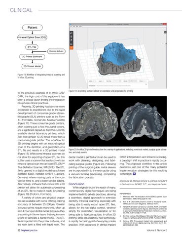
Figure 20: In-office 3D printed models allow for a variety of applications, including provisional models, surgical guide fabrication, and study models
dental model is printed and can be used to work with planning, designing, and fabricating surgical guides (Figure 20). Following printing of the surgical guide, metal sleeves are incorporated in to the resin guide using a vacuum-forming processing, completing the fabrication process.
CBCT interpretation and intraoral scanning, a paradigm shift in practice is rapidly occurring. The proposed workflow in this article describes just one of the many potential implementation strategies for this exciting technology. IP Disclosure: Dr. Michael Scherer is a clinical consultant
Conclusion While originally out of the reach of many, contemporary digital techniques are being implemented into private practices, allowing a seamless, digital approach to everyday dentistry. Intraoral scanning, especially with being able to easily export open STL files, allows for the full digital control, whether simply for restoration visualization or for being able to fabricate guides. In-office 3D printing, while still a relatively new technology, is rapidly becoming part of everyday private practice. With advanced in dental implant
to Zest Anchors, BIOMET 3i™ , and Keystone Dental.
REFERENCES 1. Mörmann WH. The evolution of the CEREC system. J Am Dent Assoc. 2006;137(suppl):7S-13S. 2. Puri S. A CAD/CAM picture is worth a thousand words. CERECdoctors.com Magazine. 2009;Q2:46-47. 3. Yamalik N, Ensaldo-Carrasco E, Cavalle E, Kell K. Oral health workforce planning part 2: figures, determinants and trends in a sample of World Dental Federation member countries. Int Dent J. 2014;64(3):117-126. 4. Dentists working. WorldMapper Web site. http://www.worldmapper.org/display.php?selected=218#. Published 2004. 5. Scherer MD. Presurgical implant-site assessment and restoratively driven digital planning. Dent Clin North Am. 2014;58(3):561-595.
Volume 8 Number 2