Behind the Cover
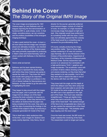
The Story of the Original fMRI Image The cover image accompanying the 1991 Science paper by Jack Belliveau and colleagues reporting the first demonstration of functional MRI is, quite simply, iconic. In that single, evocative picture, we can somehow see the endless possibilities of the emergent imaging technique. To take a peek behind the cover—to learn a bit more about how the image was conceptualized and ultimately rendered—we checked in with the two authors of the Science paper who were primarily responsible for producing it: Mark Cohen and David Kennedy, both of whom worked with Belliveau in the Martinos Center. Here’s what we learned. Belliveau and his team started thinking about ways to represent the study visually well before they knew Science was going to devote the cover to it. They knew the report and results were going to be important, says Cohen, who today is a professor in the UCLA Semel Institute for Neuroscience and Behavior, so they wanted to go the extra mile in highlighting the work.
30
director for the journal, generally preferred more textural images: a photo of a field full of rocks, for example, or a high-resolution microscopy image that played on light and dark. She originally would have preferred to do something similar with this issue, Cohen says, but ultimately agreed that the cutaway of the head would be a compelling way to represent the groundbreaking study. Of course, actually producing the image was another matter. “Back in those days, surface rendering in 3D was not common,” says Kennedy, who is now the director of the Division of Neuroinformatics, Department of Psychiatry, at the University of Massachusetts Medical Center. But the researchers had access to an advanced Sun workstation with a high-performance TAAC-1 graphics and image accelerator. The accelerator came with a number of demo videos showing surface and volume rendering, so they knew that what they wanted to do was possible. And in fact they were able to replace the data in one of the demos with their own MRI scans.
They began to play around with the images from the paper, and in particular with the image from an oblique cut of the head showing the brain activation in response to the visual stimulus. When they learned from the editors at Science that the paper was being considered for the cover, they came up with the idea of presenting the findings in the context of the subject’s head, by producing a computer-generated 3D model of the head.
They knew both the angle and the depth at which the oblique scan they were using had been acquired, and were able to cut into the 3D model at the same angle and depth, so the MRI scan in fact appeared in the right part of the head. This much was relatively straightforward. Things got slightly tricky when they had to account for the viewing angle of the head itself. “We wanted enough of the face to be recognizable [as a face], but we also wanted the exposed part of the head in view,” Kennedy says. “We probably spent days arguing over the exact angle.”
This in itself was a fairly audacious idea. At the time, cover images for Science were rarely overtly depictive. Amy Henry, the art
Once the head was turned, the MR scans no longer matched the rendering of the head. The researchers could no longer just overlay
