Non-Hodgkin’s Lymphoma-Associated Chylothorax: A Case Report and Literature Review
In neoplastic conditions, the enlarged hilar lymph nodes compress the lymphatic channels and thoracic duct, resulting in obstruction of the drainage of lymphatic fluids from the periphery of lung parenchyma and pleural surfaces. This leads to extravasation of the chyle and lymph, producing chylothorax. Another postulated mechanism for chylothorax formation is the direct infiltration of the thoracic duct by tumor cells. This infiltration by the neoplastic cells makes the duct more rigid and vulnerable to rupture following minimal or occult trauma resulting in chyle leakage and chylothorax formation.
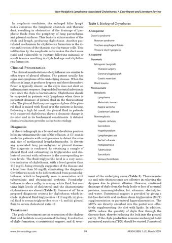
Table 1. Etiology of Chylothorax A. Congenital Down’s syndrome
Noonan syndrome
Tracheo-esophageal fistula
Thoracic duct hypoplasia
B. Acquired Traumatic
Clinical Presentation The clinical manifestations of chylothorax are similar to other types of pleural effusion. The patient usually has signs and symptoms of the underlying disease. When the effusion is large, it produces dyspnea and chest discomfort. Fever is typically absent, as the chyle does not elicit an inflammatory response. Superadded bacterial infection is rare since the chyle is bacteriostatic. Chylothorax should be suspected in patients with lymphoma when there is persistent drainage of pleural fluid in the thoracostomy tube. The pleural fluid may not appear chylous if the pleural fluid is mixed with blood or if the patient is fasting. Following a high fat meal, the pleural fluid in patients with suspected chylothorax shows a dramatic change in its color and in its biochemical constituents. A detailed clinical evaluation provides a clue to its etiology.
Iatrogenic (surgical)
Thoracic surgery
Coronary bypass graft
Gastric resection
Blunt trauma Nontraumatic
Neoplastic
Lymphoma
Metastatic tumors
Kaposi sarcoma
Castleman’s disease
Nonneoplastic
Hepatic cirrhosis
Diagnosis
Sarcoidosis
A chest radiograph in a lateral and decubitus position helps in estimating the size of the effusion. A CT scan is useful in patients with malignancies to detect the sites and size of mediastinal lymphadenopathy. It detects any associated lung parenchymal or pleural disease. The diagnosis is confirmed by obtaining a sample of pleural fluid and estimating its triglycerides and cholesterol content with reference to the corresponding serum levels. The fluid triglyceride level is a very sensitive indicator of chylothorax, with a level greater than 110 mg/dL being strongly suggestive of the disease and a level less than 50 mg/dL eliminating the diagnosis. Chylothorax needs to be differentiated from pseudochylothorax, which is frequently seen in association with tuberculosis and rheumatoid arthritis. Pseudochylothorax is also a milky to creamy white fluid, but contains high levels of cholesterol and the characteristic chylomicrons are absent (Table 2). Romero et al.8 have proposed diagnostic criteria for chylothorax which include: i) pleural fluid triglycerides >110 mg/dL; ii) pleural fluid to serum triglycerides ratio >1; and iii) pleural fluid to serum cholesterol ratio <1.
Hypothyroidism
Lymphangiectasis
Bechet’s disease
Histoplasmosis
Tuberculosis
Sarcoidosis
Venous thrombosis
ment of the underlying cause (Table 3). Thoracocentesis and tube thoracostomy are effective in relieving the dyspnea due to pleural effusion. However, continued drainage of chyle from the body leads to loss of essential proteins, immunoglobulins, fat, vitamins, electrolytes, and water. Nutritional support is provided by giving a fat-free diet with oral medium chain triglycerides (MCT) supplementation or parenteral hyperalimentation. The MCTs are directly absorbed into the portal vein effectively supplementing the diet with lipids. In addition, MCTs reduce the quantity of chyle flow through the thoracic duct, thereby reducing the leak into the pleural cavity. If the chyle production remains unchanged, total parenteral nutrition (TPN) should be started. Parenteral
Treatment The goals of treatment are a) evacuation of the chylous fluid and facilitate re-expansion of the lung; b) reduction of chyle formation; c) nutritional support; and d) treatwww.slm-oncology.com
09-0013_Kashyap.indd 99
99
APJOH 2009; 1: (1). March 2009
3/18/09 4:20:40 PM