5 minute read
Use of Phage Display to Produce Antibodies Against Campylobacter jejuni Robin Kaai and Robin Ka‘ai won the top award at the 2013 John A. Burns School of Medicine (JABSOM) Biomedical Sciences and Health Disparities Symposium. It was the first time a community col lege student had won this prestigious award

Use of Phage Display to Produce Antibodies against Campylobacter jejuni
By Robin Kaai and Thomas Premeaux (Faculty Mentors: Matthew Tuthill, Alan Garcia, and John Berestecky)
Advertisement
INTRODUCTION
Current methods for monoclonal antibody production utilize hybridoma technology. This requires repeated immunization of a mouse followed by the harvest and fusion of spleen cells with an immortalized cell line in order to produce and select for antigen-specific antibody-producing clones. Because of the cost, time, and expertise required for traditional monoclonal production, we are investigating the efficacy of using phage display to generate novel antibodies. The aim of this study is to develop proficiency in phage display, and allow for a supplemental approach to produce monoclonal antibodies at our facilities. Initial studies will target Campylobacter jejuni, a gram-negative, epsilonproteobacteria that is the most common cause of bacterial gastroenteritis.
METHODOLOGY
Phage display utilizes a library of modified M13 bacteriophages that display single-chain variable fragments of diverse specificities. Through repeated antigen-targeted biopanning, phage elution, and coinfection of host bacteria with helper phage, target phages with the desired specificity can then be amplified, isolated, and utilized in biomedical research settings.


Figure 1. M13 bacteriophage Figure 2. Phagemid containing scFv Figure 3. Biopanning The biopanning process involves the introduction of the phage library into antigen-coated microtiter wells, stringent washes and elution. Host re-infection and repeated biopanning then allows for the amplification of target-specific phages.
RESULTS
Thus far, we have been successful in amplifying the phagemid library, as well as helper phage stocks for
back up stocks and future work. In addition, we have performed various infection assays to validate the phage library, and confirm the background genotypes of the host cells involved. Primers were also designed to track the plasmids through the infection and biopanning process.
Figure 4 (left image below) Maxiprep results.
pCDisplay-3M phagemid linearized using SalI and XhoI restriction digestion (B) was electrophoresed in conjunction with the uncut control phagemid (A).
Figure 5 (right image below) Maxiprep validation.
The presence of a 0.4 kilobase band resulting from the SalI and NheI restriction digestion confirms that the VH region is present in the phagemid maxiprep (C).

Figure 6 (left image below) Phagemid primer
validation. Phagemid-specific primers were used to amplify a 1 kilobase region of the pCDisplay-3M. P.C.R. reactions were perform using annealing temperatures of 60C (A) and 55C (B).
Figure 7 (right image below)Helper phagemid
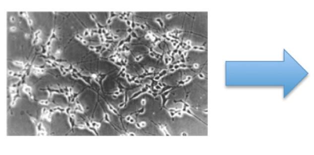
primer validation. Primers were designed and used in P.C.R. to confirm the specificity to the helper phagemid, as shown by the production of a 0.3 kilobase amplicon (C). Figure 8 (page 51). Phage viability validation E. coli TG1 host cells were uninfected, infected with the phagemid display library, the helper phage stock, or both. Uninfected, infected, or coinfected host cells were then plated as on standard media with the absence or presence of antibiotics kanamycin and/or ampicillin. The control set of plates was then scored in order to determine viability of the phages and the efficiency of cell infection. Display phages contain an ampicillin resistance cassette while helper phages contain a kanamycin resistance cassette in order to select for coinfected host cells during target phage amplification.

CONCLUSIONS
Transformation, amplification and maxiprep harvest and subsequent restriction digestion and DNA agarose gel electrophoresis confirms that the • M13 phagemid library was amplified; • M13 phagemid is the appropriate size; • upon restriction digestion, the M13 phagemid matches the published restriction map. Polymerase chain reactions performed on the phagemid or helper phage stock confirm that • the phagemid primers are specific and amplified stock is correct; • designing helper phage specific primers were successful; • both primer sets may be used to detect the presence of helper phages in various steps of library production. Plating assays of E. coli TG1 host cells with or without infection on media containing various antibiotics indicate that • uninfected TG1 host cells will not grow in the presence of ampicillin or kanamycin; • phagemid stocks are intact and contain cassettes allowing infected TG1 cells to grow in the presence of ampicillin; • helper phages are intact and contain cassettes allowing infected TG1 to grow in the presence of kanamycin; • dual infection with phagemid and helper phage is possible; AMP/KAN may be used to select for coinfected TG1.
FUTURE PLANS
With stocks established and validated, we are in the process of applying the phage display methodology to Campylobacter jejuni. Once successful phages have been isolated, amplified and banked, this technology will allow for: epitopic tagging of antigens, immunoprecipitation, antigen isolation and characterization, and recombinant modifications of antibody variable regions. Moreover, it will allow us to increase our monoclonal output, involve more students in phage antibody production and incorporate components of the process into select campus lab courses.
LITERATURE CITED
C. F. Barbas III, D. R. Burton, J. K. Scott, and G. J.
Silverman, (2001) Phage Display: A Laboratory
Manual. Cold Spring Harbor Laboratory Press,
Cold Spring Harbor, NY.

ACKNOWLEDGEMENTS
We thank C. Allen, S. Kawasaki, K. Noji and K. Noa for their ongoing laboratory and administrative support. Funding for this project was provided by the Kapiolani Community College STEM Program, the Perkins Grant and the National Center for Research Resources (5P20RR016467-11) and the National Institute of General Medical Sciences (8 P20 GM 103466) from the National Institutes of Health. The University of Hawaii RMATRIX Program is supported by award number U545MD007584 from the National Institute on Minority Health and Health Disparities, National Institutes of Health. The content is solely the responsibility of the authors and does not represent the official views of the National Institutes of Health.