
ISSN 2313-6693
OF EDUCATION AND SCIENCE OF UKRAINE The Journal of V. N. Karazin Kharkiv National University Series «MEDICINE» Issue 45 Since 2000 Вісник Харківського національного університету імені В. Н. Каразіна Cерія «МЕДИЦИНА» Випуск 45 Започаткована 2000 р. KHARKIV 2022
MINISTRY
Journal contains articles about topical issues of modern experimentalandclinicalmedicine.
Approved for publication by the Academic Council of V. N. Karazin KhNU decision (protocol №18 from 11 25.2022)
EDITORIAL BOARD
Editor-in-chief: I. V. Belozorov, MD, PhD, Prof., V. N. Karazin KhNU
Deputy Editor-in-Chief: T I Liadova, MD, PhD, Prof , V. N. Karazin KhNU,
O. I. Tsivenko , MD, PhD, Prof., V. N. Karazin KhNU.
J. Alpert, MD, PhD, Prof., University of Arizona (USA)
A. Aubert, MD, PhD, Prof., KU Leuven (Belgium)
Yu.V. Avdosyev, MD, PhD, Prof. SI «V. T. Zaitsev Institute of General and Emergency Surgery of NAMS of Ukraine»
O. M. Fedota, PhD, Prof., V. N. Karazin KhNU
M. G. Grishchenko, MD, PhD, Prof., V. N. Karazin KhNU
H. Hutten, PhD, Prof., Graz University of Technology (Austria)
I. G. Kupnovytska, MD, PhD, Prof , IFNMU
I. V. Linsky, MD, PhD, Prof., V. N. Karazin KhNU
O. V. Martynenko, PhD, Dr.Sci., Prof., V. N. Karazin KhNU
T. S. Mishchenko, MD, PhD, Prof., V. N. Karazin KhNU
Ye. Ya Nikolenko, MD, PhD, Prof., V. N. Karazin KhNU
H. Pastor, MD, PhD, Prof., University of Barcelona (Spain)
M. M. Popov, MD, PhD, Prof., V. N. Karazin KhNU
P. I. Poteiko, MD, Assoc. Prof., V. N. Karazin KhNU
O. S. Protsenko, MD, PhD, Prof., V. N. Karazin KhNU
G. Raimondi, MD, PhD, Prof., Sapienza University of Rome (Italy)
A. Wolfgang, MD, PhD, Prof , Witten/Herdecke University (Germany)
I. A. Zupanets, MD, PhD, Prof., NUPh
Executive Secretary: M. S. Matvieienko, MD, PhD, Assoс. Prof., Editorial address:
61022, Ukraine, Kharkiv, Svobody Square, 6, V.N. Karazin Kharkiv National University, School of Medicine, tel./fax (057) 702-04-55, tel. 707-54-50, E-mail: journal.medicine@karazin.ua
Articles were internal and external reviewed
Certificate about the state registration:
KV No. 21561-11461 R from 20.08.2015
The journal is a professional in the field of sciences in Ukraine, сategory «Б», 22 Health care, Specialties 222 Medicine.
MES Ukraine Order № 886 of 02.07.2020
© V N Karazin Kharkiv National University, 2022
Вісник містить статті, про актуальні питання сучасної експериментальної та клінічної медицини Затверджено до друку рішенням Вченої ради ХНУ імені В. Н. Каразіна (протокол № 18 від 25 11.2022).
РЕДАКЦІЙНА КОЛЕГІЯ
Головний редактор: І. В. Белозьоров, д. мед.н., проф., ХНУ імені В. Н. Каразіна Заступник головного редактора: Т. І. Лядова, д.мед.н., проф., ХНУ імені В. Н. Каразіна, О. І. Цівенко, д. мед.н., проф., ХНУ імені В. Н. Каразіна.
Дж. Альперт, д. мед.н., проф., Аризонский університет (США)
A Ауберт, д. мед.н., проф., КУ Лейвен (Бельгія)
Ю. В. Авдосьєв, д.мед.н., проф., ДУ «ІЗНХ імені В. Т. Зайцева НАМНУ»
О. М. Федота, д.біол.н., проф., ХНУ імені В. Н. Каразіна М. Г. Грищенко, д. мед.н., проф., ХНУ імені В. Н. Каразіна Г. Хуттен, д. техн.н., проф., Технічний університет Грац (Aвстрiя) І. Г. Купновицька, д. мед н., проф., ІФНМУ І. В. Лінський, д. мед н., проф., ХНУ імені В. Н. Каразіна О. В. Мартиненко, д.мат.н., проф., ХНУ імені В. Н. Каразіна
Т. С. Міщенко, д. мед.н., проф., ХНУ імені В. Н. Каразіна Є. Я. Ніколенко, д. мед н., проф., ХНУ імені В. Н. Каразіна Х. Пастор, д. мед н., проф., Університет Барселони (Іспанія) М. М. Попов, д. мед н., проф., ХНУ імені В. Н. Каразіна П. І. Потейко, к. мед.н., доцент, ХНУ імені В. Н. Каразіна О. С. Проценко, д. мед н., проф., ХНУ імені В. Н. Каразіна Ж. Раймонди, д. мед н., проф., Римський університет Ла Сапієнца (Італія) А. Вольфганг, д. мед н., проф., Университет ВиттенХердеке (Німеччина) І. А. Зупанець, д. мед.н., проф., НФаУ Відповідальний секретар: М. С. Матвєєнко, доктор філософії, доцент Адреса редакційної колегії: 61022, Україна, м. Харків, майдан Свободи, 6, Харківський національний університет імені В. Н. Каразіна, медичний факультет, тел./факс (057) 702-04-55; тел. 707-54-50, E-mail: journal.medicine@karazin.ua
Статті пройшли внутрішнє та зовнішнє рецензування. Свідоцтво про державну реєстрацію: КВ № 21561-11461 Р від 20.08.2015
Наукове фахове видання України, категорії «Б», галузь знань: 22 Охорона здоров’я, спеціальність: 222 Медицина. Наказ МОН України № 886 від 02.07.2020
© Харківський національний університет імені В. Н. Каразіна, 2022
CONTENTS ЗМІСТ
Fundamental researches Фундаментальні дослідження
Martynenko О., Raimondi G., Barsi L., Maliarova L.
ENTROPY OF FREQUENCY DOMAIN OF HEART RATE VARIABILITY
Martynenko O. V., Pastor X. D., Frid S. A., Rojas J. G., Maliarova L. V.
ENTROPY OF DNA SEQUENCES AND LEUKEMIA PATIENTS MORTALITY
Мартиненко О. В., Раймонді Д., Барсі Л., Малярова Л. В.
ЕНТРОПІЯ ЧАСТОТНОГО ДОМЕНУ
ВАРІАБЕЛЬНОСТІ СЕРЦЕВОГО
Мартиненко О. В., Пастор К. Д., Фрід С. А, Роджас Д. Д., Малярова Л. В. ЕНТРОПІЯ ПОСЛІДОВНОСТЕЙ ДНК І СМЕРТНІСТЬ ПАЦІЄНТІВ З ЛЕЙКЕМІЄЮ
Original researches Орігінальніні дослідження
Dorofieieva V., Fedota O.
LARGE FAMILY GENETIC ANALYSIS: EFFECTS OF VARIEGATED PORPHYRIA AND HEMOPHILIA B ON REPRODUCTIVE TRAITS
Hoshovska Alisa
FEATURES OF THE FORMATION OF PATHOGENETIC CHANGES OF THE PLACENTAL COMPLEX ON THE BACKGROUND OF INTRA-UTERONOMY INFECTION
Pokrovska Nataliia, Sklyarov Eugen ADROPIN AND RISK FACTORS OF ARTERIAL HYPERTENSION IN PATIENTS WITH EXCESS BODY WEIGHT AND OBESITY
Bil Bogdan, Chopyk Valentyna, Deeva Yulia, Dytiatkovska Yevgenia, Gogunska Inna, Popovych Vasyl, Romaniuk Lilia, Umanets Tetiana, Zabolotna Diana, Zaikov Sergii
AN ALGORITHM RECOMMENDATION FOR THE PHARMACOLOGICAL MANAGEMENT OF ALLERGIC RHINITIS IN UKRAINE: A CONSENSUS STATEMENT FROM AN EXPERT PANEL
Bogun N. Yu., Brynza M. S.
INFLUENCE OF THE LEVELS OF THYROID HORMONES ON THE RESULT OF RADIOFREQUENCY ABLATION FOR ATRIAL FIBRILLATION: LITERATURE REVIEW
Feshchenko D. I., Malyk S. L., Shevnia M. B.
HEADACHE ON THE BACKGROUND OF CORONAVIRUS INFECTION: FEATURES OF THE CLINICAL PICTURE
Kanishcheva Olena
MODERN ASPECTS OF SHORT-TERM BLOOD PRESSSURE VARIABILITY IN ARTERIAL HYPERTENSION
Дорофєєва В. Р., Федота О. М.
ГЕНЕТИЧНЕ ДОСЛІДЖЕННЯ ВЕЛИКОЇ
РОДИНИ:
ПОРФІРІЇ
ТА ГЕМОФІЛІЇ В НА РЕПРОДУКТИВНІ
ПОКАЗНИКИ
Гошовська А. В.
ОСОБЛИВОСТІФОРМУВАННЯПАТОГЕНЕТИЧНИХЗМІНПЛАЦЕНТАРНОГО КОМПЛЕКСУНАТЛІВНУТРІШНЬОУТРОБНОГО ІНФІКУВАННЯ
Огляд
Біль Богдан, Чопік Валентина, Дєєва Юлія, Дитятковська Євгенія, Гогунська Інна, Попович Василь, Романюк Лілія, Уманець
Тетяна, Заболотна Діана, Зайков Сергій РЕКОМЕНДАЦІЯ З АЛГОРИТМУ ФАРМАКО-
ЛОГІЧНОГО ЛІКУВАННЯ АЛЕРГІЧНОГО
РИНІТУ В УКРАЇНІ: КОНСЕНСУСНА ЗАЯВА ЕКСПЕРТА
Богун Н. Ю., Бринза М. С.
ВПЛИВ РІВНІВ ГОРМОНІВ ЩИТОПОДІБНОЇ
ЗАЛОЗИ НА РЕЗУЛЬТАТ РАДІОЧАСТОТНОЇ
АБЛЯЦІЇ ФІБРИЛЯЦІЇ ПЕРЕДСЕРДЬ:
ЛІТЕРАТУРНИЙ ОГЛЯД
Фещенко Д. І., Малик С. Л., Шевня М. Б.
ГОЛОВНИЙ БІЛЬ НА ФОНІ КОРОНА-
ВІРУСНОЇ ІНФЕКЦІЇ: ОСОБЛИВОСТІ
КЛІНІЧНОЇ КАРТИНИ
Каніщева О. В.
СУЧАСНІ АСПЕКТИ КОРОТКОСТРОКОВОЇ
ВАРІАБЕЛЬНОСТІ АРТЕРІАЛЬНОГО ТИСКУ
ПРИ АРТЕРІАЛЬНІЙ ГІПЕРТЕНЗІЇ
4
12
Series «Medicine». Issue 45 3
РИТМУ
ВПЛИВ ВАРІЄГАТНОЇ
24
37
Покровська
АДРОПІН ТА ФАКТОРИ РИЗИКУ АРТЕРІАЛЬНОЇ ГІПЕРТЕНЗІЇ У ПАЦІЄНТІВ З НАДЛИШКОВОЮ МАСОЮ ТІЛА ТА ОЖИРІННЯМ 44 Review
Н. К., Скляров Є. Я.
51
66
77
82
UDC: 616.12-008:004.891.3
Fundamental researches
DOI: 10.26565/2313-6693-2022-45-01
ENTROPY OF FREQUENCY DOMAIN OF HEART RATE VARIABILITY
Martynenko О. A, B, C,D ,E, F,RaimondiG.A, E, F,BarsiL. E, F,MaliarovaL.E, F A
research concept and design; B
collection and/or assembly of data; C – data analysis and interpretation; D – writing the article; E – critical revision of the article; F – final approval of the article
Introduction. The heart rate variability (HRV) is based on measuring (time) intervals between R-peaks (of RR-intervals) of an electrocardiogram (ECG) and plotting a rhythmogram on their basis with its subsequent analysis by various mathematical methods which are classified as Time-Domain (TD), FrequencyDomain (FD) and Nonlinear [1, 2]. There are a number of popular Nonlinear methods used in HRV analysis, such as entropy-based measures that mostly applied for TD. Spectral Entropy (SE) is using for FrequencyDomain: it is defined to be the Shannon entropy of the power spectral density (PSD) of the data. An important characteristic of Frequency-Domain studies is sympatho-vagal balance, which has been overlooked by entropy-based analysis. This is due to the fact that good entropy analysis restricted the number of existing HRV data, which is shrinking in FD and also in total spectrum parts.
Aim of the research. The goal of this paper is to provide a reliable formula for calculating entropy accurately for Frequency-domain of standard 5-min. HRV records and to show the advantages of such approach for analyzing of sympatho-vagal balance for healthy subjects (NSR), Congestive Heart Failure (CHF) and Atrial Fibrillation (AF) patients.
Materials and Methods. We used MIT-BIH long-term HRV records for Normal Sinus Rhythm (NSR), Congestive Heart Failure (CHF) and Atrial Fibrillation (AF).
The generalized form of the Robust Entropy Estimator (EnRE) for Frequency-domain of standard 5-min. HRV records was proposed and the key EnRE futures was shown.
The difference between means of the two independent selections (NSR and CHF, before and after AF) has been determined by a t-test for independent samples; discriminant analysis and statistical calculations have been done by using the statistical package IBM SPSS 27.
The results of the study. We calculate entropy for all valuable for HRV spectral interval, namely 0–0.4 Hz and to compare with existing results for Spectral Entropy: qualitatively we receive the same distribution number as [14] and significant difference (p < 0.001) between entropy averages for NSR and CHF or AF patients.
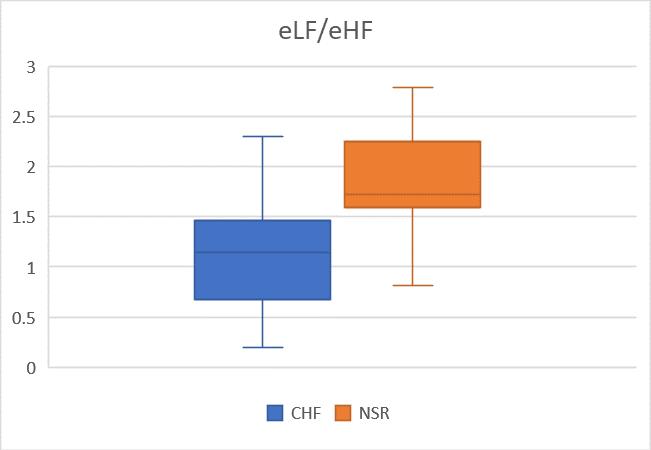
We define low-frequencies (LF) power spectrum components in the range of 0.04–0.15 Hz and highfrequencies (HF) power spectrum components in the range of 0.15–0.4 Hz [1]. The sympatho-vagal balance is a simple ratio LF/HF [1]. Then, we define an entropy eLF of the LF power spectrum components, an entropy eHF of the HF power spectrum components and entropy based sympatho-vagal balance as a ratio eLF/eHF.
The difference between NSR and CHF groups are significant in both cases LF/HF and eLF/eHF with p < 0.001, but in case of eLF/eHF the results are quite better (t = -4.8, compared to LF/HF where t = -4.4). The discriminant analysis shows total classification accuracy for eLF/eHF in 79.3 % (χ2 = 19.4, p < 0.001) and for LF/HF in 72.4 % (χ2 = 16.6, p < 0.001).
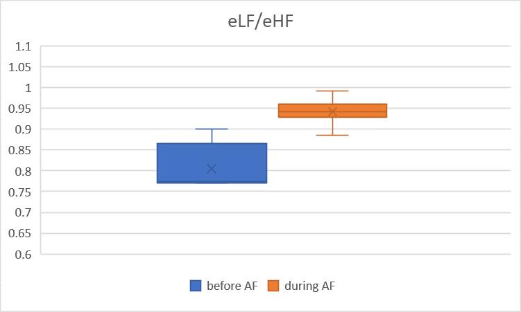
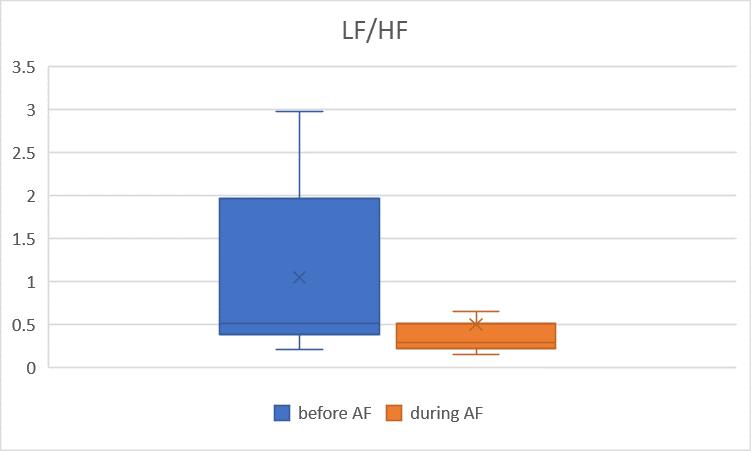
We applied entropy-based Frequencies-domain analyzing for AF patients and showed that ratio eLF/eHF is significantly higher during AF than before AF (p < 0.001). This is opposite to ordinary LF/HF where difference is insignificant due to high variation of this ratio.
Conclusion. Proposed in the article is generalized form for Robust Entropy Estimator EnRE for Frequencies-domain, which allows, for time series of a limited length (standard 5-min. records), to find entropy value of HRV power spectrum (total spectrum, low- and high- frequencies bands).
Using the proposed EnRE for MIT-BIH database of HRV records, we show for standard 5 min. HRV records the usage of EnRE of HRV power spectrum and entropy-based sympatho-vagal balance of Normal Sinus Rhythm (NSR) and Congestive Heart Failure (CHF) cases. It is demonstrated, that, entropy-based Frequencies-domain analyzing is applicable for case of Atrial Fibrillation (AF) even during AF episodes. We showed the significant difference (p < 0.001) before and during AF for entropy of total spectrum, as well as for sympatho-vagal balance in form of eLF/eHF.
KEY WORDS: hearth rate variability, entropy, frequency-domain, congestive heart failure, atrial fibrillation
The Journal of V.N. Karazin Kharkiv National University. 2022 4
–
–
Martynenko О., Raimondi G., Barsi L., Maliarova L., 2022
INFORMATION ABOUT AUTHORS
Martynenko Oleksandr Vitalyevich, D Sc., Ph D., Full Professor, Department of Hygiene and Social Medicine, School of Medicine, V. N. Karazin Kharkiv National University, 6, Svobody sq., Kharkiv, Ukraine, 61022. e-mail: Alexander.v.martynenko@karazin.ua, ORCID ID: https://orcid.org/0000-0002-0609-2220.
Gianfranco Raimondi, MD, PhD, Prof., Sapienza University of Rome (Italy), 5, Piazzale Aldo Moro, Rome, Italy, 00185; e-mail: gianfrancoraimondi@uniroma1.it
Luca Barsi, PhD, Rome, Italy, 00185; e-mail: barsiluca1@gmail.com
Maliarova Liudmila Volodimirivna, Assistant, Department of hygiene and social medicine, School of Medicine, V. N. Karazin Kharkiv National University, 6, Svobody sq., Kharkiv, Ukraine, 61022, e-mail: l.v.maliarova@karazin.ua, https://orcid.org/0000-0002-7902-7016
For citation:
Martynenko О, Raimondi G, Barsi L, Maliarova L. ENTROPY OF FREQUENCY DOMAIN OF HEART RATE VARIABILITY. The Journal of V. N. Karazin Kharkiv National University. Series «Medicine». 2022;45:4–11 DOI: 10.26565/2313-6693-2022-45-01
INTRODUCTION
The heart rate variability (HRV) is based on measuring (time) intervals between Rpeaks (of RR-intervals) of an electrocardiogram (ECG) and plotting a rhythmogram on their basis with its subsequent analysis by various mathematical methods that are classified as Time-Domain (TD), FrequencyDomain (FD) and Nonlinear [1, 2]. There are a number of popular Nonlinear methods used in HRV analysis, such as entropy-based measures, like approximate entropy (ApEn) [3] and sample entropy (SampEn) [4]. SampEn is regarded as a modified version of ApEn,intendedtosolvesuchshortcomingsas bias and relative inconsistency [4]. However, the traditional SampEn method is single-scale based and, therefore, fails to account for the multiple time scales inherent in cardiovascular systems [5–7]. Multiscale entropy (MSE) method was proposed in [7] and received much attention in the biomedical and mechanical fields [8–10]. Further MSE developing was transformed to multiscale multivariate entropy analysis [8, 10–13]. These entropy-based measures are all applied to original RRs, – that is mean their implementation for Time-Domain. Other hand the Spectral Entropy (SE) is using for Frequency-Domain: it is defined to be the Shannon entropy of the power spectral density (PSD) of the data. In article [14] the SE were estimated for healthy, thyroid and depression subjects, as well as for patients with Congestive Heart Failure (CHF) and Atrial Fibrillation (AF). It was shown the significant different of SE for all categories and ordered toincrease ofSE are: depression, thyroid, CHF, AF and healthy subjects. An important characteristic of Frequency-
Domain studies is sympatho-vagal balance, which has been overlooked by entropy-based analysis. The reason for this was that good entropy analysis restricted the number of existing HRV data, which is shrinking in FD andalsointotalspectrumparts.
Prevalence of the effective methodology of entropy analysis of FD for standard 5-min HRV records is suppressed by unsatisfactory accuracyofavailablemethodsincaseofshort records as we shown in [15]. Therefore, it appears there is a necessity for building a robust formula for calculating entropy for each part of spectrum in Frequency-Domain with required accuracy for a limited series of RR-intervals observed in a standard 5-minute HRVrecord.
MATERIALS ANDMETHODS
We used long-term HRV records by Massachusetts Institute of Technology –Boston’s Beth Israel Hospital (MIT-BIH) from [16] (http://www.physionet.org), a freeaccess, on-line archive of physiological signals. Normal Sinus Rhythm (NSR) RR Interval Database includes beat annotation files for 54 long-term ECG recordings of subjects in normal sinus rhythm (30 men, aged 28.5 to 76, and 24 women, aged 58 to 73). Congestive Heart Failure (CHF) RR Interval Database includes beat annotation files for 29 long-term ECG recordings of subjects aged 34 to 79, with congestive heart failure (NYHA classes I, II, and III). Subjects include 8 men and 2 women; gender of the remaining 21 subjects is not known. The original electrocardiography (ECG) signals for both NSR and CHF RR interval databases were digitized at 128Hz, and the beat annotations were obtained by automated analysis with manual review and correction.
Series «Medicine». Issue 45 5
The MIT-BIH Atrial Fibrillation (AF) Database[17] was used forourentropy-based analyzing with long and short RR’s subsets. This database includes 25 long-term ECG recordings of human subjects with atrial fibrillation (mostly paroxysmal). The individual recordings are each 10 hours in duration, and contain two ECG signals each sampled at 250 samples per second with 12bitresolutionoverarangeof± 10 millivolts. The original analog recordings were made at Boston’s Beth Israel Hospital (now the Beth Israel Deaconess Medical Center) using ambulatory ECG recorders with a typical recording bandwidth of approximately 0.1 Hz to40Hz.
A generalized form of the Robust Entropy Estimator (EnRE) for time series was proposed in [15] and adopted for power spectraldensity(PSD)ofRRnow:
2/ independence of EnRE from N for initialtimeseriesandforseriesaftersorting;
3/ independence of EnRE from additive changesofmean.
After numerical researches the following coefficient values had been found: l = 3, m = 1, k =2.
The difference between means of the two independent selections (NSR and CHF, before and after AF) has been determined by a t-test for independent samples; discriminant analysis and statistical calculations have been done by using the statistical package IBMSPSS27.
RESULTS ANDDISCUSSION
where MD ismedianofthesequencefor B value of PSD;
- distance between
estimated coefficients. Search conditions for coefficients A, l, m, k is thefollowing:
1/ accurate approximation for known distributionsofarandomvalue;
First of all, let us calculate entropy for all valuable for HRV spectral interval, namely 0–0.4Hz. That is give us possibility to compare with existing results for Spectral Entropy: qualitatively we receive the same distribution number as [14] and significant difference (p<0.001) between entropy averages for NSR and CHF or AF patients. Quantitatively our result is not exactly the same to [14] because we used different entropy measures: SE is based on Shannon entropy and EnRE approximated the entropy ofdistributionordifferentialentropy.
According to [1] we define lowfrequencies(LF)powerspectrumcomponents in the range of 0.04–0.15Hz and highfrequencies (HF) power spectrum components in the range of 0.15–0.4Hz. The sympatho-vagal balance is a simple ratio LF/HF [1]. We calculate an entropy of LF power spectrum components as eLF, entropy of HF power spectrum components as eHF and entropy based sympatho-vagal balance as aratioeLF/eHF.
Many authors emphasize the importance of sympatho-vagal balance measures, but statistical significance makes it difficult to estimate the effects in CHF patients: for example, in [18] showed that LF/HF is significantly lower for CHF patients compare with healthy subjects, but in compare with [19], where difference in LF/HF between CHF and NSR groups is insignificant due to p=0.175. The results of calculations of LF/HF and eLF/eHF for CHF and NSR groupsareshownontheFig.1.
The Journal of V.N. Karazin Kharkiv National University. 2022 6
EnRE = ����( �� �� ��⁄2 ∑ ∑ ((|���� ����||���� ����|) 1⁄�� (������) ��⁄2 ) �� ��=1 �� ��=1 ),
������
���� and����; A, l, m,
–
k
Entropy of all spectral interval of HRV Entropy Healthy (NSR) CHF AF EnRE 1.77 ± 0.4* 1.13 ± 0.62 1.36 ± 0.09 SE [14] 1.95 0.85 1.15
Table 1
The difference between groups are significant in both cases LF/HF and eLF/eHF with p<0.001, but in case of eLF/eHF it is something better with t=-4.8 in compare to LF/HF where is t=-4.4. The discriminant analysis shows total classification accuracy for eLF/eHF in 79.3% (χ2 =19.4, p<0.001) and for LF/HF in 72.4% (χ2 =16.6, p<0.001).
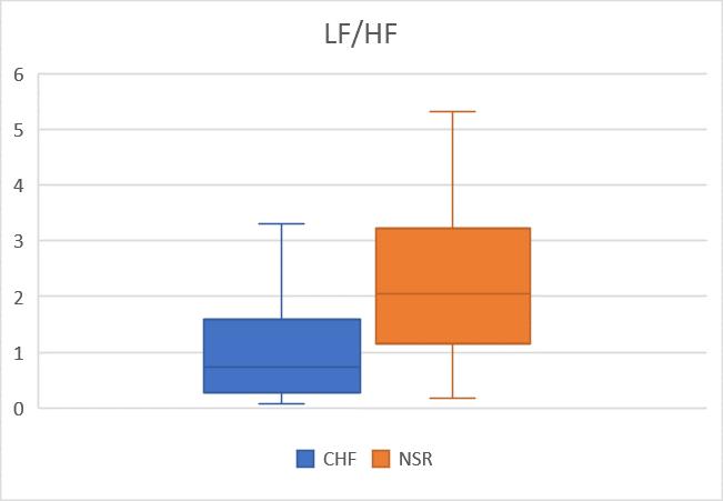
More interesting is applying such entropybased Frequencies-domain analyzing for AF patients. There is an opinion that FD analysis is unsuitable for AF and this is true in case of LF/HF, because no significant difference before and duringAF due to high variation of this ratio (see Fig.2).Theentropy-based ratio eLF/eHF is suitable much better for this case, – the eLF/eHF is significantly higher during AFthanbeforeAF(p<0.001).
Series «Medicine». Issue 45 7
Fig. 1. Box & Whiskers plots of LF/HF and eLF/eHF for NSR and CHF groups
Fig. 2. Box & Whiskers plots of LF/HF and eLF/eHF for AF patents (before and during AF episodes)
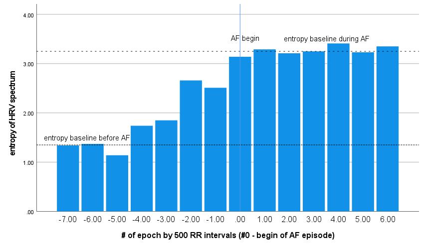
The Fig.3. shows typical pattern of entropy of HRV power spectrum evolution before and during atrial fibrillation episode: each epoch on the Fig.3. consists of short RRs records (N= 500); epoch with # ‘0’ is the beginning of AF according to MIT-BIH reference rhythm annotations. Entropy of power spectrum does not have significant difference from mean record value under Normal rhythm intervals except 4–5 epochs before and after AF episodes: entropy begin significantly growth for about 20 minutes (or 4–5 epoch by N=500 RRs) before AF and excides new maximal baseline during AF. The new baseline level is significantly different from previous one – before AF (p<0.001).
Therefore, proposed generalized form for Robust Entropy Estimator EnRE for HRV power spectrum shows significant differences (p<0.001) of total spectrum entropy and entropy-based sympatho-vagal balance for NSR and CHF groups in short records (N = 500), and presents additional advantages provided by EnRE in case of patients with atrialfibrillation.
CONCLUSIONS
Proposed in the article is generalized form for Robust Entropy Estimator EnRE for
Frequencies-domain, which allows, for time series of a limited length (standard 5-min. records), tofind entropyvalue of HRV power spectrum (total spectrum, low- and highfrequencies bands). Parameters in generalized form for EnRE have been derived from the followingcriteria:
1/ accurate approximation for known distributions of a random value in ranges that represent models of RRs for heart rate variability;
2/ independence of EnRE from N for initialtimeseriesandforseriesaftersorting;
3/ independence of EnRE from additive changesofmean.
Using the proposed EnRE for MIT-BIH database of HRV records, we show for standard 5 min. HRV records the usage of EnRE of HRV power spectrum and entropybased sympatho-vagal balance of Normal Sinus Rhythm (NSR) and Congestive Heart Failure (CHF) cases. It is demonstrated, that, entropy-based Frequencies-domain analyzing is applicable for case of Atrial Fibrillation (AF) even during AF episodes. We showed the significant difference (p<0.001) before and during AF for entropy of total spectrum, aswellasforsympatho-vagalbalanceinform ofeLF/eHF.
The Journal of V.N. Karazin Kharkiv National University. 2022 8
Fig. 3. Typical pattern of entropy of HRV power spectrum before and during atrial fibrillation episode (MIT-BIH AF Database [16]).
REFERENCES
1. Task force of the European society of cardiology and the North American society of pacing and electrophysiology. Heart rate variability – standards of measurement, physiological interpretation, and clinical use. Circulation. 1996;93(5):1043–1065. DOI: https://www.ahajournals.org/doi/10.1161/01.CIR.93.5.1043
2. Iabluchanskyi M, Martynenko A, Bydreiko N, Yabluchansky A. Heart Rate Variability for medical scientists and doctors. Kharkiv: V. N. Karazin Univer. Press; 2022. 131 p. DOI: 10.13140/RG.2.2.32435.91685/1
3. Pincus SM. Approximate entropy as a measure of system complexity. Proc. Natl. Acad. Sci. USA, 1991;88: 2297
2301. DOI: https://doi.org/10.1073/pnas.88.6.2297
4. Richman JS, Moorman JR. Physiological time-series analysis using approximate entropy and sample entropy. Am. J. Physiol. Heart Circ. Physiol. 2000; 278: H2039–H2049. DOI: 10.1152/ajpheart.2000.278.6.H2039
5. Humeau-Heurtier A. The multiscale entropy algorithm and its variants: A review. Entropy. 2015;17: 3110
3123. DOI: https://doi.org/10.3390/e17053110
6. Costa M, Goldberger AL, Peng CK. Multiscale entropy analysis of biological signals. Phys. Rev. E. 2005 71:021906. DOI: 10.1103/PhysRevE.71.021906
7. Costa M, Goldberger AL, Peng CK. Multiscale entropy analysis of complex physiologic time series. Phys. Rev. Lett. 2002;89:068102. DOI: 10.1103/PhysRevLett.89.068102
8. Gao ZK, Fang PC, Ding MS, Jin ND. Multivariate weighted complex network analysis for characterizing nonlinear dynamic behavior in two-phase flow. Exp. Therm. Fluid Sci. 2015;60:157–164. DOI: 10.1016/j.expthermflusci.2014.09.008
9. Labate D. et al. Entropic measures of eeg complexity in alzheimer’s disease through a multivariate multiscale approach IEEE Sens. J. 2013;13:3284–3292.
10.Azami H, Escudero J. Refined composite multivariate generalized multiscale fuzzy entropy: A tool for complexity analysis of multichannel signals. Physica A. 2017;465: 261–276.
11.Zhao LN, Wei SS, Tong H, Liu CY. Multivariable fuzzy measure entropy analysis for heart rate variability and heart sound amplitude variability. Entropy. 2016; 18: 430, DOI:10.3390/e18120430
12.Li P. et al. Multiscale multivariate fuzzy entropy analysis. Acta Phys. Sin. 2013;62:120512.
13.Ahmed MU, Mandic DP. Multivariate multiscale entropy analysis. IEEE Signal Proc. Lett. 2012;19: 91
94.
14.Madhavi CHR. Estimation of Spectral Entropy of HRV Data and its Application to Depression and Thyroid Subjects to Predict Cardiac Risk. Biomed Pharmacol J. 2018; 11 (3). DOI : https://dx.doi.org/10.13005/bpj/1532
15.Martynenko A, Raimondi G, Budreiko N. Robust Entropy Estimator for Heart Rate Variability. Klin. Inform. Telemed. 2019;14(15):67-73. DOI: 10.31071/kit2019.15.06
16.Goldberger A, Amaral L, Glass L, Hausdorff J, Ivanov PC, Mark R, Stanley HE. PhysioBank, PhysioToolkit, and PhysioNet: Components of a new research resource for complex physiologic signals. Circulation [Online]. 2000;101(23):e215
e220.
17.Moody GB, Mark RG. A new method for detecting atrial fibrillation using R-R intervals. Computers in Cardiology. 1983;10:227-230.
18.Tsai CH, Ma HP, Lin YT, et al. Usefulness of heart rhythm complexity in heart failure detection and diagnosis. Sci Rep. 2020;10:14916. DOI: https://doi.org/10.1038/s41598-020-71909-8
19.Liu G, Wang L, Wang Q, Zhou G, Wang Y, et al. A New Approach to Detect Congestive Heart Failure Using Short-Term Heart Rate Variability Measures. 2014;PLoS ONE 9(4): e93399. DOI:10.1371/journal.pone.0093399
ЕНТРОПІЯ ЧАСТОТНОГО ДОМЕНУ ВАРІАБЕЛЬНОСТІ СЕРЦЕВОГО РИТМУ Мартиненко О. В.A, B,C,D,E,F, Раймонді Д.A,Е,F, Барсі Л.E,F, Малярова Л. В.Е,F
A
концепція та дизайн дослідження; B
збір даних; C
аналіз та інтерпретація даних; D – написання статті; E – редагування статті; F – остаточне затвердження статті Вступ. Варіабельність серцевого ритму (ВСР) базується на вимірюванні (часових) інтервалів між R-піками (RR-інтервалів) електрокардіограми (ЕКГ) і побудови на їх основі ритмограми з подальшим її аналізом різними математичними методами, які класифікуються як Часова область (TD), частотна область (FD) і нелінійна [1, 2]. Існує ряд популярних нелінійних методів, які використовуються в
Series «Medicine». Issue 45 9
–
–
–
–
–
–
–
The Journal of V.N. Karazin Kharkiv National University. 2022
аналізі ВСР, наприклад вимірювання на основі ентропії, які в основному застосовуються для TD. Спектральна ентропія (SE) використовується для частотної області: вона визначається як ентропія Шеннона спектральної щільності потужності (PSD) даних. Важливою характеристикою частотних досліджень є симпато-вагальний баланс, який раніше не враховувався в аналізі на основі ентропії. Причиною цього було те, що якісний ентропійний аналіз обмежено кількістю існуючих даних ВСР, які зменшуються у FD, а також у частинах загального спектру.
Мета. Метою цієї статті є надання надійної формули для точного обчислення ентропії для частотної області стандартних 5 хвилин запису ВСР та показати переваги такого підходу для аналізу симпато-вагального балансу у здорових суб’єктів (NSR), пацієнтів із застійною серцевою недостатністю (CHF) та фібриляцією передсердь (AF).
Матеріали і методи. Ми використовували довгострокові записи ВСР бази даних MIT-BIH для нормального синусового ритму (NSR), застійної серцевої недостатності (CHF) і фібриляції передсердь (AF).
Була запропонована узагальнена форма надійного оцінювача ентропії (EnRE) для частотної області стандартних 5 хв. записів ВСР і показані ключові ознаки EnRE.
Різниця між середніми значеннями двох незалежних вибірок (NSR і CHF, до і після AF) була визначена t-тестом для незалежних вибірок; дискримінантний аналіз і статистичні розрахунки виконано за допомогою статистичного пакету IBM SPSS 27 Результати. Ми обчислювали ентропію для всього спектрального інтервалу ВСР, а саме 0–0,4 Гц, і порівнювали з існуючими результатами для спектральної ентропії: якісно ми отримуємо таке ж число розподілу, як у [14], і значущу різницю (p < 0,001) між середніми значеннями ентропії для NSR та пацієнтів із CHF або AF
Визначаємо низькочастотні (LF) складові спектра потужності в діапазоні 0,04–0,15 Гц і високочастотні (HF) компоненти спектра потужності в діапазоні 0,15–0,4 Гц [1]. Симпато-вагальний баланс – це просте співвідношення LF/HF [1]. Ми обчислюємо ентропію компонентів спектру потужності LF як eLF, ентропію компонентів спектру потужності HF як eHF і симпато-вагальний баланс на основі ентропії як співвідношення eLF/eHF.
Різниця між групами NSR і CHF є значною в обох випадках LF/HF і eLF/eHF з p < 0,001, але у випадку eLF/eHF це дещо краще з t = -4,8 порівняно з LF/HF, де t = -4,4. Дискримінантний аналіз показує загальну точність класифікації для eLF/eHF у 79,3 % (χ2 = 19,4, p < 0,001) і для LF/HF у 72,4 % (χ2 = 16,6, p < 0,001).
Ми застосували частотний аналіз на основі ентропії для пацієнтів з AF і показали, що співвідношення eLF/eHF значно вище під час AF, ніж до AF (p < 0,001). Це протилежно звичайному НЧ/ВЧ, де немає статистичної значущості різниці через велику варіацію цього співвідношення.
Висновки. У статті запропоновано узагальнену форму надійного оцінювача ентропії EnRE задля частотного домену ВСР, що дозволяє для часових рядів обмеженої довжини (стандартні 5-хвилинні записи) знаходити значення ентропії спектра потужності ВСР (загальний спектр, низька і висока смуги частот).
Використовуючи запропоновану формулу EnRE для MIT-BIH бази даних записів ВСР, ми показали для стандартних 5 хв. записів ВСР використання EnRE спектра потужності ВСР та симпатовагального балансу на основі ентропії у випадках нормального синусового ритму (NSR) і застійної серцевої недостатності (CHF). Продемонстровано, що ентропійний аналіз у частотній області застосований для випадків фібриляції передсердь (AF) навіть під час епізодів AF. Ми показали достовірну різницю (p < 0,001) до та під час AF для ентропії загального спектру, а також для симпатовагального балансу у формі eLF/eHF.
КЛЮЧОВІ СЛОВА: варіабельність серцевого ритму, ентропія, частотний домен, застійної серцевої недостатності, фібриляції передсердь
ИНФОРМАЦІЯ ПРО АВТОРІВ Мартиненко Олександр Віталійович, д.фіз-мат.н., професор, професор кафедри гігієни та соціальної медицини Харківського національного університету імені В. Н. Каразіна, майдан Свободи, 6, Харків, Україна, 61022, e-mail: Alexander.v.martynenko@karazin.ua, ORCID ID: https://orcid.org/0000-0002-0609-2220 Ж. Раймонди, д.мед.н., проф., Римський університет Ла Сапієнца (Італія), Пьяццале Альдо Моро, 5, Рим, Італія, 00185, e-mail: gianfrancoraimondi@uniroma1.it
Л. Барсі, доктор філософії, Рим, Італія, 00185; e-mail: barsiluca1@gmail com Малярова Людмила Володимирівна, асистент кафедри гігієни та соціальної медицини Харківського національного університету імені В. Н. Каразіна, майдан Свободи, 6, Харків, Україна, 61022, e-mail: l v maliarova@karazin ua, https://orcid.org/0000-0002-7902-7016
10
Для цитування:
Мартиненко ОВ, Раймонді ДA, Барсі ЛE, Малярова ЛВ. ЕНТРОПІЯ ЧАСТОТНОГО ДОМЕНУ
ВАРІАБЕЛЬНОСТІ СЕРЦЕВОГО РИТМУ. Вісник Харківського національного університету імені В. Н. Каразіна. Серія «Медицина». 2022;45:4–11 DOI: 10.26565/2313-6693-2022-45-01
Conflicts of interest: author has no conflict of interest to declare. Конфлікт інтересів: відсутній.
Отримано: 10 10.2022року
Прийнято до друку: 20.11.2022 року
Received: 10 10.2022
Accepted: 11 20.2022
Series «Medicine». Issue 45 11
УДК 616-006.04; 577.212.3; 57.087.1
DOI: 10.26565/2313-6693-2022-45-02
Мартиненко О. В.A,C,D,E,F, Пастор К. Д.A,B,Е,F, Фрід С. А.B,F , Гіл Д. Р.B,F , Малярова Л. В.Е,F
A
концепція та дизайн дослідження; B – збір даних; C – аналіз та інтерпретація даних; D
написання статті; E – редагування статті; F – остаточне затвердження статті
Вступ. Дезоксирибонуклеїнова кислота (ДНК) не є випадковою послідовністю чотирьох комбінацій нуклеотидів: комплексні огляди літератури переконливо показують довго- та короткодіапазонні кореляції в ДНК, періодичні властивості та кореляційну структуру послідовностей. Методи теорії інформації, зокрема інформаціїна ентропія, мають на увазі кількісну оцінку обсягу інформації, що міститься в послідовностях. Зв’язок між ентропією та виживанням пацієнтів широко поширений у деяких галузях медицини та медичних дослідженнях, таких як: кардіологія, неврологія, хірургія, травма. Таким чином, існує необхідність реалізації переваг методів теорії інформації для дослідження взаємозв'язку між смертністю певної категорії пацієнтів та ентропією їх послідовностей ДНК.
Мета. Надати надійну формулу для точного розрахунку ентропії для коротких послідовностей ДНК і показати, як використовувати запропонований аналіз ентропії для вивчення смертності хворих
на лейкемію.
Матеріали і методи. Використовувалась база даних пацієнта з лейкемією Барселонського університету (UB) з 117 знеособленими записами, які складаються з наступного: дата діагнозу пацієнта, дата смерті пацієнта, діагнози лейкемії, послідовність ДНК пацієнта. Середній час смерті пацієнта після встановлення діагнозів: 99 ± 77 місяців. Формальними характеристиками послідовностей ДНК в БД UB хворих на лейкемію є: середня кількість ДНК основ N = 496 ± 69; min (N) = 297 основ; max (N) = 745 основ.
Була запропонована узагальнена форма оцінювача ентропії (EnRE) для коротких послідовностей
ДНК та продемонстровані ключові ознаки EnRE.
Аналіз виживання був проведений за допомогою статистичного пакета IBM SPSS Statistics 27 методами Каплана-Мейєра та регресії Кокса. Результати. Точність запропонованої формули для розрахунку ентропії була перевірена для різних відрізків часових рядів і різних типів випадкових розподілів з відомими теоретичними значеннями ентропії. Показано, що у всіх випадках для N = 500 відносна похибка при розрахунку точного значення ентропії не перевищує 1 %, при цьому величина кореляції не гірше 0,995.
Код алфавіту початкової послідовності ДНК був перетворений в числовий код основ, з використанням правила оптимізації, щоб отримати тільки одне мінімальне і симетричне числове декодування близько нуля, що дає мінімум для стандартного відхилення EnRE і коефіцієнт варіації.
Ентропія EnRE була розрахована для хворих на лейкемію після оптимального цілочисельного декодування в двох спостереженнях: 2 групи, розділені медіаною EnRE = 1,47, та 2 групи, що належать до 1-го (EnRE ≤ 1.448) та 4-го квартилів (EnRE ≥ 1.490). Результат аналізу виживання Каплана-Мейєра та моделювання виживання Кокс-регресій статистично значущі з p < 0,05 для груп поділених медіаною і з p < 0,005 для груп, що уособлюють 1-й та 4-й квартілі. Небезпека смерті для пацієнта з EnRE нижче медіани в 1,556 рази більше, ніж у пацієнта з EnRE понад медіаною та небезпека смерті для пацієнта 1-го ентропійного квартиля (найнижчий EnRE) в 2,143 рази більше, ніж у пацієнта 4-го ентропійного квартиля (найвищий EnRE).
Висновки. Перехід від розширених (медіальних) до менших (квартільних) груп пацієнтів з більшою різницею у EnRE підтвердив унікальне значення ентропії послідовностей ДНК для визначення смертності пацієнтів з лейкемією. Це значення статистично доведено підвищенням небезпеки для хворих на лейкемію з меншою ентропією послідовностей ДНК: більша різниця EnRE означає збільшення риску смерті та скорочення тривалості життя після діагнозу в групах пацієнтів з меншою ентропією послідовностей ДНК.
КЛЮЧОВІ СЛОВА: ентропія, послідовності ДНК, смертність, лейкемія
© Мартиненко О. В., Пастор К. Д., Фрід С. А., Гіл Д Р., Малярова Л. В., 2022
The Journal of V.N. Karazin Kharkiv National University. 2022 12
ЕНТРОПІЯ ПОСЛІДОВНОСТЕЙ ДНК І СМЕРТНІСТЬ ПАЦІЄНТІВ З ЛЕЙКЕМІЄЮ
–
–
ІНФОРМАЦІЯ ПРО АВТОРІВ
Мартиненко Олександр Віталійович, д.фіз-мат.н., професор, професор кафедри гігієни та соціальної медицини
Харківського національного університету імені В. Н. Каразіна, майдан Свободи, 6, Харків, Україна, 61022, e-mail: Alexander.v.martynenko@karazin.ua, ORCID ID: https://orcid.org/0000-0002-0609-2220
Пастор Ксав’є Дюран, доктор медицини, професор, керівник відділення медичної інформатики, клініка Університету Барселони, вул. Віллароель 170, Барселона, Іспанія, 08036. e-mail: xpastor@clinic cat, ORCID: 00000001-8267-7151
Фрід Сантьяго Андрес, доцент кафедри фундаментальної клініки, медичний факультет, Університет Барселони, вул. Казанови 143, Барселона, Іспанія, 08036, e-mail:frid@clinic cat, ORCID: 0000-0001-8400-5770
Гіл Рожас Джессика, дата менеджер, відділення медичної інформатики, клініка Університету Барселони, вул. Віллароель 170, Барселона, Іспанія, 08036, e-mail:jegil@clinic cat ORCID: 0000-0002-7690-7288
Малярова Людмила Володимирівна, асистент кафедри гігієни та соціальної медицини Харківського
національного університету імені В. Н. Каразіна, майдан Свободи, 6, Харків, Україна, 61022, e-mail: l v maliarova@karazin ua, https://orcid.org/0000-0002-7902-7016
Для цитування:
Мартиненко ОВ, Пастор КД, Фрід СА, Гіл ДР, Малярова ЛВ. ЕНТРОПІЯ ПОСЛІДОВНОСТЕЙ ДНК І СМЕРТНІСТЬ ПАЦІЄНТІВ З ЛЕЙКЕМІЄЮ. Вісник Харківського національного університету імені
В. Н. Каразіна. Серія «Медицина». 2022;45:12–23. DOI: 10.26565/2313-6693-2022-45-02
ВСТУП
Дезоксирибонуклеїнова кислота (ДНК)
не є випадковою послідовністю чотирьох
комбінацій нуклеотидів (А – аденін, С –цитозин,Г–гуанін,Т–тимін):комплексні
огляди літератури [1,2] переконливо
показують довго- та короткодіапазонні
кореляціївДНК,періодичнівластивостіта
кореляційну структуру послідовностей.
Методи теорії інформації мають на увазі
кількісну оцінку обсягу інформації, що
міститься в послідовностях. Інформаційна
ентропія Клода Шеннона була одним з перших інформаційних заходів для досліджуванихпослідовностейДНК[3].В
даний час реалізації ентропії Шеннона продовжуютьзалишатисядужеуспішними дляаналізувірусноїРНК, такихякSARSCOV-2 [4]. Ґрунтовний огляд різних
підходів до ентропії «для виявлення
формальних звэязків між генетичним різноманіттямтапотокомінформації»був наведений в [5] і досконалий огляд [6]
демонструє сучасний стан реалізацій теорії інформації для аналізу «експресії генів та транскриптоміки, порівняння послідовностей без вирівнювання, секвенування та виправлення помилок, картування зв’язків між геномами та генами, метаболічних мереж та метаболоміка, а також аналіз послідовності білків, структуритавзаємодії».
З іншого боку, зв’язок між ентропією та виживанням пацієнтів широко поширений
у деяких галузях медицини та медичних дослідженнях.Надамодеякіприклади:
1. Кардіологія: Використовуеться
апроксімаційна ентропія на основі варіабельностісерцевогоритму(ВСР)для прогнозування раптової серцевої смерті, оцінки впливу специфічних фармакологічних засобів на ВСР [7]; застосовується ентропія на основі електрокардіограм Холтера (ЕКГ) і частоти серцевих скорочень (ЧСС) нормальної серцевої динаміки і тих, що мають різний ступінь гострих серцевих патологій [8]; пацієнти після інфаркту міокарда, які перенесли пізній гадоліній покращений магнітний резонанссерця(МР)зпохідноюентропією тканин МР-візуалізації. За пацієнтами спостерігалася відповідна імплантована кардіовертерно-дефібриляторна терапія та смертність[9].
2. Неврологія: досліджено зв'язок ентропії серцевого ритму (ЕСР) зі смертністю після внутрішньомозкової кровотечі[10];
3. Хірургія (загальна анестезія): Моніторинг ентропії передбачає використання електроенцефалографії (ЕЕГ) для оцінки глибини загальної анестезіїухірургічнихпацієнтів[11];
4. Травма: Для категоризації травми за допомогою ентропії необхідно розглянути основну ентропію хворобливості осіб, до якої додається ентропія травми, яка потім може призвести до смерті [12]; показана цілочисельна багатомасштабна ентропія (MSE) частота серцевих скорочень (HR), як показник складності, що прогнозує смерть у довго тривалому термені. MSE частоти серцевих скорочень протягом декількох годин після госпіталізації прогнозує смерть, що настає через кілька
Series «Medicine». Issue 45 13
The Journal of V.N. Karazin Kharkiv National University. 2022
днів [13]; У цьому дослідженні вимірювали ентропію Шеннона і Цалліс задля температурних сигналів у когорті критично хворих пацієнтів. Зменшені вейвлети ентропії температурних сигналів Шеннона і Цалліса може доповнюватися послідовною оцінкою органної недостатностівпрогнозуваннісмертності[14].
Таким чином, випливає, що існує необхідність реалізації переваг методів теорії інформації для дослідження взаємозв'язку
між смертністю певної категорії пацієнтів таентропієюїхпослідовностейДНК.Мета даної роботи – надати надійну формулу для точного розрахунку ентропії для коротких послідовностей ДНК і показати, як використовувати існуючий аналіз ентропії для вивчення смертності хворих налейкемію.
МАТЕРІАЛИ ТА МЕТОДИ
Ми використовували базу даних пацієнтів з лейкемією Барселонського
університету (UB) з 117 знеособленими записами, які складаються з наступного: дата діагнозу пацієнта, дата смерті
пацієнта, діагнози лейкемії, послідовність
ДНК пацієнта. Середній час смерті
пацієнта після встановлення діагнозів: 99 ± 77 місяців. Формальними характеристи-
камипослідовностейДНКвБДUBхворих
на лейкемію є: середня кількість ДНК основ N =496 ± 69; min (N)= 297 основ; max(N)= 745основ.
Статистично ДНК здебільшого близька до рівномірного розподілу, але має абсолютно різні неоднорідні частотні
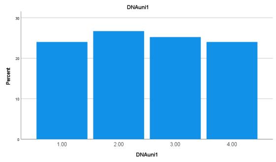
патерни, наприклад, базові фрікени мітохондріону людини (16 569 основ) становлятьА– 31 %,С– 31 %,Г– 13 %,Т – 25 % або екзони глобіну плода людини (882 основи) становлять А – 24 %, С –25 %,Г– 28 %,Т– 22%[15]. Мипоказали порівняння реальної послідовності ДНК і змодельованої рівномірним розподілом на рисунках1.a.і1.b.:
Рис. 1.a. Реальна послідовність ДНК пацієнта, N = 745 основ (UB DB пацієнтів з лейкемією).
Fig. 1а. Real patient DNA sequence, N = 745 bases (UB leukemia patient DB)

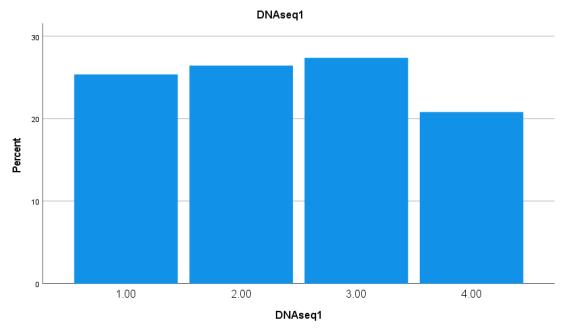
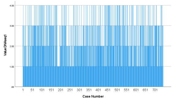
Рис. 1.b. Змодельована послідовність 4 елементів за рівномірним розподілом, N = 745
Fig. 2.b. Simulated 4 elements sequence by Uniform distribution, N = 745
Ми розрахували ентропію обмежених часовихрядівзаоригінальноюформулою, запропонованою Клодом Шенноном в
1948 році [16] і вона тут називається Емпірична ентропія (EnEmp) через обмеженнячасовихрядів:
14
����������= ∑ ��(����)����(��(����)) �� ��=1 1
Проблемою використання формули (1)
напрактиціє:
нечутливість до зміни положень
нуклеотидів в послідовності ДНК. Є
чутливістьтількидозмінибази;
низька точність для невеликої
кількості точок в ряді (наприклад, коли N < 1000);
повільна сходимість до точного значення зі збільшенням довжини послідовності
Показана в табл.2 залежність точності обчислення ентропії за формулою (1) від довжини ряду для окремих видів розподілу випадкової величини. Точні значення ентропії для пов’язаних розподілівнаведенівTab.1.
Таблиця 1 Table 1
Різні розподіли ймовірностей і відповідна ентропія [17] Various probability distributions and correspondent Entropy
Розподіл Функція ймовірності Ентропія (En, нат)
Нормальний розподіл ��(��)
Залежність від тривалості ряду точністі оцінки ентропії та кореляція за змодельованими параметрами розподілу для різних розподілів ймовірностей Dependence from the length of time series of Entropy estimation accuracy and Correlation along simulated distribution parameters for various probability distributions
Розподіл Довжина зразка Емпірична ентропія (EnImp) Робастний оцінювач ентропії (EnRE)
Точність (відносна похибка, %)
Кореляція Точність (відносна похибка, %)
Кореляція
Series «Medicine». Issue 45 15
= 1 ��√2��������( (�� ��)2 2��2 ) ���� =����(√2����2)
розподіл ��(��)= �� �� �� ���� =����(�� ��) Експоненціальний розподіл ��(��)=��������( ����) ���� =1 ����(��) Логарифмічнонормальний розподіл ��(��)= 1 ����√2��������( (����(��) ��)2 2��2 ) ���� =��+����(√2����2) Розподіл Парето ��(��)= ������ �� ����+1 ���� =����(���� �� )+1+1 �� Таблиця 2 Table 2
Рівномірний
Рівномірний розподіл (a = 0; b = 4) N = 100 6.92 0.978 4.71 0.991 N = 500 4.11 0.988 0.57 0.997 N = 1000 3.83 0.999 0.11 0.998 Нормальний розподіл (M = 1000; �� =100 200) N = 100 7.74 0.994 1.95 0.995 N = 500 1.83 0.997 0.35 0.998 N = 1000 0.91 0.999 0.16 0.999 Експоненціальний розподіл (�� =00001 0.0011) N = 100 46.24 0.452 0.77 0.993 N = 500 28.38 0.903 0.25 0.997 N = 1000 19.31 0.950 0.06 0.999 Лог-нормальний розподіл (�� =7; �� = 0.002 0.012) N = 100 3.69 0.980 3.38 0.986 N = 500 1.17 0.997 0.49 0.997 N = 1000 0.80 0.999 0.22 0.999 Розподіл Парето (�� = 2; s = 1000 – 2000) N = 100 32.68 0.589 1.01 0.997 N = 500 17.78 0.867 0.35 0.998 N = 1000 14.75 0.946 0.12 0.999
Випадок рівномірного розподілу (показаний на рис. 1.a, b) отримує особливу увагу, але інші розподіли також враховуються як необхідні приклади використання числових формул для аналізу обмеженого ряду, оскільки не завжди вдається точно зіставити спостережувану послідовність ДНК з деяким фіксованим випадковим розподілом. Можна визнати неможливість застосування формули (1) до короткого часовогоряду N <1000. Тому,здавалосяб, необхідна розробка формули для точного вимірювання ентропії для невеликої довжинипослідовностейДНК.
На початку минулого століття італійський професор статистики Коррадо Джині запропонував спосіб вимірювання нерівності між значеннями частотного розподілу(коефіцієнтДжині)[18]:
G = 1 2��2��∑ ∑ (|���� ����|) �� ��=1 �� ��=1 , (2)
де M – середнє значення x. Коефіцієнт Джині виявився дуже популярним в економіці та соціології, і є спроби застосувати його і до інших областей, включаючи аналіз ВСР [19]. Коефіцієнт Джині є екземпляром узагальненого індексу нерівності [20], а його альтернатива, як міра відхилення від балансу – узагальнений індекс ентропії –виводиться з теорії інформації як міра надмірності в даних [21]. Відомі обмеження при використанні коефіцієнта
Джині для аналізу даних: залежність від адитивної зміни середнього; малий відбір істотно зменшує величину коефіцієнта і т.і.
Тому після аналізу відомих визначень
мір відхилення від рівноваги і ступеня порядку була запропонована узагальнена форма оцінювача ентропії (EnRE) для часових рядів в [22] і наступним запропонованадляпослідовностейДНК:
EnRE = ����( �� �� ��⁄2 ∑ ∑ ((|���� ����||���� ����|) 1⁄�� (������) ��⁄2 ) �� ��=1 �� ��=1 ),
(3)
де MD медіана послідовності для чисельно закодованих основ B; ������відстань між ���� і����; A, l, m, k – оціночні коефіцієнти. Умови для пошуку коефіцієнтів A, l, m,k наступні:
1) точне наближення для відомих розподіліввипадковоївеличини;
2) незалежність EnRE від N для початковихчасовихрядівідлясерійпісля сортування;
3) незалежність EnRE від адитивної змінисередньогозначення.
Після чисельних досліджень, остаточні результати яких представлені в табл. 2, були знайдені наступні значення коефіцієнтів: l = 3, m = 1, k =2.Виділимо деякі ключові ознаки запропонованої узагальненоїформи EnRE ікоефіцієнтів:
1) форма запису (3) і знайдені коефіцієнти l, m, k забезпечують незалежність від адитивної зміни середніх рядів і від величини виділення N для базових рядів і длярядівпіслясортування;
2) значення EnRE чутливе до структурних змін рядів, таких як, наприклад, сортування, яке збільшує ступінь порядку послідовно, зменшуючи EnRE;
3) значення EnRE чутливе до зміни положення нуклеотидів у послідовності ДНК;
4) коефіцієнт переналагодження A самостійно може знадобитися для знаходження кращого значення EnRE в іншому діапазоні зміни параметрів різних випадкових розподілів, що завжди можна зробити за допомогою методу найменших квадратів. Аналіз виживання був проведений за допомогою статистичного пакета IBM SPSS27.
РЕЗУЛЬТАТИ ТА ОБГОВОРЕННЯ Точність. Перш за все, перевіримо точність запропонованої формули (3) для розрахунку ентропії: Taбл 2 надає значення EnRE для різних відрізків часових рядів і різних типів випадкових
розподілів, а для кожного з цих результатів наводяться значення похибок при обчисленні ентропії порівняно з точними значеннями при зміні параметрів розподілу.Зауважимо,щоувсіхвипадках для N = 500 відносна похибка при розрахунку точного значення ентропії не перевищує 1%, при цьому величина кореляції не гірше 0,995; при рівномірному і нормальному розподілі відносна похибка для довжини часових рядів N = 500÷1000 менше 0,6%, а кореляціястановитьблизько0,998.
16
The Journal of V.N. Karazin Kharkiv National University. 2022
Оптимальне кодування послідовності
ДНК. Код алфавіту початкової
послідовності ДНК повинен бути
перетворений в числовий код основ, але
така перестановка є довільною. Тому ми
використали принцип максимальної
ентропії, щоб уникнути такої довільності.
Одночасно оптимальне чисельне
перекодування має давати мінімальне
значення для стандартного відхилення та
коефіцієнт варіації для розрахункової
ентропії послідовностей ДНК, оскільки у
нас однорідна група пацієнтів. Крім того, це правило оптимізації зменшило самовплив дисперсії числового
декодування. У табл 3 ми наведемо різні числові розшифровки послідовностей ДНК та їх значення ентропії, стандартні відхиленнятакоефіцієнтиваріації.
Таблиця 3 Table 3
Числове кодування послідовностей ДНК і відповідне EnRE, стандартне відхилення і коефіцієнт варіації EnRE
Numerical decoding of DNA sequences and correspondent EnRE, standard deviation and coefficient of variation of EnRE
Цілочисельний
Стандартне відхилення
(трансляційна симетрії за модулем
Можна стверджувати, що за власти-
востями EnRE будь-яка симетрична зміна цілочисельного декодування дає однакове
значення EnRE (див. перші два рядки табл.3); тільки одне мінімальне і
симетричне числове декодування близько
нуля дає максимум ентропії і мінімум для стандартного відхилення EnRE і
коефіцієнт варіації (виділений жирним шрифтомвтабл.3).Такимчином,числове декодування довільності було прибрано
тільки однією можливою цілочисельною комбінацією.
Смертність пацієнтів з лейкемією.
Ентропія EnRE була розрахована для всіх
хворих на лейкемію після оптимального
цілочисельного декодування, створеного
групоюсиметріїтрансляції(A = -1, C = -2, G=1,T=2).
А. Медіанні групи. Всі пацієнти були розділені медіаною EnRE = 1,47 на 2 групи:
1. Група‘1’,58пацієнтів, EnRE нижче медіани;
2. Група ‘2’, 59 пацієнтів, EnRE вище медіани.
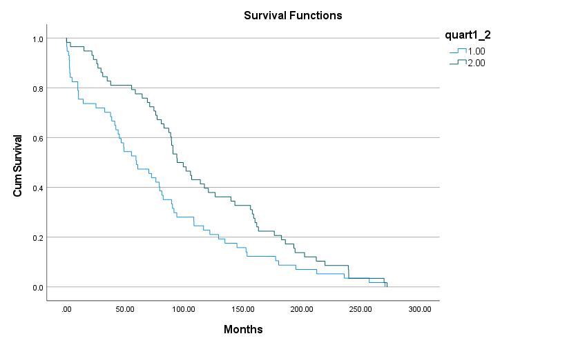
Результат аналізу виживання КапланаМейєра наведено на рис.2. Всі загальні порівняння показують статистичну значимість: p = 0,015 для Log Rank (Мантель-Кокс); p = 0,002 для Бреслоу (узагальнений Вілкоксон); p = 0,003 для Tarone-Ware. Середні та медіани для хворих на лейкемію на час виживання наведенівтаблиці4.
Series «Medicine». Issue 45 17
код ДНК Середня ентропія EnRE
EnRE Коефіцієнт варіації
A=1,C=2,G=3,T=4 or A=4,C=3,G=2,T=1 1.205 0.030 0.025 A=2,C=1,G=3,T=4 or A=3,C=4,G=1,T=2 1.254 0.036 0.029 A=3,C=4,G=2,T=1 1.241 0.034 0.027 A=1,C=4,G=3,T=2 1.235 0.043 0.035 A=1,C=3,G=4,T=2 1.221 0.040 0.033 A=1,C=3,G=2,T=4 1.211 0.033 0.027 A=1,C=2,G=4,T=3 1.223 0.039 0.032 A=-2, C=-1, G=1, T=2 (дзеркальна симетрія за модулем ) 1.430 0.023 0.016 A=-1, C=-2, G=1, T=2
1.470 0.022 0.015
(CV)
)
Рис 2. Криві виживання Каплана-Мейєра для медіанних груп Fig. 2. Kaplan-Meier survival plot for median groups
Таблиця 4 Table 4 Середні та медіани для часу виживання (медіанні групи)
a. Оцінка обмежена найбільшим часом виживання, якщо вона піддається цензурі
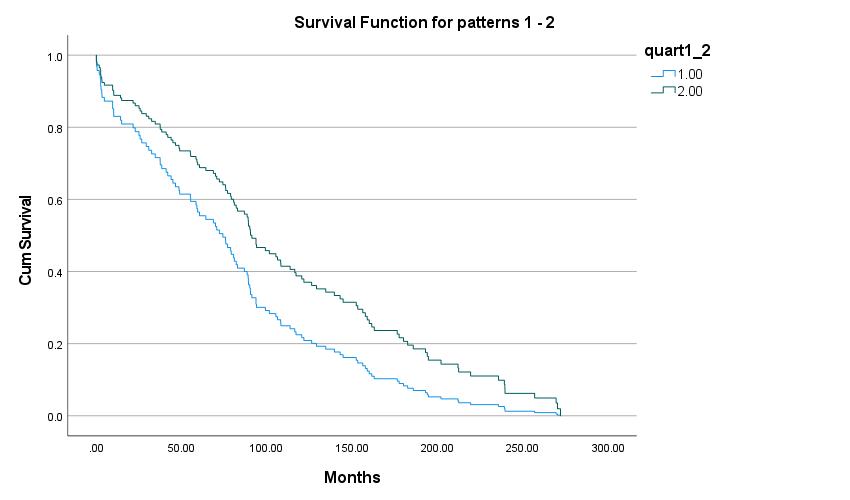
Результат моделювання виживання
Кокс-регресій наведено на рис.3. Всі загальні порівняння показують статистичну значимість: p = 0,015 для Омнібусного тесту модельних коефіцієнтів; p =0,016 для змінних у рівнянні з ‘-2 Log
Likelihood’=862.2.
Значення Exp(B) для модельної змінної показує,щонебезпекасмертідляпацієнта з EnRE нижчемедіанив1,556разибільше, ніжупацієнтаз EnRE понадмедіаною.
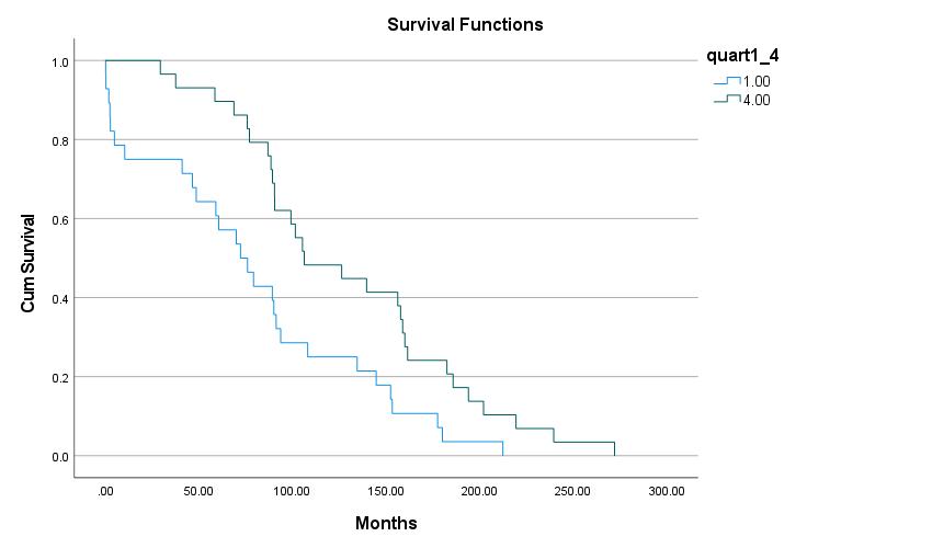
Б. 1-й і 4-й квартілі. Сформовано 2 групи хворих відповідно до їх приналежностідо1-гота4-гоквартилів:
3. Група ‘1’, 29 пацієнтів, EnRE ≤ 1.448,тобтонижче1-гоквартиля;
4. Група ‘4’, 29 пацієнтів, EnRE ≥ 1.490,тобтовище4-гоквартиля. Результат аналізу виживання КапланаМайєра наведено на рис.4. Всі загальні порівняння показують статистичну значимість: p = 0,005 для Log Rank (Мантель-Кокс); p = 0,003 для Бреслоу (узагальнений Вілкоксон); p = 0,003 для Tarone-Ware. Середні та медіани для виживанняхворихналейкеміюнаведенів таблиці 5.
The Journal of V.N. Karazin Kharkiv National University. 2022 18
quart1_2 Середні
Медіани Estimate Std. Error 95% Confidence Interval Estimate Std. Error 95% Confidence Interval Lower Bound Upper Bound Lower Bound Upper Bound 1.00 76.409 9.208 58.361 94.456 59.367 12.743 34.391 84.343 2.00 114.301 9.257 96.157 132.445 93.867 9.477 75.291 112.442 Overall 95.520 6.738 82.313 108.726 82.767 5.802 71.395 94.138
Means and Medians for Survival Time (median groups)
a
Series «Medicine». Issue 45 19
Рис. 3. Кокс-регресії виживаності для медіанних груп
Fig. 3. Cox Regressions survival plot for median groups Рис. 4. Криві виживання Каплана-Майєра (1-й і4-й квартилі)
Fig. 4. Kaplan-Meier survival plot (1st and 4th quarterlies)
Середні та медіани для виживання (1-й та4-й квартали) Means and Medians for Survival Time (1st and 4th quarterlies)
a. Оцінка обмежена найбільшим часом виживання, якщо вона піддається цензурі
Результат моделювання виживання
Кокс-регресій наведено на рис.5. Всі загальні порівняння показують статистичну значимість: p = 0,005 для
Омнібусного тесту модельних
коефіцієнтів; p = 0,005 для змінних у рівнянніз‘-2LogLikelihood‘=345.4.
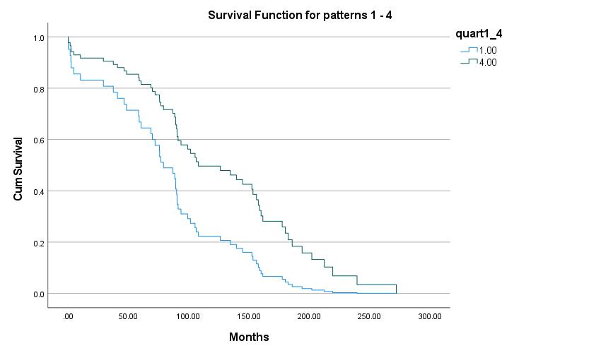
Рис. 5. Кокс-регресії криві виживання (1-й і 4-й квартилі)
Значення Exp(B) для модельної змінної показує,щонебезпекасмертідляпацієнта
1-го ентропійного квартиля (найнижчий EnRE) дорівнює 2,143 рази більше, ніж у пацієнта 4-го ентропійного квартиля (найвищий EnRE).
ВИСНОВКИ
Узагальнена форма оцінювача ентропії (3), яка була ефективно використана для обчислення значень ентропії для різноманітних випадкових розподілів (табл. 1–тaб.2), запропонована в даній роботідля послідовностей ДНКневеликої довжини(N <1000).
20 Таблиця 5 Table 5
The Journal of V.N. Karazin Kharkiv National University. 2022
quart1_4 Середніa Медіана Estimate Std. Error 95% Confidence Interval Estimate Std. Error 95% Confidence Interval Lower Bound Upper Bound Lower Bound Upper Bound 1.00 78.600 11.619 55.826 101.374 72.133 12.325 47.977 96.290 4.00 129.603 11.329 107.398 151.809 106.233 22.156 62.808 149.659 Overall 104.549 8.731 87.436 121.663 90.367 5.338 79.904 100.829
Fig. 5. Cox Regressions survival plot (1st and 4th quarterlies)
Параметривузагальненомувиглядідля
робастного оцінювача ентропії (3) були виведенізнаступнихкритеріїв:
1. точне наближення для деяких відомихфункційрозподілуймовірностей;
2. незалежність EnRE від N для початковихчасовихрядівідлясерійпісля сортування;
3. незалежність EnRE від адитивної змінисередньогозначення. Важливими характеристиками знайденої узагальненої форми EnRE і коефіцієнтівє:
1. формазапису(3)і знайденікоефіцієнти l, m, k забезпечуютьнезалежністьвід адитивної зміни середніх рядів і від величини виділення N для базових рядів і длярядівпіслясортування;
2. значення EnRE чутливе до структурних змін рядів, таких як, наприклад, сортування, яке збільшує ступінь порядку послідовно,зменшуючи EnRE;
3. значення EnRE чутливе до зміни положення нуклеотидів у послідовності ДНК;
4. коефіцієнт переналагодження A самостійно може знадобитися для знаходження кращого значення EnRE в
іншому діапазоні зміни параметрів різних
випадкових розподілів, що завжди можна зробити за допомогою методу найменших квадратів.
Використовуючи запропоновану узагальнену форму робастного оцінювача
ентропії (3) для бази даних пацієнтів з
лейкемією UB, продемонстровано використання EnRE з короткими послідовностями ДНК для аналізу смертності пацієнта:
А. Групи, розділені за медіаною. Обидва аналізи – аналіз виживання Каплана-Мейєра та моделювання вижи-
СПИСОК ЛІТЕРАТУРИ
вання Кокс-регресій, показали статистично значущі результати для p <0,05. Значення Exp(B) для змінної моделі регресій Кокса показує, що небезпека смертідляпацієнтівз EnRE нижчемедіани в1,556разивищеніждляпацієнтівз EnRE вище за медіану. Середній час після діагнозудосмертіу1,496разибільшедля пацієнтівз EnRE понадмедіанупорівняно зпацієнтамиз EnRE нижчемедіани.
Б. Групи хворих формуються з 1-го і 4-го квартилів. Обидва аналізи – аналіз виживання Каплана-Мейєра та моделювання виживання Кокс-регресій, показали статистично значущі результати для p <0,005. Значення Exp(B) для модельної змінної показує, що небезпека смертідляпацієнтів1-гоквартиляентропії (найнижчий EnRE) в 2,143 рази більше, ніж у пацієнта 4-го квартиля ентропії (найвищий EnRE). Середній час після діагнозудосмертіу1,649разибільшедля пацієнтів4-гоквартиляентропіїпорівняно зпацієнтами1-гоквартиляентропії.
Таким чином, перехід від розширених
до менших груп пацієнтів з більшою різницею у EnRE підтвердив унікальне значення ентропії послідовностей ДНК для визначення смертності пацієнтів з лейкемією. Це значення статистично доведено підвищенням небезпеки для хворих на лейкемію з меншою ентропією послідовностейДНК.
Майбутнє продовження сучасних досліджень полягає у включенні більш різних груп пацієнтів для дослідження виживання пацієнта у зв’язку з послідовністю ентропійної ДНК, а також іззалученняміншихметодівфрактального аналізу послідовностей ДНК, таких як фрактальна розмірність або оборотність послідовностей.
1. Li WT. The study of correlation structures of DNA sequences: a critical review. Comput. Chem. 1997; 21 (4): 257–271. DOI: 10.1016/s0097-8485(97)00022-3
2. Damasevicius R. Complexity estimation of genetic sequences using information-theoretic and frequency analysis methods. Informatica. 2010; 21 (1): 13–30. DOI: 10.15388/Informatica.2010.270
3. Rowe GW, Trainor LEH. On the informational content of viral DNA. J. Theoretical Biology. 1983; 101: 151–170. DOI: 10.1016/0022-5193(83)90332-6
4. Vopson MM, Robson SC. A new method to study genome mutations using the information entropy. Physica A. 2012;1-9. DOI: 10.1016/j.physa.2021.126383
5. Sherwin WB. Entropy and Information Approaches to Genetic Diversity and its Expression: Genomic Geography. Entropy. 2010;12:1765-1798. DOI:10.3390/e12071765
Series «Medicine». Issue 45 21
The Journal of V.N. Karazin Kharkiv National University. 2022
6. Chanda P, Costa E, Hu J, Sukumar S, Van Hemert J, Walia R. Information Theory in Computational Biology: Where We Stand Today. Entropy. 2020;22:627-637. DOI: 10.3390/e22060627
7. Villareal RP, Liu BC, Massumi A. Heart rate variability and cardiovascular mortality. Curr Atheroscler Rep. 2002; 4: 120–127. DOI: 10.1007/s11883-002-0035-18
8. Rodríguez J, Correa C, Ramírez L. Heart dynamics diagnosis based on entropy proportions: Application to 550 dynamics. Revista Mexicana de Cardiología. 2017; 28 (1): 10–20.
9. Androulakis AFA, Zeppenfeld K, Paiman EHM, Piers SRD, Wijnmaalen AP, Siebelink HJ, Sramko M, Lamb HJ, van der Geest RJ, de Riva M, Tao Q. Entropy as a Novel Measure of Myocardial Tissue Heterogeneity for Prediction of Ventricular Arrhythmias and Mortality in Post-Infarct Patients. JACC Clin Electrophysiol. 2019 Apr;5 (4): 480–489. DOI: 10.1016/j.jacep.2018.12.005. Epub 2019 Feb 27. PMID: 31000102.
10.Sykora M, Szabo J, Siarnik P, Turcani P, Krebs S, Lang W, Czosnyka M, Smielewski P. Heart rate entropy is associated with mortality after intracereberal hemorrhage. Journal of the Neurological Sciences. 2020: 418: 117033, ISSN 0022-510X, 1–5: DOI: 10.1016/j.jns.2020.117033
11.Matsuda E. Entropy Monitoring in Patients Undergoing General Anesthesia. Am J Nurs. 2017 Mar;117(3):62. DOI: 10.1097/01.NAJ.0000513290.22001.8d
12.Neal-Sturgess C. The Entropy of Morbidity Trauma and Mortality. Arxiv Cornell University. Med. Physics. 2010; 1–20. DOI: 10.48550/arxiv.1008.3695
13.Norris PR, Anderson SM, Jenkins JM, Williams AE, Morris JAJr. Heart rate multiscale entropy at three hours predicts hospital mortality in 3,154 trauma patients. Shock. 2008 Jul; 30 (1): 17–22. DOI: 10.1097/SHK.0b013e318164e4d0
14.Papaioannou VE, Chouvarda IG, Maglaveras NK, Baltopoulos GI, Pneumatikos IA. Temperature multiscale entropy analysis: a promising marker for early prediction of mortality in septic patients. Physiol Meas. 2013 Nov;34(11):1449-66. DOI: 10.1088/0967-3334/34/11/1449
15.Weir BS. Statistical analysis of molecular genetic data. IMA J. of Math. Applied in Medicine and Biology. 1985; 2:1–39.
16.Shannon CE. A Mathematical Theory of Communication. Bell System Technical Journal. 1948; 27 (3): 379
423. DOI:10.1002/j.1538-7305.1948.tb01338.x
17.Lazo A, Rathie P. On the entropy of continuous probability distributions. IEEE Transactions on Information Theory. 1978;24(1). DOI:10.1109/TIT.1978.1055832
18.Gini C, Ottaviani G. Università di Roma. Memorie Di Metodologia Statistica. Roma: E.V. Veschi; 1955.
19.Sánchez-Hechavarría M.E. and etc. Introduction of Application of Gini Coefficient to Heart Rate Variability Spectrum for Mental Stress Evaluation. Arq Bras Cardiol. 2019; [online].ahead print, PP.0-0. DOI: 10.5935/abc.20190185
20.Firebaugh G. Empirics of World Income Inequality. American Journal of Sociology. 1999; 104 (6): 597
1630. DOI:10.1086/210218
21.Shorrocks AF. The Class of Additively Decomposable Inequality Measures. Econometrica. 1980; 48 (3): 613–625. DOI: 10.2307/1913126
22.Martynenko A, Raimondi G, Budreiko N. Robust Entropy Estimator for Heart Rate Variability. Klin. Inform. Telemed. 2019; 14 (15): 67–73. DOI: 10.31071/kit2019.15.06
ENTROPY OF DNA SEQUENCES AND LEUKEMIA PATIENTS MORTALITY
Martynenko O V A,C,D,E,F , Pastor X D A,B,Е,F , Frid S A B,F , Gil J R.
, Maliarova L V Е,F
A – research concept and design; B – collection and/or assembly of data; C – data analysis and interpretation; D
writing the article; E – critical revision of the article; F – final approval of the article.
Introduction. Deoxyribonucleic acid (DNA) is not a random sequence of four nucleotides combinations: comprehensive reviews [1, 2] persuasively shows long- and short-range correlations in DNA, periodic properties and correlations structure of sequences. Information theory methods, like Entropy, imply quantifying the amount of information contained in sequences. the relationship between entropy and patient survival is widespread in some branches of medicine and medical researches: cardiology, neurology, surgery, trauma Therefore, it appears there is a necessity for implementing advantages of information theory methods for exploration of relationship between mortality of some category of patients and entropy of their DNA sequences.
Aim of the research. The goal of this paper is to provide a reliable formula for calculating entropy accurately for short DNA sequences and to show how to use existing entropy analysis to examine the mortality of leukemia patients.
22
–
–
B,F
–
Materials and Methods. We used University of Barcelona (UB) leukemia patient’s data base (DB) with 117 anonymized records that consists: Date of patient’s diagnosis, Date of patient’s death, Leukemia diagnoses, Patient’s DNA sequence. Average time for patient death after diagnoses: 99 ± 77 months. The formal characteristics of DNA sequences in UB leukemia patient’s DB are: average number of bases N = 496 ± 69; min (N) = 297 bases; max(N) = 745 bases
The generalized form of the Robust Entropy Estimator (EnRE) for short DNA sequences was proposed and key EnRE futures was showed.
The Survival Analysis has been done using statistical package IBM SPSS 27 by Kaplan-Meier survival analysis and Cox Regressions survival modelling
Results. The accuracy of the proposed EnRE for calculating entropy was proved for various lengths of time series and various types of random distributions. It was shown, that in all cases for N = 500, relative error in calculating the precise value of entropy does not exceed 1 %, while the magnitude of correlation is no worse than 0.995
In order to yield the minimum EnRE standard deviation and coefficient of variation, an initial DNA sequence's alphabet code was converted into an integer code of bases using an optimization rule for only one minimal numerical decoding around zero
Entropy EnRE were calculated for leukemia patients for two samples: 2 groups divided by median EnRE = 1.47 and 2 groups of patients were formed according to their belonging to 1st (EnRE ≤ 1.448) and 4th (EnRE ≥ 1.490) quartiles. The result of Kaplan-Meier survival analysis and Cox Regressions survival modelling are statistically significant: p < 0,05 for median groups and p < 0,005 for patient’s groups formed of 1st and 4th quartiles The death hazard for a patient with EnRE below median is 1.556 times that of a patient with EnRE over median and that the death hazard for a patient of 1st entropy quartile (lowest EnRE) is 2.143 times that of a patient of 4th entropy quartile (highest EnRE).
Conclusions. The transition from widen (median) to smaller (quartile) patients’ groups with more EnRE differentiation confirmed the unique significance of the entropy of DNA sequences for leukemia patient’s mortality. This significance is proved statistically by increasing hazard and decreasing of average time of death after diagnoses for leukemia patients with lower entropy of DNA sequences.
KEY WORDS: entropy, DNK sequence, patients surviving, leukemia
INFORMATION ABOUT AUTHORS
Martynenko Oleksandr Vitalyevich, D Sc., Ph D., Full Professor, Department of Hygiene and Social Medicine, School of Medicine, V. N. Karazin Kharkiv National University, 6, Svobody sq., Kharkiv, Ukraine, 61022. e-mail: Alexander.v.martynenko@karazin.ua, ORCID ID: https://orcid.org/0000-0002-0609-2220
Pastor Xavier Duran, Doctor of Medicine and Surgery, University Professor, Department of Surgery and MedicalSurgical, University of Barcelona, Chief of Medical Informatics Unit, Hospital Clinic, 170, Villarroel st. Barcelona, Spain, 08036. e-mail: xpastor@clinic.cat, ORCID: 0000-0001-8267-7151
Frid Santiago Andres, MD, M.Sc., Medical Associated Professor, Department of Clinical Foundations, School of Medicine, Universitat de Barcelona, 143, Casanova st., Barcelona, Spain, 08036. Chief of Area of Projects and Developments, Medical Informatics Unit, Hospital Clínic de Barcelona, 170, Villarroel st., Barcelona, Spain, 08036. e-mail:frid@clinic.cat, ORCID: 0000-0001-8400-5770
Gil Rojas Jessyca, MSc, Data Manager, Medical Informatics Unit, Hospital Clínic de Barcelona, 170, Villarroel st., Barcelona, Spain, 08036. e-mail:jegil@clinic.cat ORCID: 0000-0002-7690-7288
Maliarova Liudmila Volodimirivna, Assistant, Department of hygiene and social medicine, School of Medicine, V. N. Karazin Kharkiv National University, 6, Svobody sq., Kharkiv, Ukraine, 61022, e-mail: l.v.maliarova@karazin.ua, https://orcid.org/0000-0002-7902-7016
For citation:
Martynenko OV, Pastor XD, Frid SA, Gil JR, Maliarova LV. ENTROPY OF DNA SEQUENCES AND LEUKEMIA PATIENTS MORTALITY. The Journal of V. N. Karazin Kharkiv National University. Series «Medicine». 2022; 45:12
23 DOI: 10.26565/2313-6693-2022-45-02.
Conflicts of interest: author has no conflict of interest to declare. Конфлікт інтересів: відсутній.
Отримано: 12.10.2022 Прийнято до друку: 20 11.2022
Received: 10 12.2022
Accepted: 11 20.2022
Series «Medicine». Issue 45 23
–
LARGE FAMILY GENETIC ANALYSIS: EFFECTS OF VARIEGATED PORPHYRIA AND HEMOPHILIA B ON REPRODUCTIVE TRAITS
DorofieievaV. B, C, D,FedotaO. A, E, F
A – research concept and design; B – collection and/or assembly of data; C – data analysis and interpretation; D – writing the article; E – critical revision of the article; F – final approval of the article
Introduction. The relevance of the study of genetic pathologies is due to the growing prevalence in most countries, disability and mortality of persons, high costs of support and treatment. The modern classifications include various forms of porphyria and hemophilia. The study of pathologies in historical persons, when it is possible to collect information from different sources regarding members of a large family over a long period of time, is of interest for understanding the mechanisms of the development of the disease at the present time.
Aim is to analyze the genetic characteristics of variegated porphyria and hemophilia B in a large family.
Materials and methods. Data from current guidelines and clinical protocols, scientific literature and genetic databases (OMIM) on various forms of porphyria and hemophilia are analyzed. Information about 1362 people from the British royal family in 18
20th centuries was collected from open sources and scientific literature. A pedigree of 10 generations, 27 nuclear families with persons with variegated porphyria and hemophilia B has been compiled. Genealogical, segregation, linkage, statistical analysis was performed. The results were used to study reproductive traits.
Results. Genealogical analysis showed a family accumulation of porphyria – its prevalence among relatives in a large family was 1,8 %, which is three orders of magnitude higher than among the population of different countries. It was established that there is no statistically significant difference in the sex ratio among patients with the specified pathologies. Data from genealogical and segregation analysis and a penetration rate of 92 % suggest an autosomal dominant type inheritance with incomplete penetrance of disease which is consistent with the literature. The independent nature of inheritance of variegated porphyria and hemophilia B was established. It was found that in persons with porphyria reproductive traits are 3,3–4,1 times differ than the reproductive traits of persons with porphyria and hemophilia at the same time. A statistically significant difference was established between the analyzed traits of patients with porphyria, who at the same time are carriers of the mutation that causes hemophilia, and the indicators of healthy individuals.
KEY WORDS: variegated porphyria, hemophilia B, large family tree, genealogical analysis, reproductive traits
INFORMATION ABOUT AUTHORS
Valeriia Dorofieieva, Student of School of Medicine
V. N. Karazin Kharkiv National University, 6, Svobody Sq., Kharkiv, Ukraine, 61022; e-mail: valeriiadorofieieva@gmail.com, ORCID ID: 0000-0003-3463-7352
Olena Fedota, Doctor of Biology, Full Professor, Department of Obstetrics and Gynecology of School of Medicine
V. N. Karazin Kharkiv National University, 6, Svobody Sq., Kharkiv, Ukraine, 61022; е-mail: omfedota@karazin.ua, ORCID ID: 0000-0001-9659-383X
For citation:
Dorofieieva V, Fedota O. LARGE FAMILY GENETIC ANALYSIS: EFFECTS OF VARIEGATED PORPHYRIA AND HEMOPHILIA B ON REPRODUCTIVE TRAITS. The Journal of V. N. Karazin. Kharkiv National University. Series «Medicine». 2022;45:24–36 DOI: 10.26565/2313-6693-2022-45-03
INTRODUCTION
Research on genetic pathologies is especially relevant, as their prevalence is increasing in many countries, they lead to disability, mortality, require high costs for maintenanceandtreatment[1,2,3].Thereare currently more than 250 monogenic
nosological units in Ukraine that require almost constant care and significant material and moral resources. For example, the State Budget of Ukraine for 2022 provides UAH 300 million for the treatment of children with spinal muscular atrophy which is enough for 5patients, but there are more than 200 of them in Ukraine [4, 5]. As a rule, in most
The Journal of V.N. Karazin Kharkiv National University. 2022 24
УДК: 575.167:616.633.1-616.153.94:616.151.5:618-019 DOI: 10.26565/2313-6693-2022-45-03
Original researches
–
© Dorofieieva V., Fedota O , 2022
monogenic pathologies, pleiotropic effects of genes are noted with affects the cardiovascular, respiratory, endocrine, reproductive and other systems. Therefore, the research of these pathologies is especially relevantforprimarypreventionandformation of risk groups. It is expected that the number of patients with monogenic pathologies will increase over time. Among the groups of these diseases, among monogenic pathologies, modern classifications include various forms of porphyria and hemophilia [6,7].
Porphyria belongs to a group of genetic diseases associated with impaired heme biosynthesis and the accumulation of its toxic metabolites. As a result of a defect in the activity of one of the enzymes of the cycle thereisapartialblockageofacertainstageof heme synthesis, which is accompanied by accumulation in toxic concentrations of porphyrin metabolism metabolites, leading to damage to all nervous systems and anemia. The incidence in the world is 1:10,000 population. Asymptomatic carrier of the mutation
1:1000 people. More common in NorthernEurope[8–12].
The pathogenesis of clinical manifestations in acute hepatic porphyria is due to the involvement of the autonomic nervous system. Damage to the skin in porphyria is associated with increased sensitivitytosunlightduetotheaccumulation of porphyrins in the skin. There are a number of risk factors that can transport latent porphyria to the clinical stage: starvation; infections; alcohol; some drugs; cyclic changes in the hormonal profile in women; insolation[13,14,15].
Modern classification of porphyrias include 8 forms [16]. The clinic of porphyria is clear and polymorphic. According to the clinical course of porphyria can be divided into 2groups: 1. With a predominance of neurological disorders: porphyria due to deficiency of δ-aminolevulinic acid dehydratase (ALAD gene, AR-type of inheritance (TI)), acute intermittent porphyria (HMBS gene, AD-TI), hereditary coproporphyria (CPOX gene, AD-TI), variegated porphyria (HFE, PPOX genes, AD-TI); 2. With predominant skin lesions: congenital erythropoietic porphyria (UROS gene, AR-TI), erythropoietic protoporphyria (FECH gene, AD-TI), late cutaneous
porphyria (UROD, HFE genes, AD-TI) [17–21]. Diagnosis of porphyria consists of history taking, analysis of the clinical picture with 3–4main symptoms, biochemical diagnosis and DNA testing [22, 23]. To date, there are no treatments that successfully, effectively and sustainably correct impaired porphyrin metabolism. Therefore, in most cases, pathogenetic therapy is used. Modern treatment includes: excretion of toxic complexes with heavy metal ions; excretion of excess porphyrin from the person’s body; restoration of functional capacity of the erythropoietic system and liver; protection of the skin from the sun to eliminate the photodynamiceffect[24,25].
Variegated porphyria (porphyria variegate) belongs to the group of hepatic or acute porphyria. This pathology can be manifested by violations of the integrity of the skin, increased photosensitivity with blistering, increased trauma to the skin with subsequent scarring and hyperpigmentation. The incidence in the world is about 1,3:100,000 population [26]. It has been known in Great Britain since the time of Mary Stuart and James I. The most famous historical figure with this disease is George III,KingofGreatBritainandIreland[11].
Hemophilia is a genetic disease of the hemostasissystemcharacterizedbydecreased or impaired synthesis of coagulation factors VIII (hemophilia A), IX (hemophilia B), XI (hemophilia C). Hemophilia is inherited by a recessive trait linked to the sex X chromosome, the same type of hemophilia and the same severity of the disease are inherited. In the general population of patients with hemophilia 30–40% of cases are sporadic hemophilia caused by pathological gene mutations [27]. Hemophilia
A is more common occurring in 1:5000 male births, where as hemophilia B occurs in 1:30000 male births. Hemophilia is found in all ethnic groups; there is no geographic or racial predilection [28]. 2569 patients with hemophilia and Willebrand’s disease were registered in Ukraine, 667 of them (27 %) werechildren[29].
Diagnosis of hemophilia is based on the use of screening tests; confirmation of the diagnosis by determining the level of blood coagulation factors and genetic analysis [30]. The most characteristic andspecific symptom of hemophilia is hemorrhage to large joints –
Series «Medicine». Issue 45 25
–
hemarthrosis [31]. Different types of hemostatic agents and coagulation drugs are available for the treatment of hemophilia. Coagulationfactorconcentrates(CFC)arethe best treatment for patients with hemophilia [30].
The study of pathologies in historical persons, when it is possible to collect information from different sources regarding members of a large family over a long period of time, is of interest for understanding the mechanisms of the development of the diseaseatthepresenttime.
OBJECTIVE
The aim of the study is to analyze the geneticcharacteristicsofvariegatedporphyria andhemophiliaBinalargefamily.
MATERIALS ANDMETHODS
Characteristics of sources on porphyria andhemophilia
Information from scientific literature sources and the OMIM genetic database on genes and mutations associated with variegated porphyria (OMIM 176200) [32] and hemophilia B (OMIM 306900) [33] and other traits was studied. The data of modern recommendations and clinical protocols, scientific literature on various forms of porphyria and hemophilia are analyzed [20,34].
Characteristicsof theindividuals
Much historical and medical literature on members of the British royal family in 18–20th centuries has been studied [11, OMIM]. Information about the members of the large family was obtained from open sources and scientific literature Data about 1362 persons of a large family, the British royal family, was collected. A pedigree consisting of 10generations, 27 nuclear families with persons with hemophilia B and variegated porphyria was compiled and analyzed. Information on quantitative and qualitative characteristics of persons were collected: sex, years and life expectancy, data on children, diseases, gynecological history, reproductive traits.
Ethics statements. Patient consent for publication – not applicable. Ethics approval – notapplicable.
Genealogical analysis
A large genealogical tree of the ruling dynasties of Great Britain was built, starting
with George III and Charlotte MecklenburgStrelitzka (18th century). The prevalence of variegated porphyria in the studied family was assessed and the family accumulation of the disease was monitored. Data on the prevalence of variegated porphyria in European countries were obtained from the OMIMdatabase
Segregation analysis
Weinberg’s formula was used to calculate the segregation frequency (SF) and its standarderror(SSF):
SF =(A – N)/(T – N),
SSF = √����(1– ����)/(��– ��) , where A – the total number of persons in the sample; N – numberoffamilies; T – the total number of children in the sample.
Penetration index was estimated as the ratio of the number of persons in whom the phenotypic manifestations of the analyzed allele were observed to the total number of persons in whom the analyzed allele is present in the required number of copies for thephenotypicmanifestation[35].
Linkage analysis
The analysis of linkage of variegated porphyria and hemophilia was performed by study the compliance of the distribution of these traits in the analyzed pedigree with Mendelian patterns of independent inheritanceoftraits[35].
Statistical analysis andSoftware
Statistical analysis to verify the complianceofthenatureofthedistributionof traits with the Mendelian model of inheritance was performed using the criteria Fisher’s φ-transformation and criteria χ 2 Calculations were performed and databases were created using Microsoft Excel software of Windows 10 Pro operating system, AMD Ryzen 3 3200U with Radeon Vega Mobile Gfx 2.60GHz processor, conductor code: 00330-50000-00000-AAOEM.
Connectionwith scientifictopics
The work was carried out as part of the researchproject «Geneticprerequisitesforthe development and correction of genetic pathology at various stages of human and animal ontogenesis» (state registration number 0119U102493, 2019–2022) of the
The Journal of V.N. Karazin Kharkiv National University. 2022 26
SchoolofMedicine ofV.N. KarazinKharkiv NationalUniversity.
RESULTS ANDDISCUSSION
Variegated porphyria in the British royal family. Porphyria has been known in Britain since the time of Mary Stuart and James I, but the most famous case in the history of the country is George III – King of Great Britain and Ireland [11]. On the example of King George III (04.06.1738–29.01.1820) the literature presents typical manifestations of variegated porphyria. The King showed the following signs of illness: hypersensitivity to sunlight, sounds and touch, abdominal pain and colic, nausea, constipation, rheumatism, lameness, profuse sweating, frequent pulse, skin rash, red, orange, brown or purple urine, blindness, deafness, sleep problems, dementia. Attacks of the king’s illness were noted at least 4times:in50,62,65,72years.Hediedatthe ageof81[11,36,37].
HemophiliaB in the British royal family. Hemophilia is also called the royal disease because of the most famous carrier of the mutation that causes hemophilia, Queen
Victoria. She had 9 children and because of her off springs, hemophilia spread to other royal houses [38]. A number of cases of increased trauma and subsequent fatalities associated with hemophilia in males among members of the British royal family have been reported. For example, Friedrich of Hesse-Darmstadt, the grandson of Queen Victoria, died of internal bleeding after falling out of a window due to hemophilia at 2yearsold[39].
Genealogical analysis of variegated porphyria in a large pedigree. A large genealogical family of Great Britain was built, starting with George III and his wife, Charlotte Mecklenburg-Strelitz. A genealogical analysis of a large pedigree with porphyria, from 1362 people, 10 generations. Particular attention was paid to persons and probably persons with variegated porphyria, persons with hemophilia B and carriers of mutations that cause this disease. Fig 1 and fig 2 demonstrate examples inheritance in families a large pedigree with persons with variegated porphyria, persons with hemophilia B and carriers of mutations that causethisdisease.
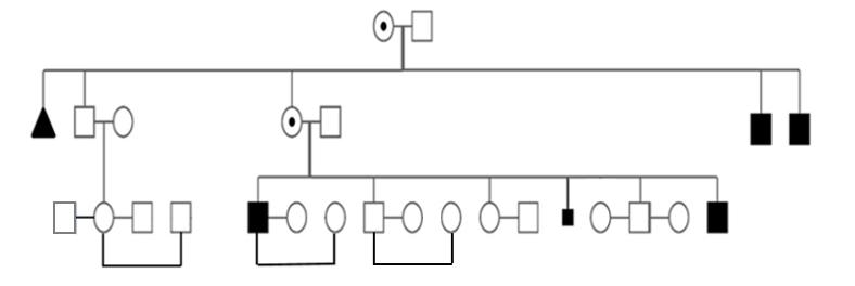
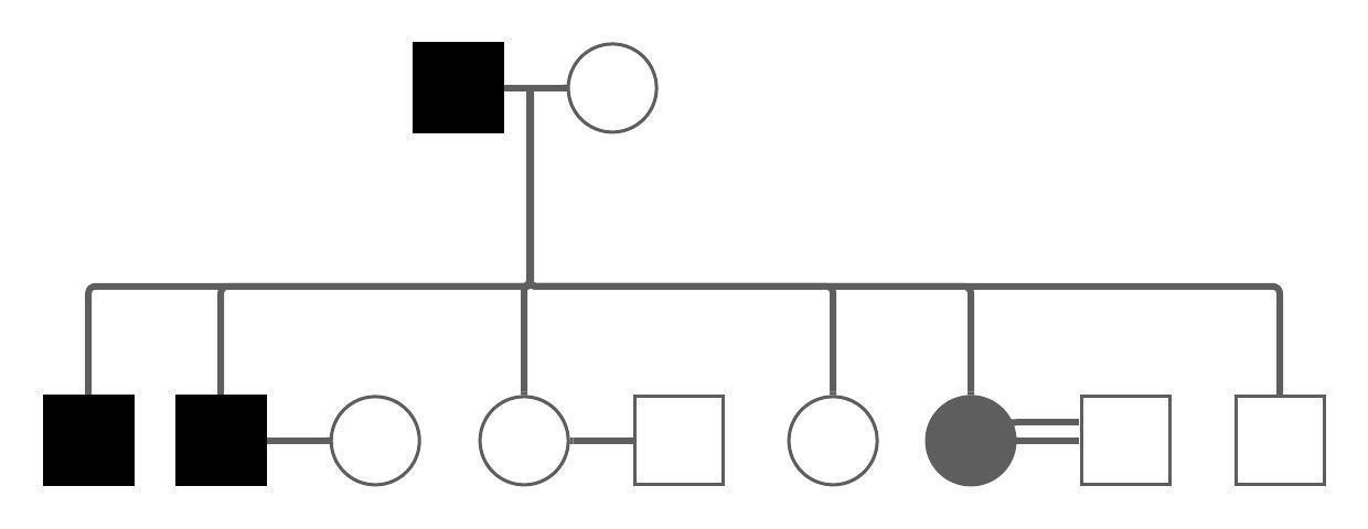
Series «Medicine». Issue 45 27
Fig. 1. Example of a fragment of a large pedigree 1
Fig. 2. Example of a fragment of a large pedigree 2
It was found that the prevalence of variegated porphyria in the studied family is 1.8%, which is 3 orders of magnitude less than in different countries, where it is 0.0013%, [40] thus in the analyzed family there is a familial accumulation of the disease. Analysis of a sex ratio among persons in a large family is demonstrated in table1.
Analysis of a sample of persons or probably persons (n=25) for variable
Analysis
porphyria revealed individuals of both sexes in a large pedigree and showed that the ratio of women to men with porphyria was 3,1%:2,9%, or 1:1, p > 0,05, which is probably in favor of the autosomal model of inheritance.
It was established that also there is no statistically significant difference in the sex ratio among patients with the specified pathologies.
Segregation analysis. The results of segregation analysis demonstrated in the table2 showed that for variegated porphyria it is possible to accept the hypothesis of multiple, both dominant and recessive types
of inheritance. According to our results of segregation and genealogical analysis, variegated porphyria has a type of inheritance – autosomal dominant with incomplete penetrance.
Notes: N - one parent is healthy, A - one parent is affected, SF - empirical segregation frequency, mSF - its standard error, SFd, SFr - theoretically expected segregation frequencies, p - significance level.
AccordingtoOMIM, variegated porphyria may be an autosomal dominant trait found in approximately half of the offspring of adult personsandcausedbymutationsinthe PPOX gene (Protoporphyrinogen oxidase, OMIM
600923), [26,41] which is localized in 1q23.3. Fig 3 demonstrates the manifestation of porphyria in a nuclear family from a large pedigree.
The Journal of V.N. Karazin Kharkiv National University. 2022 28
Table 1
Relatives Women, n (%) Men, n (%) Total, n (%) 387 (46) 453 (54) 840 (100) Of them, porphyria is sick, or probably sick 12 (3,1) 13 (2,9) 25 (3,0) Of them, porphyria is sick 4 (1,0) 10 (2,2) 14 (1,7) Among them, persons with porphyria and are carriers of the mutation that causes hemophilia at the same time 4 (1,0) 0 (0) 4 (0,5)
them are carriers of the mutation that causes hemophilia 4 (1,0) 0 (0) 4 (0,5)
them are persons with hemophilia 0 (0) 12 (2,7) 12 (1,4)
of the sex ratio among persons in large family
Among
Among
2
Table
Phenotype of parents Number of families Number of children Number of affected children SF mSF SFd SFr p N*N 2 10 4 0,25 0,15 0 0,25 > 0,05 N*A 9 45 18 0,25 0,15 0,5 0,5 > 0,05
Segregation analysis within the studied pedigree
Determination of the penetrance of the mutationthat causes variegatedporphyria
According to the analysis of a large pedigree, the penetrance of the mutation was 92%. Fig. 4 demonstrates a fragment of a large pedigree with an obligate heterozygote without clinical symptoms of porphyria in a nuclearfamilyfromalargepedigree.
In addition to the РРОХ gene, other genes that mayaffectthe development of porphyria, such as the HFE gene (Homeostatic iron
regulator, ОМІМ 613609), have been described [42,43]. Its effect may explain incomplete penetrance. The HFE gene, located in 6p22.2, affects the following phenotypes: Alzheimer disease, susceptibility to;microvascularcomplicationsofdiabetes7; porphyria cutanea tarda, susceptibility to; porphyria variegata, susceptibility to; transferrin serum level QTL2; hemochromatosis.
Estimating of linkage of variegated porphyria and hemophilia B in a large family
The results of the analysis of the association of porphyria and hemophilia B in nuclear families with 6–9 off springs from a large pedigree allowed to reject the hypothesis of their possible linkage, p = 0,918-0,608 Fig 5 demonstrates examples nuclear families and persons with porphyria
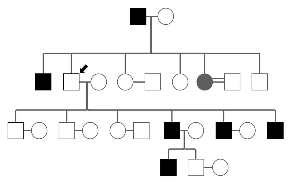
and carriers of a mutation that causes hemophilia at the same time. But the independent distribution of pathological signs does not exclude the possibility of influencing the interaction of non-allelic genes on the nature of the manifestation and degree of manifestation of clinical signs of disease, which was studied by analyzing the reproductive parameters of persons with porphyriaandhemophilia.
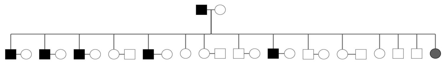
Series «Medicine». Issue 45 29
Fig. 3. The fragment shows the manifestation of porphyria in a nuclear family from a large pedigree
Fig. 4. A fragment of a large pedigree demonstrates the presence of an obligate heterozygote without clinical symptoms of porphyria
Analysis of reproductive traits of persons with variegated porphyria and hemophiliaB
The analysis of reproductive traits of 4 groups of persons from a large family with different pathologies is demonstrated in table3. Table 3
Reproductive traits of persons with porphyria, hemophilia, porphyria and hemophilia at the same time
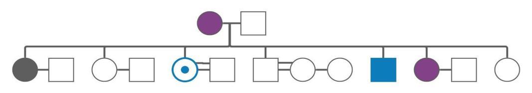
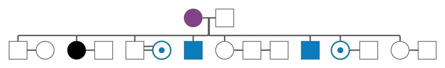
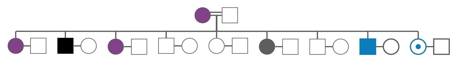
Notes: *Reproductive losses include: miscarriage, spontaneous loss of a pregnancy, stillbirth.** – mean value ± standard error.

The Journal of V.N. Karazin Kharkiv National University. 2022 30
Fig. 5. Nuclear families with persons with porphyria and hemophilia
Parameters Reproductive traits of persons with different pathologies Healthy Porphyria Porphyria + Hemophilia carries Hemophilia carrier Hemophilia Men Women Total Persons, n 10 4 14 4 4 12 12 Average life expectancy, years, ** 60,4±5,7 69,3±5,3 64,8±1,7 55,5±9,9 88,0±3,3 22,1±4,3 69,0±3,1 Pregnancies, in spouses, n 32 7 39 29 18 3 46 Pregnancies per person, in spouses, n, ** 3,2±1,5 1,8±1,1 2,5±0,7 7,3±0,9 4,5±1,0 0,25±0,2 3,8±0,7 Reproductive losses, in spouses, n * 3 0 3 0 2 0 5 Reproductive losses per person, in spouses, n, ** 0,3±0,2 0 0,2±0,2 0,0 0,5±0,3 0 0,4±0,2 Children 0–18 years, n 30 7 37 29 16 3 41 0–18 years per person, n, ** 3,0±1,5 1,8±1,1 2,6±1,1 7,3±0,9 4,0±0,7 0,25±0,2 3,4±0,7 18+ years, n 24 7 31 24 14 3 38 18+ years per person, n, ** 2,4±1,3 1,8±1,1 2,2±0,9 6,0±1,6 3,5±1,0 0,25±0,2 3,2±0,7
The analysis of reproductive traits of 14personswithporphyriafromalargefamily showed that with an average life expectancy of 64,8 ± 1,7 years of persons, the average number of pregnancies per person was 2,5 ± 0,7, reproductive losses – 0,2 ± 0,2
Number of children born – 37, children over 18 – 31, the average number of children born per person – 2,6 ± 1,1, children over 18 –2,2 ± 0,9
significantly differ by 3,3–4,1 times than the reproductive traits of persons with porphyria andhemophiliaatthesametime, p = 0,0021
A statistically significant difference was established between the analyzed traits of patients with porphyria, who at the same time are carriers of the mutation that causes hemophilia, and the indicators of healthy individuals, р = 0,022.
The analysis of reproductive traits of 4persons with porphyria and carriers of a mutation that causes hemophilia at the same time showed that with an average life expectancyof 55,5 ± 9,9 years of persons, the average number of pregnancies per person was 7,3 ± 0,9, reproductive losses
0.
Number of children born – 29, children over 18–24, the average number of children born per person – 7,3 ± 0,9, children over 18 –6,0 ± 1,6
The analysis of reproductive traits of 4persons carriers of a mutation that causes hemophilia showed that with an average life expectancyof 88,0 ± 3,3 years of persons, the average number of pregnancies per person was 4,5 ± 1,0, reproductive losses – 0,5 ± 0,3.
Number of children born – 16, children over 18 – 14, the average number of children born per person – 4,0 ± 0,7, children over 18 –3,5 ± 1,0
The analysis of reproductive traits of 4persons with hemophilia showed that with an averagelifeexpectancyof 22,1 ± 4,3 years of persons, the average number of pregnancies per person was 0,25 ± 0,2, reproductive losses – 0. Number of children born – 3, children over 18 – 3, the average number of children born per person –0,25 ± 0,2,childrenover18 – 0,25 ± 0,2.
The analysis of reproductive traits of 12healthy persons from a large family showed that with an average life expectancy of 69,0 ± 3,1 years of persons, the average number of pregnancies per person was 3,8 ± 0,7, reproductive losses – 0,4 ± 0,2.
Number of children born – 41, children over 18–38, the average number of children born per person – 3,4 ± 0,7, children over 18 –3,2 ± 0,7.
Due to the analysis of reproductive traits of persons with porphyria, hemophilia, porphyria and hemophilia simultaneously, it was found that in persons with porphyria reproductive traits are statistically
We can assume that porphyria, as a pathology of metabolism, and hemophilia, as a pathology of hemostasis, have mutually compensatory mechanisms of influence on theorganism,whichallowsindividualswitha combination of these pathologies to more successfully realize their reproductive potential, compared to individuals suffering from porphyria or who are mutation carriers, which causes hemophilia. We would like to emphasizethespecialroleofsexhormonesin triggering attacks of acute porphyria in women, since it is the change in their level in the organism that is the most frequent cause of attacks [44] According to Daphne Vassiliou and Eliane Sardh [45] the results of a 20-year study of 44 pregnant women with acute hepatic porphyria who gave birth to 44children showed that only 9% (4) of women sufferedanattackof porphyriaduring pregnancy. Authorsconcluded that pregnancy in patients with porphyria often proceeds without complications. Cyclical changes in the hormonal profile, such as the menstrual cycle, are significant triggering factors capable of realizing asymptomatic mutation carriers and turning the latent course of porphyriaintotheclinicalstage.
Therefore, in historical times, in the absence of effective treatment methods, multiple pregnancy throughout the entire fertile age became an available relief approach.
Currently, it is actual to do genetic testing before puberty to reduce the risk appearance of acute symptoms of porphyria, the cumulative risk of which among asymptomatic patients at the time of diagnosisis26,7–58,3%[46]
CONCLUSIONS
According to the results of genealogical analysis, prevalence of variegated porphyria in the studied family was 1,8%, which is three orders of magnitude higher than among the population of different countries. It was
Series «Medicine». Issue 45 31
–
established that there is no statistically significant difference in the sex ratio among patients with the specified pathologies. An autosomal dominant type of inherited variegated porphyria with incomplete penetrance 92% has been estimated. The independent nature of the inheritance of variegated porphyria and hemophilia B has been established confirmed. It was found that in persons with porphyria reproductive traits are 3,3–4,1 times differ than the reproductive traits of persons with porphyria and hemophilia at the same time. A statistically significant difference was established
REFERENCES
between the analyzed traits of patients with porphyria, who at the same time are carriers of the mutation that causes hemophilia, and theindicatorsofhealthyindividuals.
GRATITUDE
The authors expresse their sincere gratitude for discussion to the obstetrician gynecologist of the MNPE «City Maternity Hospital № 1» of the Kharkiv City Council, Kharkiv, Ukraine, candidate of medical sciences, associate professor, I.M.Merenkova
1. Nakaz MOZ Ukrainy №641/84 vid 31.12.2003 Pro udoskonalennia medyko-henetychnoi dopomohy v Ukraini. 2003. Available from: https://zakon.rada.gov.ua/rada/show/va641282-03#Text
2. Budreviciute A, Damiati S, Sabir DK, Onder K, Schuller-Goetzburg P, Plakys G, et al. Management and Prevention Strategies for Non-communicable Diseases (NCDs) and Their Risk Factors. Front Public Health. 2020 Nov 26; 8: 574111. DOI: 10.3389/fpubh.2020.574111
3. Jackson M, Marks L, May GHW, Wilson JB. The genetic basis of disease. Essays Biochem. 2018 Dec 2; 62 (5): 643–723. DOI: 10.1042/EBC20170053. Erratum in: Essays Biochem. 2020 Oct 8; 64 (4): 681.
4. Nakaz MOZ Ukrainy № 778 vid 27.10.2014 Pro zatverdzhennia pereliku ridkisnykh (orfannykh) zakhvoriuvan. 2014. Available from: https://zakon.rada.gov.ua/laws/show/z1439–14#Text
5. Zakon Ukrainy vid 02.12.2021 Pro Derzhavnyi biudzhet Ukrainy na 2022 rik. 2022. Available from: https://zakon.rada.gov.ua/laws/show/1928–20#Text
6. Kirschner J, Cathomen T. Gene Therapy for Monogenic Inherited Disorders. Dtsch Arztebl Int. 2020 Dec 21; 117 (51–52): 878–885. DOI: 10.3238/arztebl.2020.0878.
7. Pasi KJ, Rangarajan S, Mitchell N, Lester W, Symington E, Madan B, et al. Multiyear Follow-up of AAV5-hFVIII-SQ Gene Therapy for Hemophilia A. N Engl J Med. 2020 Jan 2; 382 (1): 29–40. DOI: 10.1056/NEJMoa1908490
8. Kothadia JP, LaFreniere K, Shah JM. Acute Hepatic Porphyria. 2022 May 8. In: StatPearls [Internet]. Treasure Island (FL): StatPearls Publishing; 2022 Jan. Available from: https://www.ncbi.nlm.nih.gov/books/NBK537178/
9. Yasuda M, Chen B, Desnick RJ. Recent advances on porphyria genetics: Inheritance, penetrance & molecular heterogeneity, including new modifying/causative genes Mol Genet Metab. 2019 Nov; 128 (3): 320–331. DOI: 10.1016/j.ymgme.2018.11.012
10.Gonzalez-Mosquera LF, Sonthalia S. Acute Intermittent Porphyria. StatPearls [Internet]. 2022 Jan. Available from: https://www.ncbi.nlm.nih.gov/books/NBK547665/
11.Macalpine I, Hunter R, Rimington C. Porphyria in the royal houses of Stuart, Hanover, and Prussia. A follow-up study of George 3d's illness. Br Med J. 1968 Jan 6; 1(5583):7-18. DOI: 10.1136/bmj.1.5583.7
12.Phillips JD. Heme biosynthesis and the porphyrias. Mol Genet Metab. 2019 Nov; 128 (3): 164–177. DOI: 10.1016/j.ymgme.2019.04.008
13.Usta Atmaca H, Akbas F. Porphyria cutanea tarda: a case report. J Med Case Rep. 2019 Jan 21; 13 (1): 17. DOI:10.1186/s13256-018-1956-9
14.Bonkovsky, Herbert L et al. Pathogenesis and clinical features of the acute hepatic porphyrias (AHPs). Molecular genetics and metabolism. 2019 Nov; 128 (3): 213
218. DOI: 10.1016/j.ymgme.2019.03.002
15.Cuny JF. Porphyries cutanées [The cutaneous porphyrias]. Ann Dermatol Venereol. 2019 Feb; 146 (2): 143
159. French. DOI: 10.1016/j.annder.2018.12.005
16.Fedotov VP, Diudiun AD. Porfirii (klinichna lektsiia). Dermatovenerolohiia. Kosmetolohiia. Seksopatolohiia. HZ Dnipropetrovska medychna akademiia MOZ Ukrainy. 2011;1-4:132-150. Available from: https://repo.dma.dp.ua/2527/1/25_Fedot_33.pdf
17.Stölzel U, Doss MO, Schuppan D. Clinical Guide and Update on Porphyrias. Gastroenterology. 2019 Aug; 157 (2): 365
381.e4. DOI: 10.1053/j.gastro.2019.04.050
32
The Journal of V.N. Karazin Kharkiv National University. 2022
–
–
–
18.Nehrych TI, Yur MI, Khoma A I. ta in. Problemy diahnostyky ta likuvannia hostroi porfiriinoi polineiropatii Mizhnar. nevrol. zhurn. 2015: 8 (78): 73–76 Available from: http://www.mifua.com/archive/article/42067
19.Bonnin A, Picornell A, Orfila J, Castro JA, Ramon MM. Clinic and genetic evaluation of variegate porphyria (VP) in a large family from the Balearic Islands. J Inherit Metab Dis. 2009 Dec; 32 Suppl 1. DOI: 10.1007/s10545-009-1059-2
20.Ahmed jan N, Masood S. Erythropoietic Protoporphyria. StatPearls [Internet]. 2022 Oct. Available from: https://www.ncbi.nlm.nih.gov/books/NBK563141
21.Shah A, Bhatt H. Cutanea Tarda Porphyria. StatPearls [Internet]. 2021 Sep. Available from: https://www.ncbi.nlm.nih.gov/books/NBK563209/
22.Di Pierro E, De Canio M, Mercadante R. Laboratory Diagnosis of Porphyria. Diagnostics (Basel). 2021 Oct; 11(8):1343. Available from: https://www.frontiersin.org/articles/10.3389/fgene.2022.993453/full
23.Ramanujam VS, Anderson KE. Porphyria Diagnostics-Part 1: A Brief Overview of the Porphyrias. Curr Protoc Hum Genet. 2015 Jul 1; 86: 17.20.1–17.20.26. DOI: 10.1002/0471142905.hg1720s86
24.Pischik E, Kauppinen R. An update of clinical management of acute intermittent porphyria. Appl Clin Genet. 2015 Sep 1; 8: 201–14. DOI: 10.2147/TACG.S48605
25.Hrechanina OI, Hrechanina YuB, Beletska SV, ta insh. Porfiriia: metod. nastan. z dystsypliny «Medychna henetyka» dlia pidhotovky likariv-interniv, studentiv 5 kursu, y likariv-kursantiv tsykliv pisliadyplomnoi osvity; Kharkiv, KhNMU; 2018. 28 p.
26.Singal AK, Anderson KE. Variegate Porphyria. GeneReviews. 2013 Feb. Available from: https://www.ncbi.nlm.nih.gov/books/NBK121283/
27.Doherty TM, Kelley A. Bleeding Disorders. StatPearls [Internet]. 2022 Jan. Available from: https://www.ncbi.nlm.nih.gov/books/NBK541050/
28.Castaman G, Matino D. Hemophilia A and B: molecular and clinical similarities and differences. Haematologica. 2019 Sep;104(9):1702-1709. DOI: 10.3324/haematol.2019.221093
29.Hemophilia. Situational analysis of problems in Ukraine. Quantitative research of the international research of the international research Agency IFAK Institute of Gmnh&Co; 2018. 20 p.
30.Srivastava A, Santagostino E, Dougall A, Kitchen S, Sutherland M, Pipe SW, et al. WFH Guidelines for the Management of Hemophilia panelists and co-authors. WFH Guidelines for the Management of Hemophilia, 3rd edition. Haemophilia. 2020 Aug;26 Suppl 6: 1–158. DOI: 10.1111/hae.14046
31.Vydyborets, SV, Derpak YuYu, Popovych YuYu. Hemophilia. Family Medicine. 2018 Dec;6:61–66. DOI: https://doi.org/10.30841/2307-5112.6.2018.169142
32.OMIM #176200. [Internet]. [cited 2022 Оct 26]. Available from: https://www.omim.org/entry/176200
33.OMIM #306900. [Internet]. [cited 2022 Оct 26]. Available from: https://www.omim.org/entry/306900?search=306900&highlight=306900
34.Stein P, Badminton M, Barth J, Rees D, Stewart MF; British and Irish Porphyria Network. Best practice guidelines on clinical management of acute attacks of porphyria and their complications. Ann Clin Biochem. 2013 May; 50 (3): 217-223. DOI: 10.1177/0004563212474555
35.Vogel and Motulsky's. Human Genetics: Problems and Approaches 4th ed. Springer; 2010. 1034 p.
36.Macalpine I, Hunter R. The 'insanity' of King George III: a classic case of porphyria. Brit. Med. J. 1966 Jan;1(5479):65–71. DOI: 10.1136/bmj.1.5479.65
37.Warren MJ, Jay M, Hunt DM, Elder GH, Rohl JCG. The maddening business of King George III and porphyria. Trends Biochem. 1996 Jun;21(6):229-234. DOI: https://doi.org/10.1016/S09680004(96)80022-3
38.Rogaev EI, Grigorenko AP, Faskhutdinova G, Kittler ELW, Moliaka YK. Genotype analysis identifies the cause of the 'Royal disease'. Science. 2009 Oct; 326 (5954): 817. DOI: 10.1126/science.1180660
39.Mager, Hugo. Elizabeth: Grand Duchess of Russia. Carroll and Graf Publishers; 1998. 45 p.
40.Elder G, Harper P, Badminton M, Sandberg S, Deybach JC. The incidence of inherited porphyrias in Europe. J Inherit Metab Dis. 2013 Sep;36(5):849-57. DOI: 10.1007/s10545-012-9544-4
41.OMIM #600923. [Internet]. [cited 2022 Оct 26].
Available from: https://www.omim.org/entry/600923?search=600923&highlight=600923
42.ОМІМ 613609. [Internet]. [cited 2022 Оct 26].
Available from: omim.org/entry/613609?search=613609&highlight=613609
43.Barton, James C et al. HFE gene: Structure, function, mutations, and associated iron abnormalities. Gene. 2015 Dec; 574(2): 179–192. DOI: 10.1016/j.gene.2015.10.009
44.Andersson C, Innala E, Bäckström T. Acute intermittent porphyria in women: clinical expression, use and experience of exogenous sex hormones. A population-based study in northern Sweden. J Intern Med. 2003 Aug;254(2):176–83. DOI: 10.1046/j.1365-2796.2003.01172.x
Series «Medicine». Issue 45 33
The Journal of V.N. Karazin Kharkiv National University. 2022
45.Vassiliou D, Sardh E. Acute hepatic porphyria and maternal health: Clinical and biochemical follow-up of 44 pregnancies. J Intern Med. 2022 Jan;291(1):81-94. DOI: 10.1111/joim.13376
46.Baumann K, Kauppinen R. Penetrance and predictive value of genetic screening in acute porphyria. Mol Genet Metab. 2020 May;130 (1): 87–99. DOI: https://doi.org/10.1016/j.ymgme.2020.02.003
ГЕНЕТИЧНЕ ДОСЛІДЖЕННЯ ВЕЛИКОЇ РОДИНИ: ВПЛИВ ВАРІЄГАТНОЇ ПОРФІРІЇ
ТА ГЕМОФІЛІЇ В НА РЕПРОДУКТИВНІ ПОКАЗНИКИ
Дорофєєва В. Р. B, C, D, Федота О. М. A, E, F
A – концепція та дизайн дослідження; B – збір даних; C – аналіз та інтерпретація даних; D – написання статті; E
редагування статті; F – остаточне затвердження статті
Вступ. Актуальність дослідження генетичних патологій обумовлена зростанням їхньої поширеності у більшості країн, інвалідизації та летальності хворих, великими витратами на супровід та лікування. Сучасна класифікація генетичних хвороб включає, зокрема, різні форми порфірії та гемофілії. Вивчення патологій в історичних осіб, коли є можливість зібрати інформацію з різних джерел про членів великої родини за тривалий період часу, представляє інтерес для розуміння механізмів розвитку захворювання.
Мета роботи – аналіз генетичних особливостей варієгатної порфірії та гемофілії В у великій родині.
Матеріали та методи. Проаналізовано дані сучасних рекомендацій та клінічних протоколів, наукової літератури та генетичних баз даних (OMIM) щодо різних форм порфірії та гемофілії. З відкритих джерел та наукової літератури зібрано відомості про 1362 особи з Британської королівської сім’ї, XVIII–XX ст. Складено родовід з 10 поколінь, 27 ядерних сімей з хворими на варієгатну порфірію та гемофілію В. Проведено генеалогічний, сегрегаційний, статистичний аналіз та аналіз зчеплення. Результати використано для дослідження репродуктивних ознак.
Результати. Генеалогічний аналіз показав сімейне накопичення порфірії – її поширеність серед родичів у великій родині склала 1,8 %, що на три порядки вище, ніж серед населення різних країн. Встановлено, що статистично значущої різниці у співвідношенні статей серед осіб із зазначеними патологіями немає. Дані генеалогічного та сегрегаційного аналізу, та показник пенетрантності 92 %, свідчать про аутосомно-домінантний тип успадкування з неповною пенетрантністю, що узгоджується з даними літератури. Визначено незалежний характер успадкування варієгатної порфірії та гемофілії В. Доведено, що у хворих на порфірію репродуктивні ознаки в 3,3–4,1 рази відрізняються від репродуктивних ознак у хворих на порфірію та гемофілію одночасно. Встановлено значущу різницю між проаналізованими ознаками хворих на порфірію, які водночас є носіями мутації, що обумовлює гемофілію, та показниками здорових осіб.
КЛЮЧОВІ СЛОВА: варієгатна порфірія, гемофілія В, великий родовід, генеалогічний аналіз, репродуктивні ознаки ІНФОРМАЦІЯ ПРО АВТОРІВ Дорофєєва Валерія Романівна, студентка медичного факультету Харківського національного університету імені В. Н. Каразіна, пл. Свободи, 6, Харків, Україна. 61022; e-mail: valeriiadorofieieva@gmail com, ORCID ID: 00000003-3463-7352
Федота Олена Михайлівна, д. біол. н., професор кафедри акушерства та гінекології, медичний факультет, Харківський національний університет імені В. Н. Каразіна, пл. Свободи, 6, Харків, Україна, 61022; е-mail: omfedota@karazin ua, ORCID ID: 0000-0001-9659-383X
Для цитування: Дорофєєва ВР, Федота ОМ. ГЕНЕТИЧНЕ ДОСЛІДЖЕННЯ ВЕЛИКОЇ РОДИНИ: ВПЛИВ ВАРІЄГАТНОЇ ПОРФІРІЇ ТА ГЕМОФІЛІЇ В НА РЕПРОДУКТИВНІ ПОКАЗНИКИ. Вісник Харківського національного університету імені В.Н. Каразіна. Серія «Медицина». 2022;45 24–36 DOI: 0.26565/2313-6693-2022-45-03 СПИСОК
ЛІТЕРАТУРИ
1. Наказ МОЗ України № 641/84 від 31.12.2003 Про удосконалення медико-генетичної допомоги в Україні. 2003. Available from: https://zakon.rada.gov.ua/rada/show/va641282-03#Text
2. Budreviciute A, Damiati S, Sabir DK, Onder K, Schuller-Goetzburg P, Plakys G, et al. Management and Prevention Strategies for Non-communicable Diseases (NCDs) and Their Risk Factors. Front Public Health. 2020 Nov 26;8:574111. DOI: 10.3389/fpubh.2020.574111
34
–
3. Jackson M, Marks L, May GHW, Wilson JB. The genetic basis of disease. Essays Biochem. 2018 Dec 2;62(5):643-723. DOI: 10.1042/EBC20170053. Erratum in: Essays Biochem. 2020 Oct 8;64(4):681
4. Наказ МОЗ України № 778 від 27.10.2014 Про затвердження переліку рідкісних (орфанних) захворювань. 2014. Available from: https://zakon.rada.gov.ua/laws/show/z1439-14#Text
5. Закон України від 02.12.2021 Про Державний бюджет України на 2022 рік. 2022. Available from: https://zakon.rada.gov.ua/laws/show/1928–20#Text
6. Kirschner J, Cathomen T. Gene Therapy for Monogenic Inherited Disorders. Dtsch Arztebl Int. 2020 Dec 21;117(51-52):878
885. DOI: 10.3238/arztebl.2020.0878.
7. Pasi KJ, Rangarajan S, Mitchell N, Lester W, Symington E, Madan B, et al. Multiyear Follow-up of AAV5-hFVIII-SQ Gene Therapy for Hemophilia A. N Engl J Med. 2020 Jan 2;382(1):29-40. DOI: 10.1056/NEJMoa1908490
8. Kothadia JP, LaFreniere K, Shah JM. Acute Hepatic Porphyria. 2022 May 8. In: StatPearls [Internet]. Treasure Island (FL): StatPearls Publishing; 2022 Jan. Available from: https://www.ncbi.nlm.nih.gov/books/NBK537178/
9. Yasuda M, Chen B, Desnick RJ. Recent advances on porphyria genetics: Inheritance, penetrance & molecular heterogeneity, including new modifying/causative genes Mol Genet Metab. 2019 Nov; 128(3):320–331. DOI: 10.1016/j.ymgme.2018.11.012
10.Gonzalez-Mosquera LF, Sonthalia S. Acute Intermittent Porphyria. StatPearls [Internet]. 2022 Jan. Available from: https://www.ncbi.nlm.nih.gov/books/NBK547665/
11.Macalpine I, Hunter R, Rimington C. Porphyria in the royal houses of Stuart, Hanover, and Prussia. A follow-up study of George 3d's illness. Br Med J. 1968 Jan 6; 1(5583):7-18. DOI: 10.1136/bmj.1.5583.7
12.Phillips JD. Heme biosynthesis and the porphyrias. Mol Genet Metab. 2019 Nov; 128(3):164–177. DOI: 10.1016/j.ymgme.2019.04.008
13.Usta Atmaca H, Akbas F. Porphyria cutanea tarda: a case report. J Med Case Rep. 2019 Jan 21;13(1):17. DOI:10.1186/s13256-018-1956-9
14.Bonkovsky, Herbert L et al. Pathogenesis and clinical features of the acute hepatic porphyrias (AHPs). Molecular genetics and metabolism. 2019 Nov; 128 (3): 213–218. DOI: 10.1016/j.ymgme.2019.03.002
15.Cuny JF. Porphyries cutanées [The cutaneous porphyrias]. Ann Dermatol Venereol. 2019 Feb;146(2):143
159. French. DOI: 10.1016/j.annder.2018.12.005
16.Федотов ВП, Дюдюн АД. Порфірії (клінічна лекція). Дерматовенерологія. Косметологія. Сексопатологія. ГЗ Дніпропетровська медична академія МОЗ України. 2011;1-4:132-150. Available from: https://repo.dma.dp.ua/2527/1/25_Fedot_33.pdf
17.Stölzel U, Doss MO, Schuppan D. Clinical Guide and Update on Porphyrias. Gastroenterology. 2019 Aug;157(2):365-381.e4. DOI: 10.1053/j.gastro.2019.04.050
18.Nehrych TI, Yur MI, Khoma AI. ta in. Problemy diahnostyky ta likuvannia hostroi porfiriinoi polineiropatii Mizhnar. nevrol. zhurn. 2015:8(78):73
76 Available from: http://www.mifua.com/archive/article/42067
19.Bonnin A, Picornell A, Orfila J, Castro JA, Ramon MM. Clinic and genetic evaluation of variegate porphyria (VP) in a large family from the Balearic Islands. J Inherit Metab Dis. 2009 Dec;32 Suppl 1. DOI: 10.1007/s10545-009-1059-2
20.Ahmed jan N, Masood S. Erythropoietic Protoporphyria. StatPearls [Internet]. 2022 Oct. Available from: https://www.ncbi.nlm.nih.gov/books/NBK563141
21.Shah A, Bhatt H. Cutanea Tarda Porphyria. StatPearls [Internet]. 2021 Sep. Available from: https://www.ncbi.nlm.nih.gov/books/NBK563209/
22.Di Pierro E, De Canio M, Mercadante R. Laboratory Diagnosis of Porphyria. Diagnostics (Basel). 2021 Oct; 11(8):1343. Available from: https://www.frontiersin.org/articles/10.3389/fgene.2022.993453/full
23.Ramanujam VS, Anderson KE. Porphyria Diagnostics-Part 1: A Brief Overview of the Porphyrias. Curr Protoc Hum Genet. 2015 Jul 1;86:17.20.1-17.20.26. DOI: 10.1002/0471142905.hg1720s86
24.Pischik E, Kauppinen R. An update of clinical management of acute intermittent porphyria. Appl Clin Genet. 2015 Sep 1;8:201-14. DOI: 10.2147/TACG.S48605
25.Гречаніна ОЯ, Гречаніна ЮБ, Белецька СВ, та ін. Порфірія: метод. настан. з дисципліни «Медична генетика» для підготовки лікарів-інтернів, студентів 5 курсу, и лікарів-курсантів циклів післядипломної освіти; Харьків, ХНМУ; 2018. 28 с.
26.Singal AK, Anderson KE. Variegate Porphyria. GeneReviews. 2013 Feb. Available from: https://www.ncbi.nlm.nih.gov/books/NBK121283/
27.Doherty TM, Kelley A. Bleeding Disorders. StatPearls [Internet]. 2022 Jan. Available from: https://www.ncbi.nlm.nih.gov/books/NBK541050/
Series «Medicine». Issue 45 35
–
–
–
The Journal of V.N. Karazin Kharkiv National University. 2022
28.Castaman G, Matino D. Hemophilia A and B: molecular and clinical similarities and differences. Haematologica. 2019 Sep;104(9):1702-1709. DOI: 10.3324/haematol.2019.221093
29.Гемофілія. Ситуаційний аналіз проблематики в Україні. Quantitative research of the international research of the international research Agency IFAK Institute of Gmnh&Co; 2018. 20 p.
30.Srivastava A, Santagostino E, Dougall A, Kitchen S, Sutherland M, Pipe SW, et al. WFH Guidelines for the Management of Hemophilia panelists and co-authors. WFH Guidelines for the Management of Hemophilia, 3rd edition. Haemophilia. 2020 Aug;26 Suppl 6:1–158. DOI: 10.1111/hae.14046
31.Видиборець, СВ, Дерпак Ю Ю., Попович, Ю Ю. Hemophilia. Family Medicine 2018 Грудень; 6: 61
66. DOI: https://doi.org/10.30841/2307-5112.6.2018.169142
32.OMIM #176200. [Internet]. [cited 2022 Оct 26]. Available from: https://www.omim.org/entry/176200
33.OMIM # 306900. [Internet]. [cited 2022 Оct 26]. Available from: https://www.omim.org/entry/306900?search=306900&highlight=306900
34.Stein P, Badminton M, Barth J, Rees D, Stewart MF; British and Irish Porphyria Network. Best practice guidelines on clinical management of acute attacks of porphyria and their complications. Ann Clin Biochem. 2013 May; 50 (3): 217-223. DOI: 10.1177/0004563212474555
35.Vogel and Motulsky's Human Genetics: Problems and Approaches 4th ed. Springer; 2010 1034 p.
36.Macalpine I, Hunter R. The 'insanity' of King George III: a classic case of porphyria. Brit. Med. J. 1966 Jan;1(5479):65–71. DOI: 10.1136/bmj.1.5479.65
37.Warren MJ, Jay M, Hunt DM, Elder GH, Rohl JCG. The maddening business of King George III and porphyria. Trends Biochem. 1996 Jun;21(6):229-234. DOI: https://doi.org/10.1016/S09680004(96)80022-3
38.Rogaev EI, Grigorenko AP, Faskhutdinova G, Kittler ELW, Moliaka YK. Genotype analysis identifies the cause of the 'Royal disease'. Science 2009 Oct; 326 (5954): 817. DOI: 10.1126/science.1180660
39.Mager, Hugo. Elizabeth: Grand Duchess of Russia. Carroll and Graf Publishers; 1998. 45 p.
40.Elder G, Harper P, Badminton M, Sandberg S, Deybach JC. The incidence of inherited porphyrias in Europe. J Inherit Metab Dis. 2013 Sep;36(5):849-57. DOI: 10.1007/s10545-012-9544-4
41.OMIM #600923. [Internet]. [cited 2022 Оct 26]. Available from: https://www.omim.org/entry/600923?search=600923&highlight=600923
42.ОМІМ 613609. [Internet]. [cited 2022 Оct 26]. Available from: omim.org/entry/613609?search=613609&highlight=613609
43.Barton, James C et al. HFE gene: Structure, function, mutations, and associated iron abnormalities. Gene. 2015 Dec; 574(2): 179–192. DOI: 10.1016/j.gene.2015.10.009
44.Andersson C, Innala E, Bäckström T. Acute intermittent porphyria in women: clinical expression, use and experience of exogenous sex hormones. A population-based study in northern Sweden. J Intern Med. 2003 Aug;254(2):176-83. DOI: 10.1046/j.1365-2796.2003.01172.x
45.Vassiliou D, Sardh E. Acute hepatic porphyria and maternal health: Clinical and biochemical follow-up of 44 pregnancies. J Intern Med. 2022 Jan;291(1):81–94. DOI: 10.1111/joim.13376
46.Baumann K, Kauppinen R. Penetrance and predictive value of genetic screening in acute porphyria. Mol Genet Metab. 2020 May;130(1):87–99. DOI: https://doi.org/10.1016/j.ymgme.2020.02.003
Conflicts of interest: author has no conflict of interest to declare. Конфлікт інтересів: відсутній.
Отримано: 03 10.2022
Прийнято до друку: 20 11 2022
Received: 10 03.2022
Accepted: 11 20 2022
36
–
УДК: 618.36-008.6-092-02:618.3-06:616.9
ОСОБЛИВОСТІ
DOI: 10.26565/2313-6693-2022-45-04
ПАТОГЕНЕТИЧНИХЗМІН
ПЛАЦЕНТАРНОГОКОМПЛЕКСУНАТЛІ
ВНУТРІШНЬОУТРОБНОГО ІНФІКУВАННЯ
Гошовська А. В. Анотація. Одним із основних ускладнень під час раннього ембріогенезу і плацентації є
плацентарна недостатність. Під впливом різноманітних факторів, а саме інфекційних, які в залежності
від біологічної специфіки негативно впливають на статеві клітини батьків, розвиток зародка, формування трофобласту і плаценти, що в свою чергу призводить до порушення функції плацентарного комплексу та гестаційних ускладнень під час вагітності. Завдяки розширенню діагностичних можливостей для виявлення порушень функції плаценти, а також у зв’язку з появою нових даних про механізми регуляції кровообігу в плаценті для оцінки функціонального стану фетоплацентарного комплексу на етапі формування первинної плацентарної недостатності на фоні запальних захворювань жіночих статевих органів ЖСО нами було визначено доплерометричну
картину особливостей кровотоку (яєчникова артерія, міжворсинчастий простір) у жінок основної та
контрольної груп.
Мета. Визначити доплерометричну картину особливостей кровотоку (яєчникова артерія, об′єм
хоріона, дослідження кровотоку в різних ділянках хоріона) у вагітних на тлі запальних захворювань
жіночих статевих органів в ранньому триместрі гестації. Матеріали та методи. Основну групу склали 37 жінок з ознаками запальних захворювань
жіночих статевих органів, контрольну групу склали 30 жінок практично здорові. Результати. Отримання нових даних про механізми регуляції кровообігу в яєчниковій артерії та міжворсинчастому просторі на етапі формування плацентарного комплексу при фізіологічній і ускладненій вагітності дозволить доповнити питання патогенезу розвитку та профілактики плацентарної недостатності.
Висновки. Дослідження кровотоку екстраембіональних структур на ранніх термінах гестації дозволяє діагностувати структурні зміни на етапі формування плаценти. Порушення кровотоку в І триместрі гестації на фоні інфекційного чинника, який проникає в різні судинні ланки системи матиплацента-плід з вирахуванням показників об’єму кровотоку демонструє закономірність формування порушеної гемодинаміки на етапі формування плацентарного комплексу. Дані показники можуть слугувати критерієм для відбору жінок групи ризику по формуванню первинної плацентарної недостатності.
КЛЮЧОВІ СЛОВА: плацентарна недостатність, кровотік, яєчникова артерія, міжворсинчастий простір, запальні захворювання жіночих статевих органів, індекс васкуляризації, індекс кровотоку
ІНФОРМАЦІЯ ПРО АВТОРА
Гошовська Аліса Володимирівна, к.мед.н., асистент кафедри акушерства, гінекології та перинатології, Буковинський державний медичний університет. Театральна площа, 2, Чернівці, Україна, 58002. e-mail: goshovska.alisa@bsmu.edu.ua, ORCID: http://orcid.org/0000-0002-7740-3962, Researcher ID: http://www researcherid com/rid/E-2070-2017
Для цитування:
Гошовська АВ. ОСОБЛИВОСТІ ФОРМУВАННЯ ПАТОГЕНЕТИЧНИХ ЗМІН ПЛАЦЕНТАРНОГО
КОМПЛЕКСУ НА ТЛІ ВНУТРІШНЬОУТРОБНОГО ІНФІКУВАННЯ. Вісник Харківського національного
університету імені В.Н.Каразіна Серія«Медицина». 2022:45;37–43.DOI: 10.26565/2313-6693-2022-45-04
ВСТУП
Внутрішньоутробне інфікування – це
встановлений факт внутрішньоутробного
проникнення до плідного яйця вірусів, або мікроорганізмів.
Одним із основних ускладнень під час раннього ембріогенезу і плацентації є пла-
Гошовська А. В., 2022
центарна дисфункція [1, 2, 3]. Під впливом різноманітнихфакторів,асамеінфекційних, які в залежності від біологічної специфіки негативно впливають на формування трофобласту і плаценти, відбувається порушення функції плацентарного комплексу,що,всвоючергу,призводитьдо
Series «Medicine». Issue 45 37
ФОРМУВАННЯ
The Journal of V.N. Karazin Kharkiv National University. 2022
гестаційнихускладненьпідчасвагітності[4, 5,6,7,8].
Найбільшу інформативність і цінність в діагностиці плацентарної дисфункції на ранніх термінах вагітності, безумовно, має ультразвукова оцінка стану екстраембріональнихструктур[9].
Отримання нових даних про механізми регуляції кровообігу в яєчниковій артерії, зміни об’єму хоріона, зміни кровотоку в різнихділянкаххоріонанаетапіформування плацентарного комплексу при фізіологічній
і ускладненій вагітності дозволить доповнити питання патогенезу розвитку та профілактики плацентарної недостатності [10,11,12].
Завдяки розширенню діагностичних можливостей для виявлення порушень функції плаценти, а також у зв’язку з появою нових даних про механізми регуляції кровообігу в плаценті для оцінки функціонального стану фето-плацентарного комплексу (ФПК) на етапі формування первинної плацентарної дисфункції на фоні запальних захворювань жіночих статевих органівдоцільнимєвизначеннякровотокув яєчниковій артерії та в різних ділянках хоріона[13,14,15].
Мета дослідження – визначити доплерометричну картину особливостей кровотоку(яєчниковаартерія,об’ємхоріона, дослідження кровотоку в різних ділянках хоріона) у вагітних на тлі запальних захворювань жіночих статевих органів в ранньомутриместрігестації.
МАТЕРІАЛИ І МЕТОДИ
На УЗД – апараті «Voluson Expert 730» проводиться тримірна ехографія з метою отримання об’ємного зображення хоріону. Об’ємну реконструкцію хоріального кровотокувиконуваливпрограмі VOCAL (Virtual OrganComputer-AidedAnalysis)зпобудовою гістограми судинного компоненту в заданому об’ємі хоріальної тканини. Оцінка судинного компоненту проводилася на основі визначення індексу васкуляризації (VІ)таіндексукровотоку(FI).
Визначено доплерометричну картину особливостейкровотокувяєчниковійартерії таврізнихділянкаххоріонатаоб′ємхоріона ужінокосновноїтаконтрольноїгруп.
Згідно поставленої мети було проведено ультразвукове дослідження 67 жінкам в 6–7 та 12–14 тижнів гестації. Основну групу
склали 37жінок з ознаками запальних захворювань жіночих статевих органів, контрольну групу склали 30жінок практичноздорові.
Проведення дослідження погоджене етичним комітетом Буковинського державного медичного університету, що підтверджується протоколом Комісії з питаньбіомедичноїетикищододотримання морально-правових правил проведення медичних наукових досліджень.
Дослідження здійснювали після отримання інформованоїзгодипацієнткиналікування.
РЕЗУЛЬТАТИ
Для визначення показників кровотоку в
яєчникових артеріях дослідження проводились в ділянці воронко-тазових зв’язок, при цьому контрольний об’єм поступово зміщувався по напрямку до яєчника до моментунайкращої візуалізації судини. Для вивчення кровотоку в яєчниковій артерії, спочатку оцінювалася структура яєчників, а також вимірювався об’єм з використанням формули:
V =0,523АВС(см3),
де V – об’єм яєчника, А,В,С – розміри яєчника в різних площинах, а 0,523 –постійнийкоефіцієнт.
В результаті поглибленого аналізу кровотоку в яєчникових артеріях було виявленотакіпоказники:
У переважної більшості жінок контрольної групи 28 (93,3%) визначались стандартні низькохвильові характеристики кровотоку в яєчникових артеріях з низькою систолічною швидкістю (Vmax від 0,06 до 0,14 м/с) і високим показником периферичного опору, при цьому діастолічний компонент або був слабо виражений, або взагалі був відсутнім, проте у жінок основної групи відмічався 24 (64,8%) середньошвидкісний артеріальний кровотік де(Vmax від 0,15до 0,2м/сек)іпомірними показниками периферичного опору та у 13 (35,1 %) відмічався (високошвидкісний) тип кровообігувяєчниковійартеріяхзвисокими швидкостями (Vmax від 0,14 до 0,57 м/сек) та високими показниками периферичного опору.
Слід зазначити, що найбільш вагомими прогностичними критеріями розвитку первинної плацентарної недостатності та загрози переривання вагітності на тлі запальних захворювань жіночих статевих
38
органів (основна група) являлось: зниження об’єму хоріона на 36–78 %, індекса васкуляризації на 56–78 %, висока резистентність в яєчниковій артерії на стороні жовтого тіла С/Д (систолодіастолічнеспіввідношення)>3,5,ІР(індекс резистентності) > 0,70, зменшення об’єму хоріона<2,5см.Слідзазначити,щоужінок
контрольної групи об’єм хоріона сягав > 3 мм, не відмічалось зниження індекса васкуляризації,аС/Д<3,5,ІР<0,70.
Для виявлення закономірностей
формування судинного компоненту на фоні
запальних захворювань жіночих статевих
органів та при фізіологічній вагітності
проведено визначення об’єму хоріальної
тканининаетапіформуванняплацентарного
комплексу. Встановлено, що в І триместрі
вагітностівідбувалосьпоступовезбільшення
тканини хоріона у жінок контрольної групи
від 5,5 до 15,8 см³ до 13-го тижня гестації, прицьомувперіодзавершенняформування
об’єму хоріона проходить хвилеподібний
зрістворсиннапротязі2тижнівмайжевдва разидо34,6см3 .
Проте встановлено, що у переважної
більшості жінок основної групи 32 (86,5 %)
ділянка тканини хоріона сягала в межах 2,3
до 7,4 см³ до 13-го тижня гестації, але в період формування об’єму хоріона хвилеподібного росту ворсин не було зафіксовано і показники об’єму сягали до 17,8см3
При дослідженні кровотоку в різних
ділянках хоріона використовувались такі
параметри:
● VI – індекс васкуляризації, який
відображає відсоткове співвідношення
судиннихелементівудосліджуючомуоб’ємі плацентарноїтканини;
● FI – індекс кровотоку, який відображає кількість клітин крові, які транспортуютьсявмоментдослідження.
При дослідженні кровотоку в різних ділянкаххоріонуужінокконтрольноїгрупи діагностовано,щовтермінівагітностідо11
тижнів гестації гетероморфізм (порушення)
кровообігу в периферичних і центральних
ділянках не було виявлено. Так показники
кровотоку в центральній зоні сягали VI –
15,1, FI – 36,1, а периферичній зоні VI –
12,8,FI – 30,0. Протеужінокосновноїгрупи
VI в центральній ділянці сягав 8,9, а в
периферичній зоні VI – 14,6, FI – 33. Після 12 тижнів вагітності у жінок контрольної
групи був виражений судинний гетероморфізм – індекс васкуляризації в центральній зоні значно перевищував показники кровотоку VI – 20,8, FI – 55,6 в порівнянні з периферичними ділянками хоріонуVI– 7,7, FI – 33,6. Ужінокосновної групи індекс васкуляризації в центральній зоніVI– 9,7,FI –35,1майженеперевищував показникипериферичноїділянкихоріонаVI – 6,9,FI – 31,4.
Отже, визначені нами показники кровотоку в яєчникових артеріях у жінок основної групи значно відрізнялися від показників отриманих результатів у жінок контрольної групи появою середньо швидкісного кровотоку в яєчникових артеріях де (Vmax від 0,15 до 0,2 м/сек) і помірними показниками периферичного опору. з низькою систолічною швидкістю та високошвидкісного типу кровообігу в яєчниковій артеріях з високими швидкостями (Vmax від 0,14 до 0,57 м/сек) та високими показниками периферичного опору на відміну від стандартних низькохвильових швидкостей кровотоку в яєчниковихартеріяхзнизькоюсистолічною швидкістю (Vmax від 0,06 до 0,14 м/с) і високимпоказникомпериферичногоопоруу жінокконтрольноїгрупи.
Об’єм хоральної тканини в жінок основноїгрупивідстававвдіаметрі(2,3до7,4см) проти показників жінок контрольної групи (5,5 до 15,8см³) та відсутній хвилеподібній ріст хоріона до 13 тижнів у вагітних на тлі запальних захворювань жіночих статевих органів.
Аналізуючи кровотік в різних ділянках хоріонуслідзазначити,щоужінокосновної групи був виражений гетероморфізм між центральною та периферичними ділянками якийсягавVI– 8,9, FI –21авпериферичній зоніVI– 14,6,FI – 33 впорівняннізжінками контрольної групи де порушення кровотоку в різних ділянках хоріона не були зафіксовані центральна ділянка – VI – 15,1, FI – 36,1, периферична зона VI – 12,8, FI –30,0.
Зафіксовані ехографічні зміни гемодинамічних показників дозволять спрогнозувати ранні прояви плацентарної дисфункції, здійснювати динамічний нагляд за даною групою жінок, вчасно розпочати профілактику та лікування порушень фетоплацентарного комплексу, що попередить
Series «Medicine». Issue 45 39
The Journal of V.N. Karazin Kharkiv National University. 2022
розвиток гестаційних порушень в ІІ та ІІІ триместрігестації.
ОБГОВОРЕННЯ РЕЗУЛЬТАТІВ
При визначенні кровотоку в яєчникових артеріях у жінок основної групи діагностувався середньошвидкісний артеріальний кровотік де (Vmax від 0,15 до 0,2м/сек) і середні показники периферичного опору, у 13(35,1 %) діагностувався високошвидкісний тип кровообігу в яєчникових артеріях з високими швидкостями (Vmax від 0,14 до 0,57 м/сек) та високими показниками периферичногоопору.
При визначенні об’єму хоріона у жінок основної групи було зафіксовано зниження об’єму хоріона на 36–78 %, індексу васкуляризаціїна56–78 %,високурезистентність вяєчниковійартеріїнастороніжовтоготіла С/Д (систоло-діастолічне співвідношення) > 3,5, ІР (індекс резистентності) > 0,70, зменшенняоб’ємухоріона<2,5см.
При дослідженні кровотоку в різних ділянкаххоріонаслідзазначити,щоужінок основної групи був виражений гетероморфізм між центральною та периферичними
СПИСОК ЛІТЕРАТУРИ
ділянками який сягав VI – 8,9, FI – 21, а в периферичнійзоніVI– 14,6,FI – 33.
ВИСНОВКИ
Дослідження кровотоку екстраембріональнихструктур в ранніх термінахгестації
дозволяє діагностувати структурні зміни на етапі формування плаценти. Більш значущими показниками розвитку плацентарної дисфункції у жінок на тлі запальних
захворювань жіночих статевих органів є зафіксовані зміни екстраембріональних структур, а саме: низькохвильовий та середньошвидкісний кровотік в яєчникових артеріяхзнизькоюсистолічноюшвидкістю, високим та помірним показником периферичного опору, зменшення об’єму хоріона та вираженим порушенням
кровотоку між центральною та боковими ділянками при визначенні кровотоку в різнихділянкаххоріона.
Дані показники можуть слугувати критерієм для відбору жінок групи ризику по формуванню первинної плацентарної дисфункції.
1. Кравченко ОВ., Печеряга СВ. Оцінка ефективності профілактики первинної плацентарної
дисфункції в ранні терміни гестації при аномальному розташуванні плаценти. Актуальні питання педіатрії, акушерства та гінекології. 2016;1:74-5. https://doi.org/10.11603/24116-4944.2016.1.5997
2. Анчева ИА. Клиническая характеристика плацентарной дисфункции с позиции тенденций современного акушерства (обзор литературы). Буковинський медичний вісник. 2016;20.1(77):196-9. https://doi.org/10.24061/2413-0737.XX.1.77.2016.44
3. Герман ЛВ. Оптимізація діагностики та лікування плацентарної дисфункції у вагітних з невиношуванням [автореферат]. Київ;2015;27с.
4. Бойко ВІ, Болотна МА. Діагностика та профілактика плацентарної дисфункції у юних жінок, які народжують уперше. Здоровье женщины. 2015; 8:110-1. https://medexpert.com.ua/journals/diagnostika-i-profilaktika-placentarnoj-disfunkcii-u-junyhpervorodjashhih/?link=https://med-expert.com.ua/journals/publishing-activity/zdorovezhenshchiny/zdorove-zhenshhiny-wh_%e2%84%968_2015/
5. Кравченко ОВ., Варвус ЕВ. Профілактика ускладнень в I триместрі гестації у вагітних з низькою плацентацією. Здоровье женщины.2015; 10(106):104-6. DOI 10.15574/HW.2015.106.104
6. Dall’Asta, A.; Minopoli, M.; Ghi, T.; Frusca, T. Monitoring, Delivery and Outcome in Early Onset Fetal Growth Restriction. Reprod. Med. 2021; 2: 85-94. DOI:10.3390/reprodmed2020009
7. Charlotte HJR Jansen, Arnoud W Kastelein, C Emily Kleinrouweler, Kees H De Jong, Eva Pajkrt at al. Development of placental abnormalities in location and anatomy. Acta Obstet Gynecol Scand.2020;99:983-93. DOI: 10.1111/aogs.13834
8. Ferrazzi E, Stampalija T, Monasta L, Di Martino D, Vonck S, Gyselaers W. Maternal hemodynamics: a method to classify hypertensive disorders of pregnancy. Am J Obstet Gynecol. 2018 Jan;218(1): 124.e1124.e11. DOI: 10.1016/j.ajog.2017.10.226
9. Булавенко ОВ, Мунтян ОА, Коньков ДГ, Фурман ОВ. Ультразвукова характеристика кровотоку в судинах матки в І триместрі вагітності у жінок із звичним невиношуванням в анамнезі. Вісник Вінницького національного медичного університету. 2018; 1 (22): 72. DOI
https://doi.org/10.31393/reports-vnmedical-2018-22(1)-14
40
10.Гопчук ОМ. Плацентарна дисфункція у вагітних із супутньою патологією серцево-судинної системи. Здоровье женщины. 2017;7(123):88–92. https://med-expert.com.ua/journals/placentarnajadisfunkcija-u-beremennyh-s-soputstvujushhej-patologiej-serdechno-sosudistoj-sistemy/?link=https://medexpert.com.ua/journals/publishing-activity/zdorove-zhenshchiny/zdorove-zhenshhiny-wh_07_2017/
11.Липатов ИС, Тезиков ЮВ, Линева ОИ, Азаматов АР, Мартынова НВ, Протасов АД. и др. Возможности немедикаментозной и медикаментозной терапии плацентарной недостаточности. Вопросы гинекологии, акушерства и перинатологии. 2019;18(1):53-62. DOI: 10.20953/1726-16782019-1-53-62
12.Каліновська І. Діагностика та лікування плацентарної недостатності [Інтернет]. Чернівці; 2020 [цитовано 2020 Тра 18]. Доступно: http://ag3.bsmu.edu.ua/home/pisladiplomnaosvita/kilicni-lekciie/ikalinovska-diagnostika-ta-likuvannaplacentarnoie-nedostatnos
13.Печеряга СВ, Маринчина ІМ. Профілактика виникнення порушень фетоплацентарного комплексу в ранні терміни гестації при низькій плацентації Буковинський медичний вісник. 2018;22(2):70–5. DOI: https://doi.org/10.24061/2413-0737.XXII.2.86.2018.36
14.Heazell AE, Hayes DJ, Whitworth M, Takwoingi Y, Bayliss SE, Davenport C. Biochemical tests of placental function versus ultrasound assessment of fetal size for stillbirth and small-for-gestational-age infants. Cochrane Database Syst Rev. 2019 May 14;5:CD012245. DOI: 10.1002/14651858.CD012245.pub2
15.Skeith L, Rodger M. Anticoagulants to prevent recurrent placenta-mediated pregnancy complications: is it time to put the needles away? Thromb Res. 2017; 151 (Suppl 1): S38-S42. DOI: 10.1016/S00493848(17)30065-8
FEATURES OF THE FORMATION OF PATHOGENETIC CHANGES OF THE PLACENTAL COMPLEX ON THE BACKGROUND OF INTRA-UTERONOMY INFECTION
Hoshovska Alisa
Abstract. One of the main complications during early embryogenesis and placentation is placental insufficiency. Under the influence of various factors, namely infectious factors, which, depending on the biological specificity, negatively affect the germ cells of the parents, the development of the embryo, the formation of the trophoblast and the placenta, which in turn leads to a violation of the function of the placental complex and gestational complications during pregnancy. Due to the expansion of diagnostic possibilities for detecting placental function disorders, as well as in connection with the emergence of new data on the mechanisms of blood circulation regulation in the placenta to assess the functional state of the feto-placental complex at the stage of the formation of primary placental insufficiency against the background of inflammatory diseases of the female genital organs, we were determined the dopplerometric pattern of blood flow (ovarian artery, intervillous space) in women of the main and control groups.
Goal. To determine the dopplerometric pattern of blood flow characteristics (ovarian artery, chorionic volume, blood flow study in different areas of the chorion) in pregnant women against the background of inflammatory diseases of the female genital organs in the early trimester of pregnancy.
Materials and methods. The main group consisted of 37 women with signs of inflammatory diseases of the female genital organs, the control group consisted of 30 practically healthy women.
The results. Obtaining new data on the mechanisms of blood circulation regulation in the ovarian artery and intervillous space at the stage of formation of the placental complex in physiological and complicated pregnancy will allow to supplement the question of the pathogenesis of the development and prevention of placental insufficiency.
Conclusions. The study of the blood flow of extraembryonic structures in the early stages of gestation allows to diagnose structural changes at the stage of placenta formation. Violation of blood flow in the first trimester of pregnancy against the background of an infectious factor that penetrates into various vascular links of the mother-placenta-fetus system with the deduction of blood flow volume indicators demonstrates the regularity of the formation of disturbed hemodynamics at the stage of formation of the placental complex. These indicators can serve as a criterion for the selection of women of the risk group for the formation of primary placental insufficiency.
KEY WORDS: placental insufficiency, blood flow, ovarian artery, intervillous space, inflammatory diseases of female genital organs, vascularization index, blood flow index
Series «Medicine». Issue 45 41
The Journal of V.N. Karazin Kharkiv National University. 2022
INFORMATION ABOUT AUTHOR
Alisa Hoshovska, Doctor of Medicine, Assistant of the Department of Obstetrics, Gynecology and Perinatology of Bukovinian State Medical University. Teatralna Square, 2, Chernivtsi. 58002. E-mail: goshovska.alisa@bsmu.edu.ua, ORCID: http://orcid.org/0000-0002-7740-3962, Researcher ID: http://www.researcherid.com/rid/E-2070-2017
For citation:
Hoshovska Alisa A. FEATURES OF THE FORMATION OF PATHOGENETIC CHANGES OF THE PLACENTAL COMPLEX ON THE BACKGROUND OF INTRA-UTERONOMY INFECTION. The Journal of V.N. Karazin. Kharkiv National University. Series «Medicine». 2022;45:37–43 DOI: 10.26565/2313-6693-2022-45-04
REFERENCES
1. Kravchenko OV., Pecheryaha SV Otsinka efektyvnosti profilaktyky pervynnoyi platsentarnoyi dysfunktsiyi v ranni terminy hestatsiyi pry anomalʹnomu roztashuvanni platsenty Aktualʹni pytannya pediatriyi, akusherstva ta hinekolohiyi. 2016;1:74-5. https://doi.org/10.11603/24116-4944.2016.1.5997 [in Ukrainian]
2. Ancheva YA. Klynycheskaya kharakterystyka platsentarnoy dysfunktsyy s pozytsyy tendentsyy sovremennoho akusherstva (obzor lyteratury). Bukovynsʹkyy medychnyy visnyk. 2016;20.1(77):196-9. https://doi.org/10.24061/2413-0737.XX.1.77.2016.44 [in Russian]
3. Herman LV. Optymizatsiya diahnostyky ta likuvannya platsentarnoyi dysfunktsiyi u vahitnykh z nevynoshuvannyam [avtoreferat]. Kyyiv;2015;27s [in Ukrainian]
4. Boyko VI, Bolotna MA. Diahnostyka ta profilaktyka platsentarnoyi dysfunktsiyi u yunykh zhinok, yaki narodzhuyutʹ upershe. Zdorovʹe zhenshchyny. 2015; 8:110-1. https://medexpert.com.ua/journals/diagnostika-i-profilaktika-placentarnoj-disfunkcii-u-junyhpervorodjashhih/?link=https://med-expert.com.ua/journals/publishing-activity/zdorovezhenshchiny/zdorove-zhenshhiny-wh_%e2%84%968_2015/ [in Ukrainian]
5. Kravchenko OV., Varvus EV. Profilaktyka uskladnenʹ v I trymestri hestatsiyi u vahitnykh z nyzʹkoyu platsentatsiyeyu. Zdorovʹe zhenshchyny.2015; 10(106):104-6. DOI 10.15574/HW.2015.106.104 [in Ukrainian]
6. Dall’Asta, A.; Minopoli, M.; Ghi, T.; Frusca, T. Monitoring, Delivery and Outcome in Early Onset Fetal Growth Restriction. Reprod. Med. 2021; 2: 85-94. DOI:10.3390/reprodmed2020009
7. Charlotte HJR Jansen, Arnoud W Kastelein, C Emily Kleinrouweler, Kees H De Jong, Eva Pajkrt at al. Development of placental abnormalities in location and anatomy.Acta Obstet Gynecol Scand.2020;99:983-93. DOI: 10.1111/aogs.13834
8. Ferrazzi E, Stampalija T, Monasta L, Di Martino D, Vonck S, Gyselaers W. Maternal hemodynamics: a method to classify hypertensive disorders of pregnancy. Am J Obstet Gynecol. 2018 Jan;218(1): 124.e1124.e11. DOI: 10.1016/j.ajog.2017.10.226
9. Bulavenko OV, Muntyan OA, Konʹkov DH, Furman OV. Ulʹtrazvukova kharakterystyka krovotoku v sudynakh matky v I trymestri vahitnosti u zhinok iz zvychnym nevynoshuvannyam v anamnezi. Visnyk Vinnytsʹkoho natsionalʹnoho medychnoho universytetu. 2018;1(22):72. DOI https://doi.org/10.31393/reports-vnmedical-2018-22(1)-14 [in Ukrainian]
10.Hopchuk OM. Platsentarna dysfunktsiya u vahitnykh iz suputnʹoyu patolohiyeyu sertsevo-sudynnoyi systemy. Zdorovʹe zhenshchyny. 2017;7(123):88-92. https://med-expert.com.ua/journals/placentarnajadisfunkcija-u-beremennyh-s-soputstvujushhej-patologiej-serdechno-sosudistoj-sistemy/?link=https://medexpert.com.ua/journals/publishing-activity/zdorove-zhenshchiny/zdorove-zhenshhiny-wh_07_2017/ [in Ukrainian]
11.Lypatov YS, Tezykov YUV, Lyneva OY, Azamatov AR, Martynova NV, Protasov AD. y dr. Vozmozhnosty nemedykamentoznoy y medykamentoznoy terapyy platsentarnoy nedostatochnosty. Voprosy hynekolohyy, akusherstva y perynatolohyy. 2019;18(1): 53–62. DOI: 10.20953/1726-1678-20191-53-62 [in Russian]
12.Kalinovsʹka I. Diahnostyka ta likuvannya platsentarnoyi nedostatnosti [Internet]. Chernivtsi; 2020 [tsytovano 2020 Tra 18]. Dostupno: http://ag3.bsmu.edu.ua/home/pisladiplomnaosvita/kilicni-lekciie/ikalinovska-diagnostika-ta-likuvannaplacentarnoie-nedostatnos [in Ukrainian]
13.Pecheryaha SV, Marynchyna IM. Profilaktyka vynyknennya porushenʹ fetoplatsentarnoho kompleksu v ranni terminy hestatsiyi pry nyzʹkiy platsentatsiyi. Bukovynsʹkyy medychnyy visnyk. 2018;22(2):70-5. DOI: https://doi.org/10.24061/2413-0737.XXII.2.86.2018.36 [in Ukrainian]
14.Heazell AE, Hayes DJ, Whitworth M, Takwoingi Y, Bayliss SE, Davenport C. Biochemical tests of placental function versus ultrasound assessment of fetal size for stillbirth and small-for-gestational-age infants. Cochrane Database Syst Rev. 2019 May 14;5:CD012245. DOI: 10.1002/14651858.CD012245.pub2
42
15.Skeith L, Rodger M. Anticoagulants to prevent recurrent placenta-mediated pregnancy complications: is it time to put the needles away? Thromb Res. 2017; 151 (Suppl 1): S38–S42. DOI: 10.1016/S00493848(17)30065-8
Conflicts of interest: author has no conflict of interest to declare.
Конфлікт інтересів: відсутній.
Отримано: 03.09.2022
Прийнято до друку: 20.11.2022
Received: 09.03.2022
Accepted: 11.20.2022
Series «Medicine». Issue 45 43
Покровська Н. К.A, B, C, D,Скляров Є. Я.E, F
A
концепція та дизайн дослідження; B
збір даних; C
аналіз та інтерпретація даних; D
написання статті; E – редагування статті; F – остаточне затвердження статті
Вступ. Адропін – гормон, який бере участь у патогенезі артеріальної гіпертензії і захворювань серцево-судинної системи та енергетичному гомеостазі, зокрема знижує інсулінорезистентність, шляхом контролю синтезу глюкози у печінці.
Мета. Дослідити рівень адропіну та його взаємодії з факторами ризику артеріальної гіпертензії у пацієнтів з надлишковою масою тіла та ожирінням.
Матеріали та методи. Обстежено 70 пацієнтів з артеріальною гіпертензією і надмірною масою тіла та ожирінням віком 62,4 ± 10,4 років. Для контрольної групи були відібрані 10 практично здорових осіб. Рівень адропіну визначали шляхом імуноферментного аналізу (Finetest Elisa Kit) у сироватці крові. Критеріями включення до участі у дослідженні були: вік пацієнтів 40–75 років; артеріальна гіпертензія; індекс маси тіла > 25 кг/м2
Результати. Встановлено, що середній рівень адропіну у пацієнтів з артеріальною гіпертензією та коморбідним ожирінням чи надмірною масою тіла (n = 70) був 775,5 ± 311,7 пг/мл, що є достовірно нижчим за вміст даного пептиду у практично здорових осіб (n = 10), який становив 1084,6 ± 110,2 пг/мл (p < 0,01). Відзначено тенденцію до зниження рівня гормону у чоловіків, що ймовірно пов’язано із значно більшою поширеністю тютюнопаління серед даної категорії осіб, оскільки при оцінці факторів, що сприяють розвитку артеріальної гіпертензії та погіршенню її прогнозу, знайдено достовірне зниження адропіну у осіб з анамнезом тютюнопаління (p < 0,01). У пацієнтів з ожирінням (індекс маси тіла > 30 кг/м2) рівень адропіну був достовірно нижчим (p < 0,05), як і в осіб з артеріальним тиском > 150/90 мм рт. ст. (p > 0,05).
Висновки. Рівень сироваткового адропіну достовірно знижується у осіб з АГ та надмірною масою тіла і ожирінням в порівнянні з практично здоровими особами. Факторами, що сприяють зниженню концентрації пептиду є наявність тютюнопаління, ожиріння та підвищеного рівня глюкози крові. Показники артеріального тиску не є критичними для зміни концентрації гормону крові, що ймовірно пов’язано із впливом медикаментів, котрі застосовують для лікування артеріальної гіпертензії.
КЛЮЧОВІ СЛОВА: плацентарна недостатність, кровотік, яєчникова артерія, міжворсинчастий простір, запальні захворювання жіночих статевих органів, індекс васкуляризації, індекс кровотоку
ІНФОРМАЦІЯ ПРО АВТОРІВ Покровська Наталія Костянтинівна, аспірант кафедри терапії № 1, медичної діагностики та гематології і трансплантології ФПДО, Львівський національний медичний університет імені Данила Галицького, вул. Пекарська, 69, Львів, Україна; e-mail: natapokrovska@gmail com; ORCID: https://orcid org/0000-0002-7835-5878 Скляров Євген Якович, д. мед. н., професор, завідувач кафедри терапії №1, медичної діагностики та гематології і трансфузіології ФПДО, Львівський національний медичний університет імені Данила Галицького, вул. Пекарська, 69, Львів, Україна; e-mail: depther2015@gmail com; ORCID: https://orcid org/0000-0001-9037-0969
Для цитування:
Покровська НК, Скляров ЄЯ. АДРОПІН ТА ФАКТОРИ РИЗИКУ АРТЕРІАЛЬНОЇ ГІПЕРТЕНЗІЇ
ПАЦІЄНТІВ З НАДЛИШКОВОЮ МАСОЮ ТІЛА ТА ОЖИРІННЯМ.Вісник Харківського національного університету імені В.Н. Каразіна. Серія «Медицина». 2022;45:44–50 DOI: 0.26565/2313-6693-2022-45-05
ВСТУП
Глобальною проблемою людства сьогодення є прогресуюча акумуляція жирової тканини та, як наслідок, поява надмірної маси тіла та ожиріння, з яким асоційована інсулінорезистентність.Саметомуособливу
Покровська Н. К., Скляров Є. Я., 2023
увагупредставляєадропін,якийбереучасть уенергетичномугомеостазі,зокремазнижує інсулінорезистентність, шляхом контролю синтезуглюкозиупечінці [1].
Адропін – гормон, який складається із 76 амінокислот і виділяється багатьма органамитатканинамиорганізму,включноз
The Journal of V.N. Karazin Kharkiv National University. 2022 44
УДК: 616.12-008.331.1-056.5-02-092:612.01 DOI: 10.26565/2313-6693-2022-45-05
АДРОПІН ТА ФАКТОРИ РИЗИКУ АРТЕРІАЛЬНОЇ ГІПЕРТЕНЗІЇ
У ПАЦІЄНТІВ З НАДЛИШКОВОЮ МАСОЮ ТІЛА ТА ОЖИРІННЯМ
–
–
–
–
У
головним мозком, печінкою, нирками, серцем, підшлунковою залозою та може бутиідентифікованийубіологічнихрідинах, асамегрудномумолоцітакрові [1]
У осіб з надвагою та ожирінням
спостерігається знижений рівень адропіну у порівнянні із особами з нормальною масою
тіла та зворотна кореляція між рівнем
гормону та індексом маси тіла (ІМТ),
обводом талії, діастолічним артеріальним
тиском (АТ), глюкозою, інсуліном та віком.
Вікєвагомимчинником,зякимасоційовано
зниження вмісту адропіну у крові, що
ймовірно пов’язано з формуванням
коморбідної патології у даної категорії
пацієнтів та порушенням гліколіпідного
метаболізму [1–3]
У дослідженні Lovren et al. вказано, що
адропін був виділений в ендотеліальних
клітинах і демонстрував позитивний вплив
на судинну стінку, шляхом збільшення
проліферації клітин та міграції [4]
Тканинний рівень адропіну варіює в залежностівіднаявнихстанівтапатологій,а
саме: COVID-19, цукрового діабету, метаболічного синдрому, артеріальної
гіпертензії (АГ), ожиріння, обструктивного
нічного апное, ревматоїдного артриту та інших [1].
В експериментальному дослідженні, при введенні екзогенного адропіну, у щурів знайдено зниження сироваткового рівня тригліцеридів,загальногохолестерину(ХС), ХС ліпопротеїнів низької щільності, аланінамінотрансферази (АлАТ), аспартатамінотрансферази (АсАТ), а той час як вміст ХС ліпопротеїнів високої щільності зростав [5].
Відомо, що рівень адропіну є нижчим у пацієнтів з ішемічною хворобою серця та при ознаках ремоделювання міокарда [3].
Важливим залишається протизапальний ефектадропіну.Вінмаєнеабиякезначенняу
патогенезі атеросклерозу, який, як і ожиріння, супроводжується хронічним запальним процесом, і є вагомим фактором
ризику АГ та інших захворювань серцевосудинноїсистеми [1]
Незважаючи на ряд досліджень щодо
визначення впливу адропіну на перебіг АГ, отримані результати викликають суперечності. В той час як у дітей з ожирінням не було знайдено взаємозв’язку між АТ та рівнем адропіну, у дорослих осіб з АГ концентрація пептиду знижується у порів-
няннізпрактичноздоровимиособами [6,7] Значне зниження вмісту адропіну відмічаютьуосібзАТвищимза180/110ммрт.ст. [8]. Окрім цього, в окремих дослідженнях відмічали підвищення вмісту гормону у пацієнтів з АГ, а також на фоні лікування сартанами та блокаторами кальцієвих каналів [9–11]. Не було знайдено жодної кореляції між ступенем ушкодження органів-мішенейприАГтарівнемадропіну, щовідкидаєможливістьвикористанняданої молекули у якості предиктора розвитку органічнихзмінзбокуорганів-мішеней [8]
І в цьому аспекті актуальною є оцінка рівня адропіну та його взаємодія з додатковими факторами ризику АГ у поєднаннізожиріннямтанадмірноюмасою тіла. Метою дослідження є вивчення рівня адропіну та його взаємодії з факторами ризику АГ у пацієнтів з надлишковою масоютілатаожирінням.
МАТЕРІАЛИ І МЕТОДИ
Обстежено70пацієнтівзАГінадмірною масою тіла та ожирінням віком 61,2± 9,4 років на базі Львівського обласного клінічного лікувально-діагностичного кардіологічногоцентру.Дляконтрольноїгрупи буливідібрані10практичноздоровихосіб.
Обстеження осіб було обґрунтоване підписаннямдобровільної згоди пацієнтана участь у дослідженні, яка була затверджена університетською комісією з біоетики та відповідала стандартам Гельсінкської декларації Всесвітньої медичної асоціації «Етичні принципи медичних досліджень за участю людини у якості об’єкта дослідження».
Критеріями включення до участі у дослідженнібули:вікпацієнтів40–75років; артеріальнагіпертензія;ІМТ>25кг/м2 .
З дослідження були виключені пацієнти з: супутніми захворюваннями у фазі декомпенсації;наркотичноютаалкогольною залежністю; онкологічною патологією; вікомпонад75років.
Усім особам проводили визначення базових показників біохімічного аналізу крові. Рівень адропіну визначали шляхом імуноферментногоаналізу(FinetestElisaKit) усироватцікрові.
Діагноз АГ встановлювали особам із АТ ≥ 140/90 ммрт.ст., а також пацієнтам з раніше встановленим діагнозом, які перебували на антигіпертензивній терапії
Series «Medicine». Issue 45 45
The Journal of V.N. Karazin Kharkiv National University. 2022
(2018 ESC/ESH Guidelines for the managementofarterialhypertension).
Статистична обробка результатів була проведена у ліцензійному програмному забезпеченні Microsoft Excel (2010). Результати представлені у вигляді середніх величин та середнього квадратичного відхилення для параметрів із гаусівським розподілом, та медіани і процентилей – для негаусівського розподілу. Використовували t-критерій Стьюдента та критерій МаннаУітні-Вілкоксона для проведення порівняльного міжгрупового аналізу. Рівень достовірностіприймалипри p<0,05. РЕЗУЛЬТАТИ
Оцінка рівня адропіну проводилась із врахуваннямнаступнихфакторів:статі,віку (молодші та старші за 60років), анамнезу тютюнопаління, наявності надмірної маси тіла та ожиріння, рівня глюкози венозної кровінатще(нижчетавищеза6,1ммоль/л), скоротливої здатності міокарду (фракція викиду менше та більше 55%), показників
систолічного (сАТ) і діастолічного (дАТ) артеріального тиску, стадії АГ (табл.1, рис 1). Стадію АГ встановлювали із врахуванням серцево-судинного ризику, іншихфакторівризикутаураженняорганівмішеней (серця, нирок, головного мозку, сітківки, судин). Розподіл осіб за АТ здійснювався з врахуванням медіанного значення АТ (150/95ммрт.ст) серед усіх пацієнтів. Ожиріння було діагностовано у 37 осіб,асаме:ожирінняІступеняу20осіб (ІМТ =32,0 ± 1,5 кг/м2), ІІ ступеня – у 17 (37,6 ±1,7кг/м2). Встановлено, що середній рівень адропіну у пацієнтів з АГ та коморбідним ожиріннямчинадмірноюмасоютіла(n =70) був 851,0 (502,8;1060,0) пг/мл, що є достовірнонижчимза вмістданогопептиду
у практично здорових осіб (n = 10), який становив 1084,6± 110,2 пг/мл (p <0,01), проте не було знайдено достовірних змін при порівнянні вмісту адропіну за гендерною, віковою ознакою та стадією АГ (p>0,05).
Пацієнти з АГ, коморбідним ожирінням та надмірною масою тіла (n=70) Контрольна група (n = 10)
Палять (n=33)
Не палять (n=37)
25,0 < ІМТ < 29,99 кг/м2 (n=33)
ІМТ ≥ 30 кг/м2 (n=37)
Глюкоза венозної крові < 6,1 ммоль/л (n=46)
Глюкоза венозної крові > 6,1 ммоль/л (n=23)
Всі особи* Наявність ожиріння **
Примітки: * – p < 0,01 при порівнянні груп; ** – p < 0,05 при порівнянні груп Рис. 1. Порівняння рівня адропіну у осіб з АГ та коморбідним ожирінням чи надлишковою масою тіла за наявністю анамнезу тютюнопаління, ожиріння, рівня глюкози крові, а також з практично здоровими людьми
Fig. 1 Comparison of adropin level in individuals with hypertension and comorbid obesity or excess body weight according to smoking history, obesity, and blood glucose level, and with practically healthy individuals
46
775,5 1084,6 671,0 893,0 880,6 706,4 846,7 707,3 0 200 400 600 800 1000 1200
Рвень адропіну у сироватці крові, пг/мл
Порівняння рівня сироваткового адропіну у пацієнтів з АГ та надлишковою
масою тіла і ожирінням з врахуванням окремих факторів ризику
Comparison of serum adropin levels in patients with arterial hypertension and overweight and obesity, taking into account some risk factors
Група хворих Рівень адропіну (пг/мл)
Група хворих Рівень адропіну (пг/мл)
У
нашому дослідженні спостерігаємо
тенденцію до зниження рівня адропіну у
чоловіків,атакожвікмавнегативнийвплив
на концентрацію пептиду, проте дані зміни не були достовірними (p > 0,05). Не було знайдено достовірних змін у концентрації
гормону в залежності від рівня АТ, а також стадії АГ, проте відмічено достовірне зниження концентрації пептиду у пацієнтів з підвищеним вмістом глюкози крові (p < 0,05). У пацієнтів з ожирінням (ІМТ > 30 кг/м2) рівень адропіну був достовірно
нижчим (p < 0,05), тоді як і в осіб з АТ > 150/90 спостерігали тенденцію до його зниження(p >0,05).
ОБГОВОРЕННЯ
Згідно з літературними даними відомо, що концентраціяадропінубула зниженою у
осіб з АГ в порівнянні з практично здоровими особами, що збігається з результатами нашого дослідження. Також
були знайдені зміни за гендерною ознакою, асамевищийрівеньгормонуреєструвавсяу осіб чоловічої статі та знижувалась його концентрація з віком [1–3]. Це може бути пов’язано з обтяженим коморбідним фоном пацієнтів, та додатковим впливом супутніх патологійнаконцентраціюадропінувкрові. У нашому дослідженні спостерігаємо тенденцію до зниження рівня гормону у чоловіків, що ймовірно пов’язано із значно більшоюпоширеністютютюнопаліннясеред даної категорії осіб, оскільки ми виявили достовірне зниження пептиду у людей з анамнезомтютюнопаління.
Результати дослідження продемонстрували, що рівень АТ не є критичним показником для оцінки рівня адропіну, як і ураження органів-мішеней, оскільки стадія АГ не була репрезентативною для встановлення достовірних змін вмісту адропінувкрові.Цеможебутипов’язаноіз додатковим впливом антигіпертензивних
Series «Medicine». Issue 45 47 Таблиця 1
1
Table
Р Чоловіки (n = 40) 768,0 (574,5; 1003,8) Жінки (n = 30) 871,0 (554,0; 1066,8) > 0,05 Вік < 60 років (51,9 ± 7,4 років; n = 26) 850,0 (590,3; 1113,5) Вік ≥ 60 років (68,6 ± 6,0; n = 44) 830,0 (522,3; 957,8) > 0,05 Палять (n = 33) 603,0 (430,0; 948,0) Не палять (n = 37) 893,0 ± 236,6 < 0,01 25,0 < ІМТ < 29,99 кг/м2 (27,6 ± 1,5 кг/м2; n = 33) 880,6 ± 287,6 ІМТ ≥ 30 кг/м2 (34,3 ± 3,1 кг/м2; n = 37) 610 (447,0; 915,0) < 0,05 Глюкоза < 6,1 ммоль/л (5,2 ± 0,7 ммоль/л; n = 46) 884,0 (605,5; 1108,8) Глюкоза ≥ 6,1 ммоль/л (8,4 ± 2,5 ммоль/л; n = 23) 707,3 ± 220,9 < 0,05 ФВ < 55 % (43,8 ± 7,0 %; n = 19) 888,0 (603,5; 1133,0) ФВ ≥ 55% (59,4 ± 2,4 %; n = 47) 806,0 (456,5; 949,0) > 0,05 АТс ≤ 150 мм рт.ст. (145,9 ± 5,7 мм рт. ст.; n = 39) 837,2 ± 254,3 АТс > 150 мм рт.ст. (167,4 ± 10,9 мм рт. ст.; n = 31) 720,0 (431,0; 1072,5) > 0,05 АТд ≤ 95 мм рт. ст. (88,8 ± 5,0 мм рт.ст.; n = 40) 841,5 ± 262,9 АТд > 95 мм рт.ст. (101,0 ± 4,3 мм рт.ст.; n = 30) 661,5 (448,3; 999,0) > 0,05 АГ ІІ стадії (n = 52) 817,0 (522,3; 983,3) АГ ІІІ стадії (n = 18) 878,0 (604,8; 1100,0) > 0,05
The Journal of V.N. Karazin Kharkiv National University. 2022
медикаментів, котрі сприяли зміні концентрації гормону та нівелювали первинний вплив АТ, оскільки були доведенізміниконцентраціЇадропінунатлі лікування [10]
У попередніх дослідженнях не було знайдено взаємозв’язку між рівнем сироваткового адропіну та концентрацією глюкози крові, проте, у осіб з АГ та надмірною масою тіла і ожирінням відмічено зниження рівня гормону із зростанням вмістуглюкози (p < 0,05). В той же час, був знайдений взаємозв’язок між адропіном та інсулінорезистентністю, що важливо у патогенезі метаболічних порушень [9].
Ожиріння сприяє розвитку хронічного запального процесу та виділенню прозапальних цитокінів, котрі відіграють важливу роль у патогенезі атеросклерозу та захворювань серцево-судинної системи. Зниження концентрації адропіну, яке ми спостерігаємо у осіб з АГ та ожирінням збігається з результатами інших досліджень та доводить протизапальний ефект даного гормонутанегативнуасоціаціюйоговмісту з надмірною масою тіла та ожирінням [1,7,8]
відмовою від шкідливих звичок ймовірно можутьпокращуватипрогнозпацієнтівзАГ та знижувати ймовірність розвитку ускладнень.
ВИСНОВКИ
певну роль в метаболічнихпроцесах,пов’язанихзенергетичнимгомеостазом,гліколіпіднимобміном та володіє цитопротективними ефектами.
Адропін відіграє
Показники АТ не є критичними для зміни концентрації гормону крові, що ймовірно пов’язано із впливом лікарських засобів, котрізастосовуютьдлялікуванняАГ.
Рівень сироваткового адропіну достовірно знижується у осіб з АГ та надмірною масою тіла і ожирінням в порівнянні з контрольною групою практично здорових осіб.Факторами,щосприяютьдостовірному зниженню концентрації пептиду є тютюнопаління, ожиріння та підвищеного рівняглюкозикрові.
ФІНАНСУВАННЯ
Виконання досліджень було виконано за відсутностізовнішньогофінансування
ПОДЯКА
Висловлюємо подяку керівництву ЛОКЛДКЦ за надану можливість обстеження пацієнтів з артеріальною гіпертензією, надлишковою масою тіла та ожирінням СПИСОК ЛІТЕРАТУРИ
Адропін та його асоціація з факторами ризику АГ має неабияке значення у патогенезі серцево-судинної патології, оскільки модифікація способу життя, включно зі зниженням маси тіла та
1. Ali II, D’Souza C, Singh J, Adeghate E. Adropin’s Role in Energy Homeostasis and Metabolic Disorders. Int J Mol Sci 2022; 23: 8318. https://doi.org/10.3390/IJMS23158318.
2. Erman H, Ozdemir A, Sitar ME, Cetin SI, Boyuk B. Role of serum adropin measurement in the assessment of insulin resistance in obesity. J Investig Med 2021;69:1318–23. https://doi.org/10.1136/JIM2021-001796.
3. Wang B, Xue Y, Shang F, Ni S, Liu X, Fan B, et al. Association of serum adropin with the presence of atrial fibrillation and atrial remodeling. J Clin Lab Anal 2019;33:33. https://doi.org/10.1002/JCLA.22672.
4. Lovren F, Pan Y, Quan A, Singh KK, Shukla PC, Gupta M, et al. Adropin is a novel regulator of endothelial function. Circulation 2010;122. https://doi.org/10.1161/CIRCULATIONAHA.109.931782.
5. Akcılar R, Koçak FE, Şimşek H, Akcılar A, Bayat Z, Ece E, et al. The effect of adropin on lipid and glucose metabolism in rats with hyperlipidemia. Iran J Basic Med Sci 2016;19:245.
6. Altincik A, Sayin O. Evaluation of the relationship between serum adropin levels and blood pressure in obese children. J Pediatr Endocrinol Metab 2015;28:1095–100. https://doi.org/10.1515/JPEM-20150051/MACHINEREADABLECITATION/RIS.
7. Gu X, Li H, Zhu X, Gu H, Chen J, Wang L, et al. Inverse correlation between plasma adropin and ET-1 levels in essential hypertension a cross-sectional study. Med (United States) 2015;94. https://doi.org/10.1097/MD.0000000000001712.
8. Gulen B, Eken C, Kucukdagli OT, Serinken M, Kocyigit A, Kılıc E, et al. Adropin levels and target organ damage secondary to high blood pressure in the ED. Am J Emerg Med 2016;34:2061–4. https://doi.org/10.1016/J.AJEM.2016.04.014.
48
9. Maciorkowska M, Musiałowska D, Małyszko J. Adropin and irisin in arterial hypertension, diabetes mellitus and chronic kidney disease. Adv Clin Exp Med 2019; 28. https://doi.org/10.17219/ACEM/104551.
10.Çelik HT, Akkaya N, Erdamar H, Gok S, Kazanci F, Demircelik B, et al. The effects of valsartan and amlodipine on the levels of irisin, adropin, and perilipin. Clin Lab 2015;61:1889–95. https://doi.org/10.7754/CLIN.LAB.2015.150420.
11.Bozic J, Kumric M, Kurir TT, Males I, Borovac JA, Martinovic D, et al. Role of Adropin in Cardiometabolic Disorders: From Pathophysiological Mechanisms to Therapeutic Target. Biomedicines 2021;9. https://doi.org/10.3390/BIOMEDICINES9101407.
ADROPIN AND RISK FACTORS OF ARTERIAL HYPERTENSION IN PATIENTS WITH EXCESS BODY WEIGHT AND OBESITY
Pokrovska Nataliia A, B, C, D , Sklyarov Eugen E, F A – research concept and design; B – collection and/or assembly of data; C – data analysis and interpretation; D –writing the article; E – critical revision of the article; F – final approval of the article
Introduction. Adropin is a hormone that is involved in the pathogenesis of arterial hypertension (AH) and diseases of the cardiovascular system and energy homeostasis, in particular, it reduces insulin resistance by controlling glucose synthesis in the liver.
Aim. To investigate the level of adropin and its interaction with risk factors for AH in overweight and obese patients.
Materials and methods. 70 patients with AH and overweight or obesity aged 62.4 ± 10.4 years were examined. 10 practically healthy people were selected for the control group. The level of adropin was determined by enzyme immunoassay (Finetest Elisa Kit) in blood serum. The inclusion criteria for participation in the study were: age of patients 40–75 years; arterial hypertension; BMI > 25 kg/m2.
Results. It was established that the average level of adropin in patients with AH and comorbid obesity or excess body weight (n = 70) was 775.5 ± 311.7 pg/ml, which is significantly lower than the content of this peptide in practically healthy individuals (n = 10), which was 1084.6 ± 110.2 pg/ml (p < 0.01). A tendency towards a decrease in the level of the hormone in men was noted. It is probably associated with a much higher prevalence of smoking among this category of people, since when assessing the factors contributing to the development of AH and worsening its prognosis, a significant decrease in adropin was found in persons with a history of smoking (p < 0.01). In obese patients (BMI > 30 kg/m2), the level of adropin was significantly lower (p < 0.05), as well as in individuals with blood pressure > 150/90 mm Hg (p > 0.05).
Conclusions. The level of serum adropin is significantly reduced in persons with AH and overweight and obesity compared to practically healthy persons. Factors contributing to a decrease in peptide concentration include smoking, obesity, and elevated blood glucose levels. Blood pressure indicators are not critical for changes in blood hormone concentration, which is probably due to medications used to treat AH.
KEY WORDS: arterial hypertension, adropin, obesity, comorbid pathology, metabolic syndrome
INFORMATION ABOUT AUTHORS
Pokrovska Nataliia, PhD-student of the department of therapy № 1, medical diagnostics and hematology and transfusiology FPGE; Danylo Halytsky Lviv National Medical University; 69, Pekarska street, Lviv, Ukraine; e-mail: gradst_pokrovska_nataliia@meduniv.lviv.ua; ORCID: https://orcid.org/0000-0002-7835-5878
Eugen Sklyarov, MD, PhD, Professor, head of the department of therapy № 1, medical diagnostics and haematology and transfusiology FPGE; Danylo Halytsky Lviv National Medical University; 69, Pekarska street, Lviv, Ukraine; e-mail: depther2015@gmail.com; ORCID: https://orcid.org/0000-0001-9037-0969
For citation:
Pokrovska NK, Sklyarov EY. ADROPIN AND RISK FACTORS OF ARTERIAL HYPERTENSION IN PATIENTS WITH EXCESS BODY WEIGHT AND OBESITY. The Journal of V. N. Karazin Kharkiv National University. Series «Medicine». 2022;45:44
50 DOI: 10.26565/2313-6693-2022-45-05
REFERENCES
1. Ali II, D’Souza C, Singh J, Adeghate E. Adropin’s Role in Energy Homeostasis and Metabolic Disorders. Int J Mol Sci 2022;23:8318. https://doi.org/10.3390/IJMS23158318.
2. Erman H, Ozdemir A, Sitar ME, Cetin SI, Boyuk B. Role of serum adropin measurement in the assessment of insulin resistance in obesity. J Investig Med 2021;69:1318–23. https://doi.org/10.1136/JIM-
Series «Medicine». Issue 45 49
–
The Journal of V.N. Karazin Kharkiv National University. 2022
2021-001796.
3. Wang B, Xue Y, Shang F, Ni S, Liu X, Fan B, et al. Association of serum adropin with the presence of atrial fibrillation and atrial remodeling. J Clin Lab Anal 2019;33:33. https://doi.org/10.1002/JCLA.22672.
4. Lovren F, Pan Y, Quan A, Singh KK, Shukla PC, Gupta M, et al. Adropin is a novel regulator of endothelial function. Circulation 2010;122. https://doi.org/10.1161/CIRCULATIONAHA.109.931782.
5. Akcılar R, Koçak FE, Şimşek H, Akcılar A, Bayat Z, Ece E, et al. The effect of adropin on lipid and glucose metabolism in rats with hyperlipidemia. Iran J Basic Med Sci 2016;19:245.
6. Altincik A, Sayin O. Evaluation of the relationship between serum adropin levels and blood pressure in obese children. J Pediatr Endocrinol Metab 2015;28:1095–100. https://doi.org/10.1515/JPEM-20150051/MACHINEREADABLECITATION/RIS.
7. Gu X, Li H, Zhu X, Gu H, Chen J, Wang L, et al. Inverse correlation between plasma adropin and ET-1 levels in essential hypertension a cross-sectional study. Med (United States) 2015;94. https://doi.org/10.1097/MD.0000000000001712.
8. Gulen B, Eken C, Kucukdagli OT, Serinken M, Kocyigit A, Kılıc E, et al. Adropin levels and target organ damage secondary to high blood pressure in the ED. Am J Emerg Med 2016;34:2061–4. https://doi.org/10.1016/J.AJEM.2016.04.014.
9. Maciorkowska M, Musiałowska D, Małyszko J. Adropin and irisin in arterial hypertension, diabetes mellitus and chronic kidney disease. Adv Clin Exp Med 2019; 28. https://doi.org/10.17219/ACEM/104551.
10.Çelik HT, Akkaya N, Erdamar H, Gok S, Kazanci F, Demircelik B, et al. The effects of valsartan and amlodipine on the levels of irisin, adropin, and perilipin. Clin Lab 2015;61:1889–95. https://doi.org/10.7754/CLIN.LAB.2015.150420
11.Bozic J, Kumric M, Kurir TT, Males I, Borovac JA, Martinovic D, et al. Role of Adropin in Cardiometabolic Disorders: From Pathophysiological Mechanisms to Therapeutic Target. Biomedicines 2021;9. https://doi.org/10.3390/BIOMEDICINES9101407
Conflicts of interest: author has no conflict of interest to declare. Конфлікт інтересів: відсутній.
Отримано: 12.09.2022 Прийнято до друку: 20 11 2022
Received: 09 12.2022
Accepted: 11 20 2022
50
AN ALGORITHM RECOMMENDATION FOR THE
PHARMACOLOGICAL
MANAGEMENT OF ALLERGIC RHINITIS IN UKRAINE: A CONSENSUS STATEMENT FROM AN EXPERT PANEL
BilBogdanA, B, C, D, E, F,ChopykValentynaA, C, E, F,DeevaYuliaC, F , DytiatkovskaYevgeniaB, C, F , GogunskaInnaA,B, E, F,PopovychVasylA, B, D, E, F,RomaniukLiliaC, F,UmanetsTetianaC, F , ZabolotnaDianaB, F,ZaikovSergiiA, E, F
A – research concept and design; B – collection and/or assembly of data; C – data analysis and interpretation; D – writing the article; E – critical revision of the article; F – final approval of the article
Summary. Allergic rhinitis (AR) is increasing at an alarming rate in Ukraine. The clinical picture of AR in modern conditions is changing towards more severe and mixed forms. Allergic rhinitis, especially moderate to-severe, has a negative impact on patient quality of life, productivity, direct, and indirect costs. Achieving adequate symptom control is essential for successful AR management, and relies mostly on pharmacotherapy. Most patients use multiple medications to control symptoms faster and better, but symptoms may persist. With the advent of new combination therapies, such as the intranasal formulation of azelastine hydrochloride and fluticasone propionate in a single device (MP-AzeFlu) like Dymista®, most AR symptoms can be treated effectively. MP-AzeFlu acts synergistically and blocks two important pathophysiological pathways involved in the early- and late-phase reactions of the disease, providing rapid relief from all AR-associated symptoms. A total of 13 experts from Ukraine, Germany, and India participated in the development of this consensus statement. The lead author drafted the questions pertaining to diagnosis, management, treatment adherence, and real-life evidence of AR in Ukraine, and was agreed with the co-authors and expert panel. This consensus is obtained through guiding statements and recommendations based on literature evidences (recent research outcomes, randomized, and comparative studies), clinical practices and personal experience of using MPAzeFlu in AR by allergist/ immunologists/ otolaryngologists from Ukraine. This consensus statement aimed to assist practitioners in selecting the appropriate treatment strategies, facilitate optimum use of MP-AzeFlu and provide symptomatic relief for patients with AR in the in Ukraine
KEY WORDS: allergic rhinitis, azelastine hydrochloride, fluticasone propionate, disease management, Ukraine
INFORMATION ABOUT AUTHORS
Bil Bogdan, ENT, Kyiv City Clinical Hospital-9, 1, Riga Street, Kyiv, Ukraine, 04201; e-mail: bohdanbill@gmail.com
Chopyk Valentyna, Allergologist, Medical Center of Clinical Immunology and Allergology, 69 Pekarska str., Lviv, Ukraine, e-mail: chopyakv@ukr.net
Deeva Yulia, ENT, Bogomolets National Medical University, 13, T. Shevchenko Blvd., Kyiv, Ukraine, 01601; e-mail: deyeva@bigmir.net
Dytiatkovska Yevgenia, Allergologist, Communal Institution «Dniprovsk Clinical Association of Emergency Medical Care», 26, Shmidta str , Dnipro, Ukraine, 49006; e-mail: Dneprallergy@i.ua
Gogunska Inna, Allergologist, Institute of Otolaryngology of National Academy of Medical Sciences, 3, Zoologichna str , Kyiv, Ukraine, e-mail: innagogunskaya@gmail.com
Popovych Vasyl, ENT, Ivano-Frankovsky National Medical University, 2, Galitska str , Ivano-Frankivsk Ukraine, 76000; e-mail: popovychvasyl@gmail.com
Romaniuk Lilia, Allergologist, Kyiv City Clinical Hospital, 8, Kondratiuka str , Kyiv, Ukraine, e-mail: lilia.rmnk@yahoo.com
Umanets Tetiana, Allergologist, Institute of Pediatrics, Obstetrics and Gynecology, 8, P. Mayborodi str , Kyiv, Ukraine, 04050; e-mail: tetiana.umanets@gmail.com
Zabolotna Diana, ENT, SI Institute of Otolaryngology n.a. Prof. O.S. Kolomiychenko of NAMS, 3, Zoolohichna str , Kyiv, Ukraine, 03680; e-mail: dianazab@ukr.net
Zaikov Sergii, Allergologist, Shupyk National University of Health of Ukraine, 9, Dorogozhytska str., Kyiv, Ukraine, 04112; e-mail: zaikov1960@gmail.com
For citation:
Bil B, Chopyk, Deeva Yu, Dytiatkovska Ye, Gogunska I, Popovych V, Romaniuk L, Umanets T, Zabolotna D, Zaikov S. AN ALGORITHM RECOMMENDATION FOR THE PHARMACOLOGICAL MANAGEMENT OF Bil B., Chopyk V., Deeva Yu , Dytiatkovska Ye., Gogunska I., Popovych V., Romaniuk L., Umanets T , Zabolotna D , Zaikov S , 2022
Series «Medicine». Issue 45 51 REVIEW УДК 616.211-002-021.5-085.218.3-085.276:615.451.35 DOI: 10.26565/2313-6693-2022-45-06
INTRODUCTION
Allergic rhinitis (AR) is an immunoglobulin E (IgE)-mediated inflammatory disorder of the nasal mucosa, induced by airborne allergens [1,2]. Prevalence is high globally and affects >400 million people worldwide [3], and is increasing at an alarming pace in Ukraine, ranging from 6 % to24.7%[4,5].Despiteasubstantialpsychosocio-economic burden, AR remains underdiagnosed, underestimated and undertreated in Ukraine [3]. Patients often tend to ignore the disease till it progresses to moderate-to-severe form, while those with mild AR might not report at all. Patients also prefer to self-manage the disease with overthe-counter drugs and depend on internet and advice from neighbors and family doctors. Even if they reach the specialty clinic, a low physician to patient ratio (107 physicians per one million patients), keeps the need unmet. Condition is worsened with polypharmacy and discredit of topical corticosteroids, with no symptomatic relief. About 80% of the patients in Ukraine develop polysensitization and progress to moderate-to-severe AR, upon missing timely monotherapy [6]. Therefore, optimization of treatment modalities is critical as suboptimal management increases morbidity and impairs patient’s functional capacity. The local data presented in this section is solely based on expert opinions in the backdrop of lack of official statistical records.
The key unmet needs in AR are poor efficacy, lack of 24 h coverage for symptommatic relief, breakthrough symptoms, lack both nasal and ocular symptom control, and sideeffects[7–9].
MP-AzeFlu (Dymista®), is a new addition to the AR therapeutic armamentarium, comprising an intranasal formulation of antihistamine (INAH; azelastine hydrochloride [AZE], 137 µg/spray) and an intranasal corticosteroid (INCS; fluticasone propionate [FP], 50 µg/spray) in a single device [10]. It offers broad pathophysiological coverage by incorporating two different drug classes with different yet complementary modes of action. The combination antagonizes both early- and late-phase allergic responses, is convenient to
use, and has the potential to be considered as the drug of choice for the management of patients with AR in Ukraine. MP-AzeFlu is indicated for symptomatic relief from moderate and severe seasonal AR (SAR) and perennial AR (PAR) in patients aged ≥ 12years), considering monotherapy with eitherINAHsorINCSisnotsufficient[11].
With a steadily increasing disease prevalence coupled with lack of awareness around disease and treatments, a patientcentered approach to AR management is warranted. Therefore, a consensus guideline was developed to optimize the usage of MP–AzeFlu for the management of patients with ARinUkraine.
METHODS: CONSENSUS PROCESS
The current consensus reviews the recent evidence on AR and presents evidence-based recommendations on the use of MP-AzeFlu for the management of AR in Ukraine. An expert panel of 13authors from Ukraine, Germany, and India with prior national and international publications in AR participated in this consensus statement development. In order to impart the highest possible evidence base for the use of MP-AzeFlu in the management of AR, a systematic review of the literature was initiated. Existing guide-lines, meta-analyses, systematic reviews, randomized controlled trials (RCTs), non-RCTs, and key cited articles relating to management of AR were identified by conducting a literature search from electronic database using PubMed, The Cochrane Library, Google, Google Scholar, and trial registers at Clinical Trials (http://www.clinicaltrials.gov/). These articles were screened, publications considered relevant to the topic were included, and inputs from all the participants were sought. The lead author drafted the questions pertaining to diagnosis, management, treatment adherence, and reallife evidence of AR in Ukraine and was agreed with the co-authors and expert panel. All the co-authors also provided their perspectives on unmet needs in surgical treatment of patients with AR. The lead author collated all the responses and disagreement were resolved via e-mail correspondence. Thereafter, the panel and the
The Journal of V.N. Karazin Kharkiv National University. 2022 52
Journal
N.
Series «Medicine». 2022; 45: 51–65 DOI: 10.26565/2313-6693-2022-45-06
ALLERGIC RHINITIS IN UKRAINE: A CONSENSUS STATEMENT FROM AN EXPERT PANEL The
of V.
Karazin. Kharkiv National University.
authors met virtually on December 03, 2020, andthedatawasreviewedanddiscussed.The lead author then drafted and finalized the consensus statement following discussion with the co-authors and shared with the expert panel for the final review. In case of little or no evidence, the expert panel relied on logical empiricism and consensus to make theirrecommendations.
DIAGNOSIS
The definition, etiology and classification of AR presented in Table1 [12–15]. The diagnosis of AR is mainly clinical, although symptoms do not establish either the cause or
type of AR. Therefore, clinical examination combined with diagnostic tests are necessary for a complete disease characterization. The diagnosis is confirmed on the basis of family history, social history (exposure to animal dander, pollens and dust, pets, and possible triggers) and visual examination [15]. In-vivo diagnostic test includes the skin prick test (SPT) and in-vitro include serum IgE assay. Serum IgE testing is done either when SPT is not available or it is not feasible due to eczema, dermatographism, urticaria, or if the patients are taking antihistamine (AH) or other medications that interferes with test results[16].
Table 1
Definition, etiology, classification, triggers, history and physical examination of allergic rhinitis
Definition of AR
IgE-mediated inflammation of the nasal mucosa, induced by exposure to airborne allergens
Etiology
Th2-mediated inflammation
Defining symptoms
Rhinorrhea, sneezing, nasal itching, and nasal congestion
Early-phase reaction
Rapid IgE-mediated degranulation of mast cell and mediator release
Occurs in sensitized individuals within minutes of exposure to the allergen
Classifications of AR
Seasonal: In response to a seasonal allergen (e.g., tree pollen)
According to duration
Intermittent: Symptoms are present for <4 days/week OR for <4 consecutive weeks
According to severity
Mild: All of the following
Normal sleep
Normal daily activities
Normal work and school
No trouble symptoms
Trigger type
Mites
Perennial: In response to an allergen present all year round (e.g., house dust mite)
Persistent: Symptoms are present for >4 days/week AND for >4 consecutive weeks
Moderate/severe: One or more factors are present
Common triggers
Characteristics
Symptoms are reversible spontaneously or with treatment
Late-phase reaction
Predominantly inflammatory in nature, characterized by inflammatory cellular influx comprising of T-lymphocytes, basophils and eosinophils
Prolongation of symptoms (e.g., sneezing, rhinorrhea), nasal congestion lasts for 18–24 h
Factors
Abnormal sleep
Impairment of daily activities, leisure, sport
Impairment of school/work
Troublesome symptoms
Origin/specific example of trigger Types of AR caused
House dust mite, storage mites, allergen in mite fecal pellets
Perennial
Pollens Trees, grasses, shrubs, weeds
Cats, dogs, horses, mice rats
Fungi (moulds) Alternaria, Cladosporium, Aspergillus
Occupational induced
Flour, latex, laboratory animals, wood dust, chlorine, chloramine, enzymes, other airborne proteins
Perennial
Seasonal Animals
Seasonal and/or perennial
Reversible with early diagnosis and avoidance but becomes chronic and irreversible if exposure is prolonged; may progress to asthma
Series «Medicine». Issue 45 53
Occupation aggravated Smoke, cold air, formaldehyde, sulphur dioxide, ammonia, glues, solvents
Continued of table 1
Pre-existing rhinitis can be aggravated by work-place irritants
Components of complete history and physical examination
History Physical examination
Personal: congestion, nasal itch, rhinorrhea, sneezing, eye involvement, seasonality, triggers
Family: allergy, asthma
Environmental: pollens, animals, mould, humidity, tobacco use
Medication/drug use: betablockers, ASA, NSAIDs, ACE inhibitors, hormone therapy
Comorbidities: asthma, otitis media, nasal polyps, sinus involvement, conjunctivitis
Response to previous interventions: avoidance measures, saline nasal rinses, second-generation oral antihistamines, intranasal corticosteroids
Outward signs: mouth breathing, rubbing the nose/transverse nasal crease, frequent sniffling and/or throat clearing, allergic shiners (dark circles under eyes)
Nose: mucosal swelling, bleeding, pale, thin secretions, polyps or other structural abnormalities
Ears: generally normal, pneumatic otoscopy to assess for Eustachian tube dysfunction, Valsalva’s maneuver to assess for fluid behind the ear drum
Sinuses: palpation of sinuses for signs of Tenderness, maxillary tooth sensitivity
Posterior oropharynx: postnasal drip, lymphoid hyperplasia («cobblestoning»), tonsillar hypertrophy
Chest and skin: atopic disease, wheezing
Abbreviations: ACE, angiotensin-converting enzyme; AR, allergic rhinitis; ASA, acetylsalicylic acid; Ig, immunoglobin; NSAIDs, non-steroidal anti-inflammatory drugs Adapted from Scadding et al. [10]; Bousquet et al. [11]; Pawankar et al. [12]; Small et al. [13]
UNMETNEEDS INTHE MANAGEMENTOF AR Allergensavoidance
Thefirst-linetreatmentofARinvolvesthe avoidance of allergens including house dust mites, animal dander, molds, pets, pollens, and other triggers. Allergen-impermeable beddingcovers,removingpetsfromthehome and the use of high-efficiency particulate air (HEPA) filters may also reduce symptoms [13].
Education
Patients or parents of children should be educated on the nature of allergens, allergen avoidance, potential side effects of available agents, alternative treatment options, and be apprised with realistic expectations of the results of therapy [10,17,18]. Patients should be informed about the negative effects of AR on their quality of life (QoL) and benefits of complying with therapeutic recommen-
dations. Patients should also be motivated to attend AR educational programs in order to become active partners in managing their disease. Appropriate training is essential regarding the importance of the correct use of intranasal sprays and drops to ensure maximum adherence to the therapy [19]. AR education program needs to be better targeted to otolaryngologist’s, family doctors, pharmacists and healthcare professionals for better management of AR symptoms [10,20]. Standardized allergy education of healthcare professionals has shown to improve diseasespecificQoLinpatientswithPAR[21].
Mobilehealth
Mobile health (mHealth), including apps running on smart devices (i.e., smartphones and tablets) has the potential to profoundly impact health-related services, screening of undiagnosed patients, data and information flow, and self-management of AR patients
The Journal of V.N. Karazin Kharkiv National University. 2022 54
[22–24]. mHealth technologies may assist physicians in making well-informed diagnostic and therapeutic decisions. The recent position papers from European Academy of Allergy and Clinical Immu-nology (EAACI) [22]andtheAmerican College ofAllergyand Immunology (ACAAI) [25] recommends the use of mHealth apps for high-quality care to patients with AR. In Ukraine, there is an unmet need for well-informed decision making and hence warrants the implementtation of mHealth for better management of patients with AR. Several e-diaries are also available for management of allergic diseases in other European countries, and some leveraged in clinical studies or observational studies[23,26–28].
Pharmacologicaltreatment
If symptoms persist despite avoidance strategies, second generation oral AHs (OAHs) are the first-line therapies for management of AR, have been used commonly and demonstrated acceptable safety and efficacy profile. Compared with first generation OHAs, second-generation OAHs are less sedating, with the additional benefit of a faster onset of action within minutes and longer duration of action [29]. The therapeutic effect of INAH is superior to that of OHAs, with faster onset of action within <30mins [30]. INAHs ensure drug delivery directly to the nasal mucosa, thus enhancing its local anti-allergic and antiinflammatory effects while minimizing systemic exposure to therapy [31]. Responsive patients may be recommended to continue INAH therapy during periods of allergen exposure, while those symptomatic after two weeksoftherapyshouldbestepped-up.
INCSs are also recommended as first-line therapy, with better efficacy than AHs, but slower onset of action as their maximum efficacy is only reached after two weeks of treatment [29, 32
34]. The combination of INCSs and OAHs offer no advantage over INCSs alone [35]. In case of INCSs intolerance or in case of non-preference, adding a leukotriene receptor antagonists (LTRAs) to an oral AH may be considered [36–38]. To achieve quicker and more profound symptom relief, MP-AzeFlu can be administered immediatelyinpatientswithAR.
Combination treatment with INAHs and INCSs may provide greater relief for patients with SAR [39]. Indeed, the sequential use of
INCS and INAH has shown benefits over monotherapy in patients with moderate-tosevere SAR [39]. However, there are some disadvantages,includinganegativeimpacton concordance [40], increased run-off both posteriorly and anteriorly [41], and nonhomogeneousdistributionofactiveagents on the nasal mucosa [42]. Besides, it is also inconvenientforapatienttousetwospraysin turn, within an interval of 20minutes, which calls for a better administration, i.e., a single spraywithboththeagents.
MP-AzeFlu, is currently the only combination therapy, is available for the treatment of AR and has the potential for broad disease coverage and symptom control. This combination treatment extends an additive advantage culminating from the different primarymechanisms of action ofthe two active agents (AZE and FP), and adherence is enhanced as both the agents are delivered in a single spray packaged in a single device [41]. Single spray enhances the delivery by providing a more uniform distribution, larger spray pattern diameter and area coverage, greater retention in the nasal cavity and no runoff versus sequential dosing of AZE and FP [42–44]. A summary of guidelines recommending MP-AzeFlu in combination therapy for the management of ARisdepictedinTable2[17,45–47].
Two pharmacokinetic (PK) studies (randomized, three-period, six-sequence three-treatment crossover design) were conducted to determine if there were any drug-drug interactions between the active components of MP-AzeFlu, and to evaluate the bioavailability of these components versus commercially available formulations of AZE and FP [43]. Results from the PK assessment showed no drug-drug interactions between AZE and FP in the MP-AzeFlu formulation. The bioavailability of AZE was comparable to the MP-AzeFlu formulation and marketed AZE (Astelin®) but FP bioavailability differed between the marketed and MP-AzeFlu-FP-mono formulations. Maximum and total FP exposure was higher for the MP-AzeFlu formulation than marketed FP, indicating differences in how it is constituted (formulation) and how it is delivered (device) [43]. This unique PK profile of the FP component within the MPAzeFlu formulation suggests that MP-AzeFlu is more than just two drugs in the same
Series «Medicine». Issue 45 55
–
device [43]. Four MP-AzeFlu efficacy and safety studies were conducted over two weeks in ≥ 4000 patients (USA), ≥ 12 years of age with moderate-to-severe SAR [48–51]. The design of all four of these 2-week SAR studies was similar. However, for three of the studies (MP 4002, MP 4004, and MP4006) [50,51], MP-AzeFlu was compared with noncommercially available formulations of AZE and FP which were reformulated and
prepared in the same vehicle and delivery device as MP-AzeFlu, in order to observe the pure pharmacological differences between active groups, without the contribution of formulation and device. For the fourth study (MP4001) [48,49], MP-AzeFlu was compared with commercially available AZE (i.e., Allergodil®) and a generic FP nasal spray(i.e.,Flixonase®).
Summary of guidelines recommending MP-AzeFlu for management of allergic rhinitis
Guideline Summary
MACVIA [45]
EUFOREA AR Pocket Guide 2020 guidelines [17]
The American Academy of Otolaryngology-Head and Neck Surgery Foundation (AAO-HNSF) guidelines [46]
2020 ARIA NEXT generation guidelines [47]
Table 2
Combination of intranasal AZE and intranasal FP in a single intranasal formulation (MP-AzeFlu) is recommended more effective than monotherapy, and as a first-line option for moderate-to-severe AR
Recommends fixed combination of INCSs and INAHs (MP-AzeFlu) as the first-line treatment option for those with difficult to treat AR
MP-AzeFlu is the only step up option recommended by EUFOREA for patients already on INCS with VAS score ≥5/10 cm
Recommends symptom-targeted add-on treatments to MP-AzeFlu
Recommends combination pharmacologic therapy in patients with AR uncontrolled with monotherapy
The specific recommendation is given in tiers, tier 1: AH, LTRA or cromones; tier 2: INCS; tier 3: MP-AzeFlu; tier 4: oral corticosteroid (short course); tier 5: allergen immunotherapy
In treatment initiation, MP-AzeFlu is recommended as a first-line treatment option in patients with moderate-to-severe, intermittent AR, and in patients with persistent AR regardless of its severity
It is necessary to consider the patient's preferences in order to improve compliance, because adherence to treatment is the main unresolved issue in AR
Before intensifying therapy (step-up), it is necessary to check again the diagnosis, adherence to treatment, the impact of comorbidities, the presence or absence of anatomical abnormalities of the nasal cavity
MP-AzeFlu is recommended as a first-line step-up treatment option in all scenarios of AR.
Strengthening of pharmacotherapy with the transition to a fixed-dose combination is appropriate a) for all patients who are already receiving AR treatment with VAS score ≥5; b) for those who are already taking a fixed-dose combination of AZE and FU and have a VAS score ≥5, it is possible to add oral glucocorticosteroids as a short course; c) if the assessment of AR by VAS remains ≥5/10, the use of ASIT should be considered
When taking a fixed-dose combination of AZE and FP: step down to INCS or INAH, depending on the predominant symptoms (nasal congestion or rhinorrhea)
Switching treatments during step-up and step-down has the potential to reduce compliance, and patients may prefer agents that is more effective and fast acting, like MP-AzeFlu
Abbreviations: AH, antihistamine; ARIA, allergic rhinitis and its impact on asthma; AR, allergic rhinitis; ASIT, allergenspecific immunotherapy; AZE, azelastine hydrochloride; EUFOREA, European forum for research and education in allergy and airway diseases; INCS, intranasal corticosteroid, INAH, intranasal antihistamine; LTRA, leukotriene receptor antagonist; MACVIA, Contre les MAladies Chroniques pour un VIeillissement Actif; MP-AzeFlu, azelastine–fluticasone propionate combination; PAR, perennial allergic rhinitis; SAR, seasonal allergic rhinitis; VAS, visual analogue scale
observed between MP-AzeFlu and commercially available FP (i.e., MP4001 study) [48,49] versus MP-AzeFlu and reformulated
The Journal of V.N. Karazin Kharkiv National University. 2022 56
The impact of MP-AzeFlu’s formulation/device on clinical efficacy is suggested from the larger treatment differences
FP (i.e., MP4002/MP4004/MP4006 studies) [50,51], since in the latter the effect of formulation and device have been eliminated. This greater treatment effect versus a comer-
cial FP occurred for each efficacy parameter assessed, including change from baseline in rTNSS, rTOSS, and each of the individual nasalandocularsymptoms[48–50](Table3).
Table 3
Contribution of MP-AzeFlu’s formulation to its superior efficacy of over marketed fluticasone propionate
Abbreviations: CI, confidence interval; FP, fluticasone propionate; LS, least squares; MP-AzeFlu, a novel formulation of an intranasal antihistamine, azelastine hydrochloride, and an intranasal corticosteroid, fluticasone propionate, in a single spray; rTNSS, reflective total nasal symptom score; rTOSS, reflective total ocular symptom score
Percentage of patients exhibiting ≥ 50% improvement in rTNSS by treatment day showed more patients with MP-AzeFlu achieved rTNSS reductions and did so up to six days faster than either AZE or FP, highlighting the fact that FP contained within MP-AzeFlu has a different clinical efficacy profile to marketed FP. Thus, the time advantage of MP-AzeFlu over re-formulated FP (i.e., ≤ 3 days) was half of that observed versus the time advantage over commercial FP (i.e., ≤ 6days) [50–51]. This rapid improvement could also improve compliance. Taken together, these results confirm MPAzeFlu as a new product for management of AR. MP-AzeFlu provided superior symptomatic relief to FP and AZE in patients with moderate-to-severe AR, even when the effect of formulation and device had been eliminated. This provides a sound evidence for a purely pharmacological and additive benefit over either monotherapy [50]. MPAzeFlu also demonstrated a significant and speedy reduction in overall nasal symptoms versus marketed FP in a randomized, openlabel, active-controlled, parallel-group, longterm (52-week) study of 612patients with
chronic AR including nonallergic rhinitis (NAR) and PAR [52]. Statistical superiority of MP-AzeFlu over FP wasnoted fromDay1 with consistent statistical significance maintained for up to 28 weeks (-2.88 vs. -2.53; p=0.0048), with treatment difference sustainedfor52weeks,representinga>75% reduction in symptom score in the MPAzeFlu group. Approximately, seven of 10 patients achieved symptom relief in the first month of treatment and did so a median of nine days faster than patients treated with FP (p=0.0024). Over 52weeks, patients treated with MP-AzeFlu experienced 26 more symptom-free days than FP-treated patients (8.4% more, p=0.0005). A similar pattern wasobservedinthePARsubpopulation[52].
In a highly controlled environmental exposure chamber study, MP-AzeFlu demonstrated significantly faster onset of action versus the combination of INFP and oral loratadine (LORA) (5vs. 150min, p<0.05), with a difference of almost 2.5 h 53]. For the first time, the significant onset of action after 5min already reached the minimal clinically important difference (MCID, clinically relevant size). MP-AzeFlu
Series «Medicine». Issue 45 57
Parameter MP 4001 (effect of formulation included [48] MP 4002/MP 4004/ MP 4006 (effect of formulation eliminated) [50] Formulati on effect MPAzeflu (n = 153) Markete d FP (n = 151) MPAzeFlu-FP MPAzeflu (n = 848) Reformula ted-FP (n = 846) MPAzeFlu-FP LS mean rTNSS (95 % CI) -5.31 -3.84 -1.47 (-2.44, -0.50), p = 0.0031 -5.7 -5.1 -0.8 (-1.18, -0.34), p=0.001 0.67 points LS mean rTOSS (95 % CI) -3.33 -2.17 -1.17 (-1.91, -0.42), p = 0.0022 -3.2 -2.8 -0.47 (-0.78, -0.16), p = 0.003 0.7 points Patients at day 14, % No. days advantage over FP Patients at day 14, % No. days advantage over FP ≥ 50% reduction from baseline rTNSS 49.1 38.2 ≤ 6 days, p = 0.0284 ~50 ~45 ≤ 3 days, (significanc e not reported) ≤ 3 days
also showed significantly (p=0.005) greater nasal symptom relief versus the combination of INFP and loratadine, which did not differ from placebo during the 4h study period (p=0.182) [53]. Thus, MP-AzeFlu has the potential to be the preferred therapeutic option for patients requiring a rapid symptomaticimprovement.
Real-lifedata
The controlled clinical studies don’t represent the real-life scenarios, and hence real-world studies are required to better understand how treatment works and which patient population benefits most in daily in routine care. The efficacy of MP-AzeFlu in real-life setting was assessed in a panEuropean noninterventional study conducted in Germany, Scandinavia (Sweden, Norway, and Denmark), the United Kingdom, and Romania usinga visual analogue scale (VAS) in line with EAACI and MACVIA-ARIA initiatives [45,54–57]. VAS is a simple, intuitive to use (requiring no training), reproducible, sensitive, has wide score ranging from 0 mm (not at all bothersome) to 100mm(extremelybothersome).VAScanbe usedtoassesstotalsymptomsscoreas wellas individual nasal or ocular symptoms [58]; however, it is not used widely in Ukraine. Results of a real-life assessment of MPAzeFlu by using a VAS, in a large panEuropean population of 2988patients (≥ 12years) with ARIA-defined moderatesevere AR demonstrated a significant reduction of VAS scores (0 mm-100 mm = not at all bothersome-very bothersome) from baseline to Day 14 (73.7mm vs. 23.4 mm; p<0.001), a clinically relevant shift of 50.4mm [59]. A VAS score changes of 23mmfor AR symptoms followingtreatment was considered as clinically relevant improvement.
MP-AzeFlu-treated patients also experienced rapid and sustained symptom control, with one in two patients (50.3%) reporting that their AR was well-controlled by Day3 and achieving the ARIA define control level within 3 days after treatment initiation. Proportion of patients treated with MP-AzeFlu who had «well-controlled» (i.e., VASscore≤ 38mm) were 18.2%, on Day 1, 40.0% on Day 3, 66.6% on Day7, and 75.9% on Day14. After This effect was consistent, irrespective of disease severity, phenotype (SAR, PAR, SAR+PAR and
unknown phenotype), age (12–17, 18–65 and >65years) or previous treatment (with monotherapy or multiple therapies) underline the clinically relevant response that patients expectfromanewARtreatment[59].
Kaulsayetal.assessedtheeffectivenessof 6weeks of MP-AzeFlu treatment for relieving AR symptom severity in 53 Irish patients with persistent allergic rhinitis (PAR), demonstrating a rapid VAS score reduction from 73.4 mm at baseline to 31.5mm at Day28 (p<0.0001) and to 28.1mm at Day42 (p<0.0001), which corresponds to a 57% and 62% change from baseline, respectively [60]. Over half of the patients exhibited a clinically significant improvement (∼ 23 mm) on Day3 and approximately 75% on the last day of treatment. Using the ARIA-defined VAS score cut-off of 50mm for controlled symptoms, patients achieved this reduction prior to Day7, on average. Endoscopy was used to assess edema, discharge, and redness of the nasal mucosa. After treatment with MPAzeFlu, the total endoscopy scores significantly decreased, from 7.5 at baseline to 3.5 at Day 28 (p<0.0001). Reductions were observed in the proportion of patients with severe edema (53.1% vs. 3.8%), thick mucus discharge (28.3% vs. 4.8%), and severe redness (34.9% vs. 0%). Increased proportion of patients withvery good or good sleep quality from baseline (25%) through Day 28 (78.4%) were seen with MP-AzeFlu treatment, which further increased to 85.7% onDay42.Decreasingproportionsofpatients reported fair, bad, or very bad sleep quality from Day7 through Day42. Symptom improvement was seen in all patient subpopulations irrespective of type of AR (PAR only, SAR, and PAR), age (adolescents, adults), baseline symptom severity, or sex. The mucosal appearance also improvedafter28daysoftreatmentwithMPAzeFlu[60].
Overall, the treatment met patients’ expectations for a clinically relevant response expected from a new AR treatment. This supportsMP-AzeFlu asthedrugofchoicefor the management of moderate-to-severe SAR and/or PAR. A recent multinational, multicenter, prospective, noninterventional study of 1103 participants evaluated the reallife effect of MP-AzeFlu on symptoms of AR and asthma usinga VAS for 14 days. In total,
The Journal of V.N. Karazin Kharkiv National University. 2022 58
24% of patients with AR reported comorbid asthma and 81.8% of patients with comorbid asthma responded to AR therapy (AR-VAS <50mm on at least 1 study day). Among patients with AR and comorbid asthma, MPAzeFluimprovedVASscoresacrossallstudy parameters, including AR symptoms, sleep quality, daily work or school activities, daily social activities, and daily outdoor activities. Asthma symptom severity improved by 24.8mm on the VAS along with reduction in usageofasthmarelievermedication[61].
Safetyandtolerability
In general, MP-AzeFlu was well-tolerated in clinical [52,62] and real-life studies [9,61], with no safety findings that would preclude its long-term use. Most of the adverse events (AEs) were mild-to-moderate in severity and unrelated to treatment. The most common AEs with MP-AzeFlu were headache, epitaxis and dysgeusia (bitter taste). No patient discontinued therapy due to serious or unexpected AEs. Furthermore, in a long-term (52-week) study of 4022 patients (≥ 12years) with chronic rhinitis, the incidence of treatment-related adverse events (TRAEs) was low, with no evidence of accumulation of TRAEs over time. Additionally, nasal mucosal ulceration or perforations were not seen, and ocular examination findings wereunremarkable[63].
RECOMMENDATIONS
MP-AzeFlu is more effective than INAH or INCS alone and is indicated for patients when monotherapy with either an INAH or INCS is considered inadequate with moderate-to-severe AR or for patients who requirequickreliefofsymptoms.
MP-AzeFlu is has a faster onset of action, as early as Day 1, and could be a drug ofchoiceforthemanagementofmoderate-tosevereSARand/orPAR.
MP-AzeFlu delivers the two drugs (AZE and FP) in a single spray using a single device, and enhances the administration in terms of spread, coverage, and uniform deliveryofthedrugs.
MP-AzeFlu has a comparable safety profilethanitsmono-componentsintheusual dosage.
ALLERGEN-SPECIFIC IMMUNOTHERAPY FOR AR
UNCONTROLLED BY PHARMACOLOGIC AGENTS
Allergen-specific immunotherapy may be appropriate for those patients with AR in whom symptoms remain uncontrolled by pharmacotherapies [17]. Immunotherapy may be considered in patients with persistent symptoms predominantly due to grass pollen allergen in Europe [64. Although some patients experience symptomatic relief, but some patients still remain symptomatic and show signs of allergic sensitization [65, 66]. These patients should receive symptomatic treatment in addition to immunotherapy for persistent therapeutic benefits. Relapse may occur even after a successful immunotherapy [67], and hence further treatment options including symptomatic treatment may be considered.
SURGICALMANAGEMENTOF AR
Chronic AR patients may require comprehensive otolaryngologic care for the presence of any anatomical deviations. Often, coordination between allergists/immunologists and otolaryngologists is required to maximize control of nasal congestion. In rare cases, adjunctive surgerymaybe necessaryto alleviate nasal blockage due to architectonic or architectural changes. Hence, conchotomy, septoplasty, and vasectomy might be indicated in affected patients [68–70]. Symptom control using postoperative medications also play an important role for better health outcomes. MP-AzeFlu would be of great help in these patients, as it has the potential to reduce pharmaceutical burden, which has been already high in the operated patients. Also, it can help manage AR effectively by reducing multiple prescriptions, number of doctor visits, referrals to secondary care, and potentially reducing the number of patients referred for allergenspecific immunotherapy. This is in-line with the UK National Health Service (NHS) costsaving initiatives for managing more chronic conditionsintheprimarycaresetting[71].
ADHERENCETO TREATMENT
Adherence to pharmacotherapy is an essential determinant of optimal AR management as patients need often long-term therapy. Medication non-adherence lowers the effectiveness of treatment and also increases the cost of therapy. The patients expect faster relief, facilitating of nasal breathing immediately, with a sustained
Series «Medicine». Issue 45 59
benefit. MP-AzeFlu has shown the both, faster onset of action and sustained benefit, which tends to improve adherence. Although there is lack of data on the impact of health education on adherence in AR, it is believed that physician-patient relationship is essential inimprovingadherencerates.
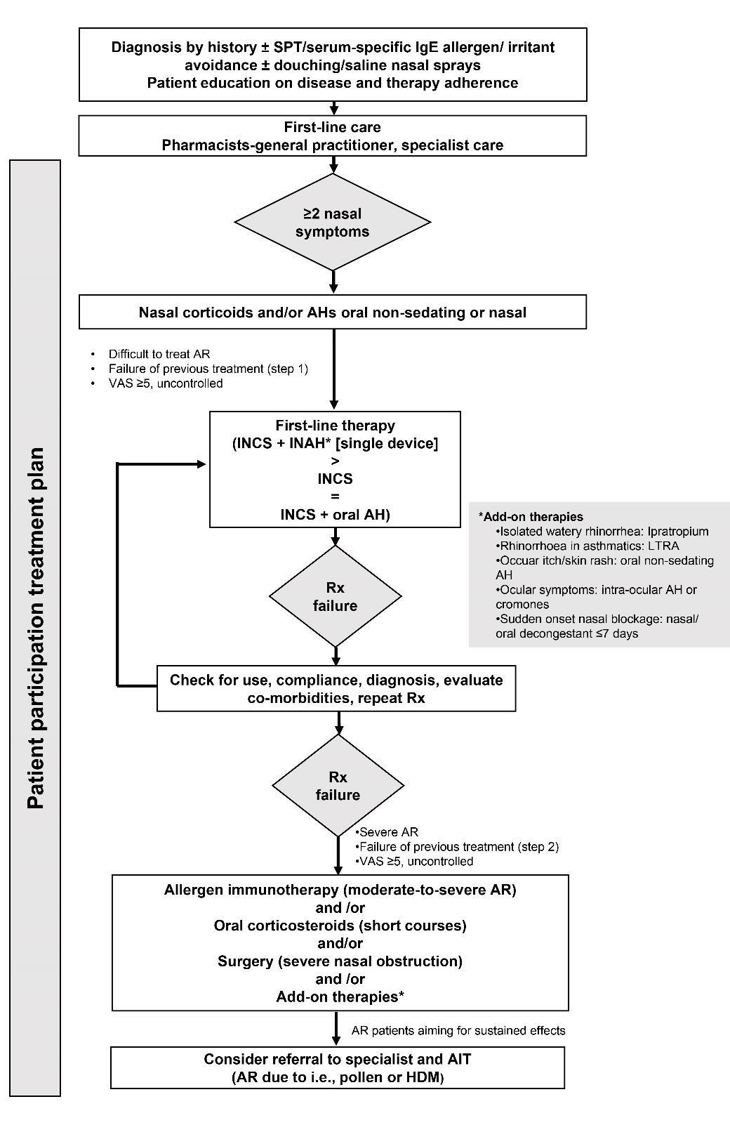
CONCLUSIONS
MP-AzeFlu, an intranasal unique formulation of AZE and FP in single device, is easy to use, effective, and safe in patients with moderate-to-severe AR. MP-AzeFlu provides faster and sustained symptom control versus first-line treatments in phase 3 studies and in real-life settings, making it an
ideal treatment consideration for AR. It offers a better symptomatic relief versus an INAH and INCS irrespective of disease severity, age, response criteria or phenotype. MPAzeFlu represents a first-line therapy in patients with moderate-to-severe AR. Reallife data obtained via mHealth technologies should offer new insights into the phenotypes and management of AR. These consensus statements provide clarity on essential practical issues like indications for use of MP-AzeFlu dose, efficacy and safety. These statements are likely to provide guidance to physicians on optimizing the use of MPAzeFlu for patients with AR in clinical practice (Fig.1).
Abbreviations: AH, anti-histamine; AR, allergic rhinitis; Ig, immunoglobin; INCS, intranasal corticosteroid, INAH, intranasal antihistamine; LTRA, leukotriene receptor antagonist; SPT, skin prick test; VAS, visual analogue scal
The Journal of V.N. Karazin Kharkiv National University. 2022 60
Fig. 1. Treatment algorithm for the management of allergic rhinitis in Ukraine. This figure has been adapted from Hellings et al.[17]
ACKNOWLEDGEMENTS
The authors thank Dr. Palash Kumar Das (Ph.D.) for providing medical writing and Gaurav Anand (both Tata Consultancy Services, India) for additional editorial
REFERENCES
assistance towards the development of this article,fundedbyViatrisInc.
FUNDING
ThisworkwassponsoredbyViatrisInc.
1. Watts AM, Cripps AW, West NP, Cox AJ. Modulation of allergic inflammation in the nasal mucosa of allergic rhinitis sufferers with topical pharmaceutical agents. Front Pharmacol. 2019; 10: 294. doi: 10.3389/fphar.2019.00294.
2. Shamji MH, Sharif H, Layhadi JA, Zhu R, Kishore U, Renz H. Diverse immune mechanisms of allergen immunotherapy for allergic rhinitis with and without asthma. J Allergy Clin Immunol. 2022; 149 (3): 791801. doi: 10.1016/j.jaci.2022.01.016.
3. World Allergy Association. In-depth review of allergic rhinitis. Available from: https://www.worldallergy.org/education-and-programs/education/allergic-disease-resourcecenter/professionals/in-depth-review-of-allergic-rhinitis; 2020 [Last accessed 30 December 2021].
4. Puhlik B, Buleza B, Yakovenko O, Chonka Y. The state of allergic service and spread of allergic diseases in Ukraine. Balneo Research Journal. 2013; 4 (1): 46–48. doi:10.12680/balneo.2013.1041.
5. Duda L, Okhotnikova O, Sharikadze O et al. Epidemiological characteristics of allergic rhinitis in children in Ukraine. Accessed from: http://eapcongress.com/e-poster///files/DudaLyudmyla_1000.pdf; 2019 [Last accessed 30 December 2021].
6. Umanets TR, Buratynska AA, Rudnev OO, Lapshyn VF, Antipkin YG. Sensitization profile in children with allergic respiratory diseases. Аsthma and Allergy. 2020; 3: 29–34. doi: 10.1186/s12948-019-0112-4.
7. Nathan RA. The burden of allergic rhinitis. Allergy Asthma Proc. 2007; 28 (1): 3–9. doi: 10.2500/aap.2007.28.2934.
8. Valovirta E, Myrseth SE, Palkonen S. The voice of the patients: allergic rhinitis is not a trivial disease. Curr Opin Allergy Clin Immunol. 2008; 8 (1): 1–9. doi: 10.1097/ACI.0b013e3282f3f42f.
9. Ciprandi G, Incorvaia C, Scurati S et al. Patient-related factors in rhinitis and asthma: the satisfaction with allergy treatment survey. Curr Med Res Opin. 2011; 27 (5): 1005–1011. doi: 10.1185/03007995.2011.559580.
10.Dymista® Summary of product characteristics (SmPC) and patient information leaflet (PIL). Accessed from: https://www.dymista.co.uk/en-gb/hcp/dymista/smpc-and-pil; 2019 [Last accessed 14 December 2021].
11.Smoke. Fluticasone, combinations. Accessed from: https://mozdocs.kiev.ua/likiview.php?id=38764; 2016 [Last accessed 26 December 2021].
12.Scadding GK, Kariyawasam HH, Scadding G et al. BSACI guideline for the diagnosis and management of allergic and non-allergic rhinitis (Revised Edition 2017; First edition 2007). Clin Exp Allergy. 2017; 47 (7): 856
889. doi: 10.1111/cea.12953.
13.Bousquet J, Khaltaev N, Cruz AA et al. World Health Organization; GA(2)LEN; AllerGen. allergic rhinitis and its impact on asthma (ARIA) 2008 update (in collaboration with the World Health Organization, GA(2)LEN and AllerGen). Allergy. 2008; 63 (Suppl 86): 8–160. doi: 10.1111/j.13989995.2007.01620.x.
14.Pawankar R, Mori S, Ozu C, Kimura S. Overview on the pathomechanisms of allergic rhinitis. Asia Pac Allergy. 2011; 1 (3): 157–167. doi: 10.5415/apallergy.2011.1.3.157.
15.Small P, Keith PK, Kim H. Allergic rhinitis. Allergy Asthma Clin Immunol. 2018; 14 (Suppl 2): 51. doi: 10.1186/1710-1492-7-S1-S3.
16.Heinzerling L, Mari A, Bergmann KC et al. The skin prick test - European standards. Clin Transl Allergy. 2013; 3 (1): 3. doi: 10.1186/2045-7022-3-3.
17.Hellings PW, Scadding G, Bachert C et al. EUFOREA treatment algorithm for allergic rhinitis. Rhinology 2020; 58 (6): 618
622. doi: 10.4193/Rhin20.246.
18.Scadding GK, Durham SR, Mirakian R et al. BSACI guidelines for the management of allergic and nonallergic rhinitis. Clin Exp Allergy. 2008; 38 (1): 19–42. doi: 10.1111/j.1365-2222.2007.02888.x.
19.Gani F, Pozzi E, Crivellaro MA et al. The role of patient training in the management of seasonal rhinitis and asthma: clinical implications. Allergy. 2001; 56 (1): 65–68. doi: 10.1034/j.1398-9995.2001.00794.x.
20.Canonica GW, Triggiani M, Senna G. 360 degree perspective on allergic rhinitis management in Italy: a survey of GPs, pharmacists and patients. Clin Mol Allergy. 2015; 13: 25. doi: 10.1186/s12948-015-0029-5.
Series «Medicine». Issue 45 61
–
–
21.Sheikh A, Khan-Wasti S, Price D, Smeeth L, Fletcher M, Walker S. Standardized training for healthcare professionals and its impact on patients with perennial rhinitis: a multi-center randomized controlled trial. Clin Exp Allergy. 2007; 37 (1): 90–99. doi: 10.1111/j.1365-2222.2006.02619.x.
22.Matricardi PM, Dramburg S, Alvarez-Perea A et al. The role of mobile health technologies in allergy care: An EAACI position paper. Allergy. 2020; 75 (2): 259–272. doi: 10.1111/all.13953.
23.Bousquet J, Caimmi DP, Bedbrook A et al. Pilot study of mobile phone technology in allergic rhinitis in European countries: the MASK-rhinitis study. Allergy. 2017; 72 (6): 857–865. doi: 10.1111/all.13125.
24.Bousquet J, Ansotegui IJ, Anto JM et al. Mobile technology in allergic rhinitis: Evolution in management or revolution in health and care? J Allergy Clin Immunol Pract. 2019; 7 (8): 2511–2523. doi: 10.1016/j.jaip.2019.07.044.
25.Elliott T, Shih J, Dinakar C, Portnoy J, Fineman S. American college of allergy, asthma & immunology position paper on the use of telemedicine for allergists. Ann Allergy Asthma Immunol. 2017; 119 (6): 512
517. doi: 10.1016/j.anai.2017.09.052.
26.Tripodi S, Giannone A, Sfika I et al. Digital technologies for an improved management of respiratory allergic diseases: 10 years of clinical studies using an online platform for patients and physicians. Ital J Pediatr. 2020; 46 (1): 105. doi: 10.1186/s13052-020-00870-z.
27.Florack J, Brighetti MA, Perna S et al. Comparison of six disease severity scores for allergic rhinitis against pollen counts a prospective analysis at population and individual level. Pediatr Allergy Immunol. 2016; 27 (4): 382–390. doi: 10.1111/pai.12562.
28.Di Fraia M, Tripodi S, Arasi S et al. Adherence to prescribed e-diary recording by patients with seasonal allergic rhinitis: Observational study. J Med Internet Res. 2020; 22 (3): e16642. doi: 10.2196/16642.
29.Bousquet J, Van Cauwenberge P, Bachert C et al. Requirements for medications commonly used in the treatment of allergic rhinitis. European Academy of Allergy and Clinical Immunology (EAACI), Allergic Rhinitis and its Impact on Asthma (ARIA). Allergy. 2003; 58 (3): 192–197. doi: 10.1034/j.13989995.2003.00054.x.
30.Bousquet J, Anto JM, Bachert C et al. Allergic rhinitis. Nat Rev Dis Primers. 2020; 6 (1): 95. doi: 10.1038/s41572-020-00227-0.
31.Horak F, Zieglmayer UP. Azelastine nasal spray for the treatment of allergic and nonallergic rhinitis. Expert Rev Clin Immunol. 2009; 5 (6): 659–669. doi: 10.1586/eci.09.38.
32.Kaszuba SM, Baroody FM, deTineo M, Haney L, Blair C, Naclerio RM. Superiority of an intranasal corticosteroid compared with an oral antihistamine in the as-needed treatment of seasonal allergic rhinitis. Arch Intern Med. 2001; 161: 2581–2587. doi: 10.1001/archinte.161.21.2581.
33.Brożek JL, Bousquet J, Agache I et al. Allergic Rhinitis and its Impact on Asthma (ARIA) guidelines2016 revision. J Allergy Clin Immunol. 2017; 140 (4): 950–958. doi: 10.1016/j.jaci.2017.03.050.
34.Weiner JM, Abramson MJ, Puy RM. Intranasal corticosteroids versus oral H1 receptor antagonists in allergic rhinitis: systematic review of randomised controlled trials. BMJ. 1998; 317 (7173): 1624–1629. doi: 10.1136/bmj.317.7173.1624.
35.Nielsen LP, Dahl R. Comparison of intranasal corticosteroids and antihistamines in allergic rhinitis: a review of randomized, controlled trials. Am J Respir Med. 2003; 2 (1): 55–65. doi: 10.1007/BF03256639.
36.Erdoğan BA, Sanlı A, Paksoy M, Altın G, Aydın S. Quality of life in patients with persistent allergic rhinitis treated with desloratadine monotherapy or desloratadine plus montelucast combination. Kulak Burun Bogaz Ihtis Derg. 2014; 24 (4): 217–224. doi: 10.5606/kbbihtisas.2014.48108.
37.Cingi C, Oghan F, Eskiizmir G, Yaz A, Ural A, Erdogmus N. Desloratadine-montelukast combination improves quality of life and decreases nasal obstruction in patients with perennial allergic rhinitis. Int Forum Allergy Rhinol. 2013; 3 (10): 801–806. doi: 10.1002/alr.21185.
38.Lipworth B, Newton J, Ram B, Small I, Schwarze J. An algorithm recommendation for the pharmacological management of allergic rhinitis in the UK: a consensus statement from an expert panel. NPJ Prim Care Respir Med. 2017; 27 (1): 3. doi: 10.1038/s41533-016-0001-y.
39.Ratner PH, Hampel F, Van Bavel J et al. Combination therapy with azelastine hydrochloride nasal spray and fluticasone propionate nasal spray in the treatment of patients with seasonal allergic rhinitis. Ann Allergy Asthma Immunol. 2008; 100 (1): 74–81. doi: 10.1016/S1081-1206(10)60408-5.
40.Bjermer L, Westman M, Holmström M, Wickman MC. The complex pathophysiology of allergic rhinitis: scientific rationale for the development of an alternative treatment option. Allergy Asthma Clin Immunol. 2019; 15: 24. doi: 10.1186/s13223-018-0314-1.
41.D’Addio A, Ruiz N, Mayer M, Murray R, Bachert C. Deposition characteristics of a new allergic rhinitis nasal spray (MP29-02*) in an anatomical model of the human nasal cavity. Clin Transl Allergy. 2015a; 5 (Suppl 4): P40. doi: 10.1186/2045-7022-5-S4-P40.
2022 62
The Journal of V.N. Karazin Kharkiv National University.
–
42.D’Addio A, Ruiz N, Mayer M, Berger WE, Meltzer EO. Quantification of the distribution of azelastine HCl/fluticasone propionate nasal spray in an anatomical model of the human nasal cavity. J Allergy Clin Immunol. 2015b; 135: AB218. doi: https://doi.org/10.1016/j.jaci.2014.12.1648.
43.Derendorf H, Munzel U, Petzold U et al. Bioavailability and disposition of azelastine and fluticasone propionate when delivered by MP29-02, a novel aqueous nasal spray. Br J Clin Pharmacol. 2012; 74 (1):125–133. doi: 10.1111/j.1365-2125.2012.04222.x.
44.Meltzer EO, Bachert C, Mayer MJ et al. Deposition characteristics of a novel intranasal formulation of azelastine hydrochloride plus fluticasone propionate in an anatomic model of the human nasal cavity. Allergy Asthma Proc. 2020; 41 (4): 265–270. doi: 10.2500/aap.2020.41.200028.
45.Bousquet J, Schünemann HJ, Hellings PW et al. MACVIA clinical decision algorithm in adolescents and adults with allergic rhinitis. J Allergy Clin Immunol. 2016; 138 (2): 367–374.e2. doi: 10.1016/j.jaci.2016.03.025.
46.Seidman MD, Gurgel RK, Lin SY et al. Clinical practice guideline: allergic rhinitis executive summary. Otolaryngol Head Neck Surg. 2015; 152 (2): 197–206. doi: 10.1177/0194599814562166.
47.Bousquet J, Schünemann HJ, Togias A et al. Next-generation allergic rhinitis and its impact on asthma (ARIA) guidelines for allergic rhinitis based on grading of recommendations assessment, development and evaluation (GRADE) and real-world evidence. J Allergy Clin Immunol. 2020; 145 (1): 70–80.e3. doi: 10.1016/j.jaci.2019.06.049.
48.Meltzer E, Ratner P, Bachert C et al. Clinically relevant effect of a new intranasal therapy (MP29-02) in allergic rhinitis assessed by responder analysis. Int Arch Allergy Immunol. 2013; 161 (4): 369–377. doi: 10.1159/000351404.
49.Hampel FC, Ratner PH, Van Bavel J et al. Double-blind, placebo-controlled study of azelastine and fluticasone in a single nasal spray delivery device. Ann Allergy Asthma Immunol. 2010; 105 (2): 168–173. doi: 10.1016/j.anai.2010.06.008.
50.Carr W, Bernstein J, Lieberman P et al. A novel intranasal therapy of azelastine with fluticasone for the treatment of allergic rhinitis. J Allergy Clin Immunol. 2012; 129 (5): 1282–1289.e10. doi: 10.1016/j.jaci.2012.01.077.
51.Meltzer EO, LaForce C, Ratner P, Price D, Ginsberg D, Carr W. MP29-02 (a novel intranasal formulation of azelastine hydrochloride and fluticasone propionate) in the treatment of seasonal allergic rhinitis: a randomized, double-blind, placebo-controlled trial of efficacy and safety. Allergy Asthma Proc. 2012; 33 (4): 324–332. doi: 10.2500/aap.2012.33.3587.
52.Price D, Shah S, Bhatia S et al. A new therapy (MP29-02) is effective for the long-term treatment of chronic rhinitis. J Investig Allergol Clin Immunol. 2013; 23 (7): 495–503.
53.Bousquet J, Meltzer EO, Couroux P et al. Onset of Action of the Fixed Combination Intranasal AzelastineFluticasone Propionate in an Allergen Exposure Chamber. J Allergy Clin Immunol Pract. 2018; 6 (5): 1726
1732.e6. doi: 10.1016/j.jaip.2018.01.031.
54.Dollner R, Lorentz LP, Sheyauldeen S, Kuhl H, Steinsvag S. Real-life effectiveness of a new allergic rhinitis therapy (MP29-02) in Norway. Allergy. 2015; 70 (502). doi: 10.1186/2045-7022-5-S4-P37.
55.Haahr P, Jacobsen C, Blegvad S, Christensen M, Kuhl H, Nielsen K. Real-life effectiveness of a new allergic rhinitis therapy (MP29-02) in Denmark. Allergy. 2015; 70 (500). doi: 10.1002/alr.22258.
56.Klimek L, Bachert C, Mosges R et al. Effectiveness of MP29-02 for the treatment of allergic rhinitis in real-life: Results from a noninterventional study. Allergy Asthma Proc. 2015; 36 (1) :40–47. doi: 10.2500/aap.2015.36.3823.
57.Stjarne P, Strand V, Theman K, Kuhl H, Ehngage A. Real-life effectiveness of a new allergic rhinitis therapy (MP29-02) in Sweden. Clin Transl Allergy. 2015; 5 (Suppl 4): P37. doi:10.1186/2045-7022-5-S4P37.
58.Klimek L, Bergmann KC, Biedermann T et al. Visual analogue scales (VAS): Measuring instruments for the documentation of symptoms and therapy monitoring in cases of allergic rhinitis in everyday health care: Position Paper of the German Society of Allergology (AeDA) and the German Society of Allergy and Clinical Immunology (DGAKI), ENT Section, in collaboration with the working group on Clinical Immunology, Allergology and Environmental Medicine of the German Society of Otorhinolaryngology, Head and Neck Surgery (DGHNOKHC). Allergo J Int. 2017; 26 (1): 16–24. doi: 10.1007/s40629-0160006-7.
59.Klimek L, Bachert C, Stjarne P et al. MP-AzeFlu provides rapid and effective allergic rhinitis control in real life: A pan-European study. Allergy Asthma Proc. 2016; 37 (5): 376–386. doi: 10.2500/aap.2016.37.3979.
60.Kaulsay R, Nguyen DT, Kuhl HC. Real-life effectiveness of MP-AzeFlu in Irish patients with persistent allergic rhinitis, assessed by visual analogue scale and endoscopy. Immun Inflamm Dis. 2018; 6 (4): 456–464. doi: 10.1002/iid3.237.
Series «Medicine». Issue 45 63
–
61.Price D, Klimek L, Gálffy G et al. Allergic rhinitis and asthma symptoms in a real-life study of MPAzeFlu to treat multimorbid allergic rhinitis and asthma. Clin Mol Allergy. 2020; 18: 15. doi: 10.1186/s12948-020-00130-9.
62.Berger W, Sher E, Gawchik S, Fineman S. Safety of a novel intranasal formulation of azelastine hydrochloride and fluticasone propionate in children: A randomized clinical trial. Allergy Asthma Proc. 2018; 39 (2): 110–106. doi: 10.2500/aap.2018.39.4116.
63.Price D, Bousquet J, Hellings P et al. Short and long-term safety of MP29-02*: a new therapy for the treatment of allergic rhinitis. Clin Transl Allergy. 2013; 3 (Suppl 2): O15. doi: 10.1517/14740338.2016.1122755.
64.Cappella A, Durham SR. Allergen immunotherapy for allergic respiratory diseases. Hum Vaccin Immunother. 2012; 8 (10):1499–1512. doi: 10.4161/hv.21629.
65.Larsen JN, Broge L, Jacobi H. Allergy immunotherapy: the future of allergy treatment. Drug Discov Today. 2016; 21 (1): 26–37. doi: 10.1016/j.drudis.2015.07.010.
66.Alvaro-Lozano M, Akdis CA, Akdis M et al. EAACI allergen immunotherapy user's guide. Pediatr Allergy Immunol. 2020; 31 (Suppl 25): 1–101. doi: 10.1111/pai.13189.
67.Campisi L, Yong PFK, Kasternow B, Karim MY. Illustrative case series and narrative review of therapeutic failure of immunotherapy for allergic rhinitis. Allergy Rhinol (Providence). 2020; 11: 2152656720943822. doi: 10.1177/2152656720943822.
68.Chhabra N, Houser SM. Surgical options for the allergic rhinitis patient. Curr Opin Otolaryngol Head Neck Surg. 2012; 20 (3): 199–204. doi: 10.1097/MOO.0b013e328352b821.
69.Karatzanis AD, Fragiadakis G, Moshandrea J, Zenk J, Iro H, Velegrakis GA. Septoplasty outcome in patients with and without allergic rhinitis. Rhinology. 2009; 47 (4): 444–449. doi: 10.4193/Rhin08.126.
70.Ivanovich KA, Bondareva GP, Thao NPT. Allergic rhinitis complicated by hypertrophy of the mucous membrane of nasal turbinates in patients of Northern Vietnam. Biomedical Research and Therapy. 2020; 7: 3813–3818. doi: 10.15419/bmrat.v7i6.609.
71.Scadding G, Price D, El-Shanawany T, Ahmed S, Ray J, Sargur R, Kumar N. Multicentre, noninterventional study to assess the profile of patients with uncontrolled rhinitis prescribed a novel formulation of azelastine hydrochloride and fluticasone propionate in a single spray in routine clinical practice in the UK. BMJ Open. 2017; 7 (4): e014777. doi: 10.1136/bmjopen-2016-014777.
РЕКОМЕНДАЦІЯ З АЛГОРИТМУ ФАРМАКОЛОГІЧНОГО ЛІКУВАННЯ
АЛЕРГІЧНОГО РИНІТУ В УКРАЇНІ: КОНСЕНСУСНА ЗАЯВА ЕКСПЕРТА
Біль Богдан, Чопік Валентина, Дєєва Юлія, Дитятковська Євгенія, Гогунська Інна, Попович Василь, Романюк Лілія, Уманець Тетяна, Заболотна Діана, Зайков Сергій
Алергічний риніт в Україні зростає загрозливими темпами. Клінічна картина алергічного риніту в сучасних умовах змінюється в бік більш важких і змішаних форм. Алергічний риніт, особливо від середнього до тяжкого, негативно впливає на якість життя пацієнтів. Досягнення адекватного контролю симптомів має важливе значення для успішного лікування алергічного риніту в основному залежить від фармакотерапії. Більшість пацієнтів використовують кілька ліків, щоб швидше та краще контролювати симптоми, але симптоми можуть зберігатися. З появою нових комбінованих методів лікування, таких як інтраназальний препарат азеластину гідрохлориду та флутиказону пропіонату в одному пристрої (MP-AzeFlu), як-от Dymista®, більшість симптомів алергічного риніту можна ефективно лікувати. MP-AzeFlu діє синергетично та блокує два важливі патофізіологічні шляхи, залучені в ранню та пізню фазу реакцій захворювання, забезпечуючи швидке полегшення всіх симптомів, пов’язаних з алергічним ринітом. Загалом у розробці цієї консенсусної заяви взяли участь 13 експертів з України, Німеччини та Індії. Провідний автор підготував питання, що стосуються діагностики, лікування, прихильності до лікування та реальних доказів алергічного риніту в Україні, і було узгоджено зі співавторами та групою експертів. Цей консенсус досягнуто через керівні твердження та рекомендації, засновані на літературних доказах (результати останніх досліджень, рандомізованих та порівняльних досліджень), клінічній практиці та особистому досвіді використання
MP-AzeFlu при АР алергологами/імунологами/отоларингологами з України. Ця консенсусна заява мала на меті допомогти лікарям-практикам у виборі відповідних стратегій лікування, полегшити оптимальне використання MP-AzeFlu та забезпечити симптоматичне полегшення для пацієнтів з алергічним ринітом в Україні.
КЛЮЧОВІ СЛОВА: алергічний риніт, азеластину гідрохлорид, флутиказону пропіонат, лікування захворювань, Україна
64
The Journal of V.N. Karazin Kharkiv National University. 2022
ІНФОРМАЦІЯ ПРО АВТОРІВ
Біль Богдан Назарович, ЛОР, Київська міська клінічна лікарня-9, вул. Ризька, 1, Київ, Україна, 04201; e-mail: bohdanbill@gmail com
Чопик Валентина Николаевна, лікар-алерголог, Медичний центр клінічної імунології та алергології, вул. Пекарська, 69, Львів, Україна, e-mail: chopyakv@ukr net
Дєєва Юлія Валеріївна, ЛОР, Національний медичний університет імені Богомольця, бульвар Т. Шевченка, 13, Київ, 01601, Україна, e-mail: deyeva@bigmir net
Дітятківська Євгенія Михайлівна, лікар-алерголог, КУ «Дніпровське клінічне об'єднання швидкої медичної
допомоги», вул. Шмідта, 26, Дніпро, 49006, Україна, e-mail: Dneprallergy@i ua
Гогунская Инна Володимирівна, врач-аллерголог, Інститут отоларингології НАМН, вул. Зоологічна, 3, Київ, Україна, e-mail: innagogunskaya@gmail com
Попович Василь Іванович, ЛОР, Івано-Франківський національний медичний університет, вул. Галицька, 2, Івано-Франківськ, Україна, 76000; e-mail: popovychvasyl@gmail.com
Романюк Лілія Іванівна, лікар-алерголог, Київська міська клінічна лікарня, вул. Кондратюка, 8, Київ, Україна, e-mail: lilia.rmnk@yahoo.com
Уманець Тетяна Миколаївна, лікар-алерголог, Інститут педіатрії, акушерства та гінекології, вул. П. Майбороді, 8, Київ, Україна, 04050; e-mail: tetiana umanets@gmail com
Заболотна Діана Дмитрівна, ЛОР, ГУ Інститут отоларингології імені проф. О. С. Коломійченка НАМН, вул.
Зоологічна, 3, Київ, Україна, 03680; e-mail: dianazab@ukr net
Зайков Сергій Вікторович, лікар-алерголог, Національний університет здоров’я України імені Шупика, вул. Дорогожицька, 9, Київ, Україна, 04112; e-mail: zaikov1960@gmail com
Для цитування:
Біль Б, Чопік В, Дєєва Ю, Дитятковська Є, Гогунська І, Попович В, Романюк Л, Уманець Т, Заболотна Д, Зайков С. РЕКОМЕНДАЦІЯ З АЛГОРИТМУ ФАРМАКОЛОГІЧНОГО ЛІКУВАННЯ АЛЕРГІЧНОГО РИНІТУ В УКРАЇНІ: КОНСЕНСУСНА ЗАЯВА ЕКСПЕРТА. Вісник Харківського національного університету імені В.Н. Каразіна. Серія «Медицина». 2022;45:51–65 DOI: 0.26565/2313-6693-2022-45-06
Conflicts of interest: author has no conflict of interest to declare. Конфлікт інтересів: відсутній. Отримано: 03 09.2022 Прийнято до друку: 20 11.2022
Received: 09 03.2022
Accepted: 11 20.2022
Series «Medicine». Issue 45 65
Богун Н. Ю. B, C, D, Бринза М. С. A, E, F A – концепція та дизайн дослідження; B – збір даних; C – аналіз та інтерпретація даних; D –написання статті; E – редагування статті; F – остаточне затвердження статті
Вступ. Фібриляція передсердь є однією з найбільш розповсюджених, клінічно значущих серцевих аритмій і пов’язана з підвищеним ризиком смертності, госпіталізацій та погіршення якості життя. Поширеність фібриляції передсердь в середньому становить 1 % і має тенденцію до зростання з віком. Загальновідомо, що аберації гормонів щитоподібної залози мають проаритмогенний ефект і підвищують ризик серцево-судинних захворювань. На даний час для лікування фібриляції передсердь активно застосовується радіочастотна абляція, яка в більшості випадків має високу ефективність і стійкий ефект. Однак, рецидив фібриляції передсердь може спостерігатися у деяких пацієнтів навіть після процедури радіочастотної абляції. Відомо, що функція щитоподібної залози тісно пов’язана з успіхом радіочастотної абляції та ризиком рецидиву фібриляції передсердь після процедури.
Метою цього літературного огляду є визначення того, що вже відомо про вплив рівнів тиреоїдних та тиреотропного гормону на результат процедури радіочастотної абляції фібриляції передсердь, та проаналізувати попередні дослідження.
Матеріали та методи. Було проведено повний огляд літератури про фібриляцію передсердь та взаємозв’язок між тиреоїдними та тиреотропним гормонами та рецидивом фібриляції передсердь після радіочастотної абляції, щоб вивчити вплив рівнів гормонів на успіх радіочастотної абляції та рецидив фібриляції передсердь після процедури.
Результати. Дані значної кількості досліджень свідчать про те, що частота рецидивів фібриляції передсердь залишається високою після радіочастотної абляції, та гормони щитоподібної залози навіть у межах референтних значень впливають на успіх радіочастотної абляції. Також дані свідчать про те, що перед процедурою у пацієнтів із дисфункцією щитоподібної залози, навіть за наявності субклінічних форм, може знадобитися кращий моніторинг функції щитоподібної залози, що може знизити частоту рецидивів фібриляції передсердь.
Висновки. Слід зазначити, що оцінка і корекція всіх модифікуючих факторів ризику перед виконанням радіочастотної абляції може запобігти рецидивам надшлуночкових пароксизмальних аритмій, поліпшити прогноз і загальну якість життя у пацієнтів даної групи.
КЛЮЧОВІ СЛОВА: фібриляція передсердь, радіочастотна абляція, щитоподібна залоза, рецидив, огляд ІНФОРМАЦІЯ ПРО АВТОРІВ Богун Наталія Юріївна, асистент кафедри пропедевтики внутрішньої медицини і фізичної реабілітації медичного факультету, Харківський національний університет імені В. Н. Каразіна, майдан Свободи, 6, Харків, Україна, 61022, e-mail nbogun@karazin.ua. ORCID ID: https://orcid.org/0000-0002-8176-8171
Бринза Марія Сергіївна, к. мед. н., доцент, завідувачка кафедри пропедевтики внутрішньої медицини і фізичної реабілітації медичного факультету, Харківський національний університет імені В.Н. Каразіна, майдан Свободи, 6, Харків, Україна, 61022, e-mail m.brynza@karazin.ua. ORCID ID: https://orcid.org/0000-0002-8260-3600
Для цитування: Богун НЮ, Бринза МС. ВПЛИВ РІВНІВ ГОРМОНІВ ЩИТОПОДІБНОЇ ЗАЛОЗИ НА РЕЗУЛЬТАТ РАДІОЧАСТОТНОЇ АБЛЯЦІЇ ФІБРИЛЯЦІЇ ПЕРЕДСЕРДЬ: ЛІТЕРАТУРНИЙ ОГЛЯД. Вісник Харківського національного університету імені В. Н. Каразіна. Серія «Медицина». 2022; 45:66–76 DOI: 10.26565/2313-66932022-45-07
ВСТУП
Відповідно до твердження Європейського товариства кардіологів фібриляція передсердь (ФП) визначається, як надшлуночкова тахіаритмія з некоординованою електричною активацією
Богун Н. Ю., Бринза М. С., 2022
передсердь і, отже, неефективним їхскороченням[1].
ФП є однією з найбільш розповсюджених, клінічно значущих серцевих аритмій. Поширеність ФП в середньомустановить 1% імаєтенденцію до зростання з віком: 10% населення у
The Journal of V.N. Karazin Kharkiv National University. 2022 66 УДК
DOI: 10.26565/2313-6693-2022-45-07
616.12-008.313-085.846:577.175.44.03
ВПЛИВ РІВНІВ ГОРМОНІВ ЩИТОПОДІБНОЇ ЗАЛОЗИ НА РЕЗУЛЬТАТ РАДІОЧАСТОТНОЇ АБЛЯЦІЇ ФІБРИЛЯЦІЇ ПЕРЕДСЕРДЬ: ЛІТЕРАТУРНИЙ ОГЛЯД
віці старше 80 років страждає на ФП, і
приблизно 70% випадків ФП у пацієнтів
вікомвід 65 до 85 років.Очікується,щов
міру старіння населення кількість
пацієнтів з ФП збільшиться на 150% до
2050 року, при цьому більше 50%
пацієнтівзФПбудутьстарше 80 років [2].
Частота ФП більша у чоловіків, ніж у жінок,алецярізницязменшуєтьсязвіком [3].
ФП може мати несприятливі наслідки, пов'язанізізменшеннямсерцевоговикиду, а також з утворенням тромбів. ФП
залишається однією з основних причин
інсульту, серцевої недостатності, раптової
смерті та інших серцево-судинних подій [4].Встановлено,щопацієнтизФПмають
в п’ять-сім разів більший ризик інсульту,
ніж населення в цілому. Більш того, інсульти,вторинніповідношеннюдоФП, маютьгіршийпрогноз,ніжупацієнтівбез ФП[2].ПацієнтизФПмаютьпідвищений
ризик смертності, госпіталізацій та
погіршення якості життя. За даними літератури, ризик смертіу пацієнтів з ФП
збільшується в 5 разів внаслідок серцевосудинних захворювань порівняно з загальноюпопуляцією[6].
Серед чисельних факторів, що є
необхідними для підтримки нормальної
структури та функціювання серцево-судинноїсистеми(ССС),однимізвизначних єрівеньтиреоїднихгормонів(ТГ).
В Україні близько 44% загальної
ендокринної патології складають
захворювання щитоподібної залози (ЩЗ) [7].ПоширеністьдисфункціїЩЗзалежить
від віку, статі, географічних факторів та етнічної приналежності. У Сполучених
Штатах та Європі гіпотиреоз більш розповсюджений ніж гіпертиреоз: поширеність явного гіпертиреозу коливається в межах від 0,2 до 1,3%, тоді як поширеність гіпотиреозу становить від 1–2 % до 7 % у осіб у віці від 85 до 89 років [8]. У всьому світі дефіцит йоду залишається
однією з лідируючих причин гіпотиреозу, поступаючись аутоімунним захворюванням.Такожцепитанняєвкрайактуальним
для нашої країни: дефіцит йоду в тій чи
іншій мірі відчуває практично все
населення України – лідирують в цьому
списку західні області [9]. Тим часом аутоімунний тиреоїдит зустрічається у біля5%населеннявУкраїні[7].
Встановлено, що ТГ відіграють фундаментальне значення в підтримці морфології і функцій ССС за допомогою геномних (ядерних) і негеномних ефектів. Численні геномні ефекти ТГ на серце опосередковуються шляхом взаємодії зі специфічними рецепторами до ТГ (TRs), які пов’язані з диференціюванням і зростанням клітин, скорочувальною функцією, пейсмекерською активністю і провідною системою. Негеномні ефекти ТГ на кардіоміоцити і загальний периферичний судинний опір не передбачають зв’язування з ядерними рецепторами. Зазначені ефекти розвиваються досить швидко й обумовлені впливом на транспорт іонів кальцію, натрію й калію через клітинну мембрану, а також транспорт глюкози, на функції мітохондрій і різні внутрішньоклітинні сигнальнішляхи [10,11].
Рівень тиреотропного гормону (ТТГ) дуже чутливий до дисфункції гормонів ЩЗ. Навіть незначні зміни рівнів тиреоїдних гормонів (ТГ) можуть призводити до коливань показників ТТГ. ЦепідтверджуєрольТТГ(привідсутності захворювань гіпоталамо-гіпофізарної системи) як найбільш чутливого маркера функціїЩЗ [12].
Пацієнти з дисфункцією ЩЗ (гіпертиреозом чи гіпотиреозом) мають підвищенийризиксерцево-судиннихподій[13].
Гіпертиреоз є добре відомим проаритмогенним фактором, частота ФП у пацієнтівзпідтвердженимгіпертиреозомв анамнезі складає 16–60 % за даними Rossetal.(2016)[14]. Роль гіпотиреозу в аритмогенезі передсердь менш визнана і докінцяневивчена.
Гіпертиреоз характеризується посиленим синтезом та секрецією ТГ. Явний гіпертиреоз викликає гіпердинамічний стан ССС (високий серцевий викид з низьким системним судинним опором), який асоціюється з прискореним серцебиттям, посиленням систолічної та діастолічної функції лівого шлуночка та збільшенням поширеності надшлуночковихтахіаритмій [12].
Гіпотиреоз діагностується при низькому рівні ТГ, що призводить до підвищення рівню ТТГ, тоді як субклінічний гіпотиреоз діагностується при підвищені рівню ТТГ вище верхньої межі
Series «Medicine». Issue 45 67
The Journal of V.N. Karazin Kharkiv National University. 2022
референтного діапазону з нормальним рівнемТГ.Гіпотиреоз,однак,асоціюється з брадикардією, дисліпідемією, гіпертензією, атеросклерозом, зниженням частоти серцевих скорочень та збільшенням ризику інфаркту міокарда. Візуальні дослідження пацієнтів із субклінічним гіпотиреозом показали незначні зміни серцевоїдіяльності[15].
Трьома основними цілями терапії у пацієнтів з ФП є полегшення симптомів, попередження тахікардії-опосередкованої кардіоміопатії та зменшення ризику інсульту(антикоагулянтнатерапія).Перші
дві цілі досягаються за допомогою стратегії контролю частоти (контроль частоти серцевих скорочень) або контролю ритму (відновлення синусового ритму), та покращення якості життя [16]. Для пацієнтів, у яких обрана стратегія контролю ритму, радіочастотна абляція (РЧА) та антиаритмічна терапія є двома основними терапевтичними стратегіями для зниження частоти або усунення епізодівФП [17].
Стратегія вибору методу лікування залежить від наступних факторів: ступень вираженості симптомів, ймовірність успішної кардіоверсії, наявність супутніх захворювань та бажання пацієнта проводитиабляціюФП.
На даний час РЧА – це добре зарекомендований метод лікування, який активно використовується при ФП для пацієнтів із симптоматичною, стійкою до лікарських засобів ФП [4]. Згідно з консенсусним висновком експертів HRS / EHRA/ECAS/APHRS/ SOLACEвід2020 року, катетерна абляція рекомендована в якості терапії першої лінії при симптоматичної пароксизмальної або персистуючоїформахФП[18].
ЗначнимиперевагамиРЧАвпорівнянні з медикаментозною антиаритмічною терапією є: збільшення ймовірності збереження синусового ритму, поліпшення якості життя, у пацієнтів із СН знижується кількість госпіталізацій і смертність від різних причин, зменшується питома вага непароксизмальних формФПіймовірністьїхвиникнення[19].
Метою РЧА ФП є усунення тригерів, які її ініціюють та/або модифікація субстрату для ФП. Перш за все, це досягається за допомогою ізоляції
легеневих вен (ЛВ), які зазвичай функціонують як джерела ФП, викликаючи передчасніскорочення[20].
Проте, незважаючи на те, що ця методика широко використовується у
всьому світі, великий відсоток пацієнтів
може не підтримувати синусовий ритм і
мати рецидив після РЧА ФП [21]. Слід зазначити, що навіть у разі рецидиву ФП
після процедури РЧА, переносимість ФП
значно краща в порівняні з рецидивом
післяфармакологічноїкардіоверсії.
Раннім рецидивом (РР) вважається
будь-яка надшлуночковатахіаритмія (ФП, тріпотіння передсердь або надшлуночкова
тахікардія), що триває щонайменше 30
секунд і виникає впродовж перших 3 місяців після абляції. РР після РЧА ФП є досить поширеним явищем; ризик виникнення РР частіше спостерігається
незабаром після процедури, та зменшується протягом наступних днів [22].
Наразі вважається, що РР, на відміну від пізнього рецидиву (ПР), які відбуваютьсячерез3–12місяцяпісляРЧА, є минущим феноменом, пов’язаним із постабляційними запальними реакціями й автономноюдисфункцієюсерця.Оскільки більшість РР закінчуються відновленням синусового ритму без жодних медичних втручань, 3місяці після РЧА називають «сліпим періодом» (blanking period). Хоча РР не завжди супроводжується ПР протягом тривалого періоду спостереження, результати багатьох досліджень підтвердили, що РР є незалежним фактором ризику ПР [23]. Це також підтверджується даними нещодавно опублікованого мета-аналізу (2021 р.), що пацієнти, які не відчували ФП під час «сліпого періоду», мали набагато нижчу поширеністьФПпісля[24].
Основними предикторами РР ФП є
наявність судинних захворювань у пацієнтів та кардіоверсія під час процедури РЧА, тривалість зубця Р була довшою у
пацієнтів із рецидивуючою ФП [25].
Дослідники YunGi Kim та ін. (2021р.)
відмітили, що пацієнти, у яких
спостерігався РР, мали більше ліве
передсердя (ЛП), гіршу гемодинаміку в
придатку ЛП та більшу поширеність
непароксизмальної форми ФП (коли вона
прогресуєдоперсистуючої)таСН [26].
68
Кількадослідженьзтривалимперіодом
спостереження (>5 років) запропонували
та запровадили новий підтип рецидивів
ФП – дуже пізній рецидив (ДПР).
Рецидиви ФП через довгий час після РЧА
єрезультатомпосиленняструктурнихзмін
міокарду передсердь. Прогресування
фіброзу передсердь, збільшення лівого
передсердятанесприятливеелектричнета
молекулярне ремоделювання тканини
міокарда беруть участь у цих типах
рецидивів.Такимчином,ДПРєпричиною
останньої стадії електричної хвороби
передсердь, хоча необхідні подальші
дослідженняуцьомунапрямку [22].
Було показано, що РЧА приблизно до
80 % є ефективною для пароксизмальної
форми ФП та до 60% для персистуючої
форми ФП. РЧА забезпечує кращу якість
життя, ніж антиаритмічна терапія, зменшуючи симптоми, частоту і тривалість ФП у пацієнтів, які погано
переносять ФП, знижуючи клас EHRA [27]. РЧА є ефективним та безпечним
втручанням для відновлення та підтримки
синусового ритму, однак незважаючи на нововведення в технології РЧА, частота
рецидивів ФП, за різними даними, коливаєтьсявід20%до50 %[28].
Отже, актуальним є вивчення модифікуючих факторів, що впливають на рецидив ФП та можуть виступати предикторамиуспішностіРЧАпривідборі пацієнтів до процедури. На сьогоднішній день існує значна кількість предикторів рецидивуФПпісляРЧА,наприклад,збільшення правого передсердя, більше двох
спроб проведення РЧА, тривалість ФП та збільшення лівого передсердя (> 43 мм) [29]. Також жіноча стать, вік > 60 років, тип ФП, а саме тривало персистуюча форма, наявність супутніх захворювань, таких як серцева недостатність та порушення функції нирок були включені до списку предикторів рецидиву після
РЧА[30].
Існує ряд кількість досліджень, які
демонструють зв’язок ТГ із ризиком
виникнення ФП. Однак також цікаво
вивчити їх вплив на рецидив ФП.
Загальновідомо, що функція ЩЗ тісно
пов'язаназуспіхомРЧАтарецидивомФП
після процедури; проте складний
взаємозв’язок між гормонами ЩЗ та
рецидивом ФП усе ще потребує
дослідження. Даних стосовно взаємозв’язкурівняТТГ,якмаркерафункціїЩЗ, таефективностіРЧАвкраймало,томуцеє актуальним напрямком подальшого вивчення.
Усі змінні моделі (наприклад, рівень ТТГ) можна виміряти перед абляцією, і тому це може бути використано до процедур з метою прогнозування ймовірності ризику рецидиву. Напевно, ці зусилля допомогли б ретельніше оцінити прогностичні показники щодо повторних епізодівФПпісляРЧА[31].
Нами було знайдено декілька досліджень стосовно впливу рівнів ТГ та ТТГ на результат хірургічної процедури РЧА у пацієнтів з ФП. Однак дані суперечливі.
У Нью-Йорку Юхуа Чжан та ін. (2013 р.)провелипершеекспериментальне дослідження, що вивчало вплив різних рівнівТГ,відгіпотиреозудогіпертиреозу, на аритмогенез ФП на моделі щурів після тиреоїдектомії. Результати цього дослідження продемонстрували, що як гіпотиреоз, так і гіпертиреоз пов'язані з підвищеною вразливістю ФП порівняно з щурами, які мали нормалізований рівень ТГ. Дані цього дослідження чітко продемонстрували, що гіпотиреоз призводить до збільшення сприйнятливості до ФП,подібнодогіпертиреозу.
Хоча гіпотиреоз і гіпертиреоз збільшують частоту ФП, вони по-різному впливають на інші електрофізіологічні параметри передсердь. Наприклад, гіпертиреоз збільшує частоту серцевих скорочень (ЧСС) і скорочує ефективний рефрактерний період (ЕРП) передсердь, тоді як гіпотиреоз зменшує ЧСС і подовжує ЕРП передсердь. Також дослідники виявили, що інтерстиціальний колаген передсердь у гіпотиреоїдних щурів був підвищений, тоді як вміст колагену не збільшився у гіпертиреоїдних щурів. Цей висновок узгоджується з повідомленнями про те, що гіпотиреоз сприяє розвитку фіброзу міокарда. Підвищений фіброз у гіпотиреоїдних щурів може сприяти збільшенню ЕРП передсердь. Фіброз передсердь може призвести до повільної провідності та збільшення неоднорідності провідності, тимсамимсприяючиформуваннюre-entry тапідвищеннювразливостіФП.
Series «Medicine». Issue 45 69
The Journal of V.N. Karazin Kharkiv National University.
Отже,отриманіданідемонструють,що гіпотиреоз, як і гіпертиреоз, збільшує ризик ФП та підкреслюють потенційне клінічнезначеннякорекціїдисфункціїЩЗ для запобігання ФП та інших серцевих аритмій[32].
Bruereтаін.(2015р.)визначилизв’язок між наявності в анамнезі дисфункції ЩЗ та фактором ризику тромбоемболічних ускладнень (системна емболія, інсульт) у пацієнтів з ФП. У дослідженні брало участь 8962 пацієнтів з ФП: у 141 був гіпертиреоз в анамнезі, тоді як у 540
відмічався гіпотиреоз в анамнезі, решта пацієнтів не мали дисфункції ЩЗ. Наявність в анамнезі гіпотиреозу, таким чином, на 300% перевищувала показник гіпертиреозу у пацієнтів з ФП протягом 10 років. У своєму дослідженні автори зробили висновок, що гіпертиреоз в анамнезі не є незалежним фактором ризику розвитку інсульту / системної емболії при ФП, тоді як гіпотиреоз асоціювався з вищим ризиком розвитку кровотеч. Докази, викладені в цій статті, свідчать про те, що не можна залишати позаувагоюзв'язокгіпотиреозузФП[33].
WanwarangWongcharoenтаін.(2015р.) вивчали вплив гіпертиреозу на віддалені результатикатетерноїабляціїупацієнтівз ФП, резистентною до фармакологічного лікування. Пацієнти з гіпертиреозом в анамнезі мали значно більшу кількість ектопій ЛВ та більш високу поширеність ектопічних вогнищ нелегеневих вен порівняно з пацієнтами з нормальною функцією ЩЗ. Як результат, це призводило до більш високої частоти рецидивів ФП після одноразової процедурикатетерноїабляції[34].
Ще раніше Ri-Bo Tang та ін. (2010 р.) досліджували вплив високого нормального рівню тироксину (Т4) на рецидив ФП після катетерної абляції. 244 пацієнти з пароксизмальною формою ФП були поділені на чотири квартилі.
Рецидив ФП частіше спостерігався у пацієнтів, які належали до найвищого четвертого квартилю. Отже, можна зробити висновок, що пацієнти навіть з нормальною функцією ЩЗ мають підвищений ризик рецидиву ФП після катетерної абляції у разі рівню тироксину наверхніймежіреферентнихзначень [35].
Pedro A Sousa та ін. (2015р.) також вивчали вплив вільного тироксину на результати процедури катетерної абляції
ЛП. ТТГ та вільний тироксин вимірювалися у пацієнтів, однак ТТГ не був предиктором рецидиву ФП в той час, як вільний тироксин впливав на успішність
процедури абляції ЛП, навіть якщо він знаходитьсявмежахреферентнихзначень [36].
Shao-Bin Wei та ін. (2018р.) досліджували вплив вільного трийодтироніну
(Т3) на рецидив ФП після РЧА. Були оцінені рівні вільного T3, вільного Т4 і
ТТГ в сироватці. Пацієнти були розділені
на п'ять квартилів залежно від рівнів T3, T4 і ТТГ відповідно. Результатами цього
дослідженнябуливпливяквисокого,такі
низького рівню вільного Т3 на рецидив
ФП після РЧА; також рівень вільного Т4
на верхній межі референтного значення
був пов’язан з рецидивом ФП; однак не було виявлено зв’язку між нормальним рівнемТТГірецидивомФП[37]. ЯпонськедослідженняItsuroMorishima та ін. (2018р.) вивчало зв’язок між рівем ТТГ та рецидивом надшлуночкової тахіаритміїпісляРЧА. Це перше дослідження, яке вивчало довгостроковий результат РЧА ФП залежно від рівню ТТГ. Основними висновкамидослідженнябулитакі:
1) у пацієнтів з діагнозом явного та субклінічного гіпотиреозу спостерігалась
значно більша частота рецидивів надшлуночкової тахіаритмії, ніж у пацієнтів із нормальним ТТГ після процедурРЧА;
2) у пацієнтів з високим нормальним рівнем ТТГ(квартіль4)в межах нормальногодіапазонуТТГспостерігаєтьсязначно більшачастотарецидивівнадшлуночкової тахіаритмії, ніж у нижчих квартилів ТТГ (квартіль1–3);
3) ця тенденція виявилася більш вираженою у підгрупі пароксизмальної ФП,ніжунепароксизмальнійФП.
Усукупностіціданівказуютьнате,що пацієнти з успішною процедурою РЧА можуть піддаватися більшому ризику рецидиву ФП, якщо рівень ТТГ знаходиться у верхній межі референтних значень та у пацієнтів з субклінічним або явним (маніфестним) гіпотиреозом в анамнезі[38].
70
2022
–
Основними недоліками цього
дослідження було те, що пацієнти з
захворюваннямиЩЗванамнезіаботі,які
приймали ліки від захворювань ЩЗ не
були виключені. Хоча ТТГ є важливим
параметром для оцінки функції ЩЗ і
зазвичай є першим кроком у діагностиці
захворювань ЩЗ, ряд лікарських засобів
та нетиреоїдні захворювання можуть
впливатинайогорівень.
Крімтого,тривалістьспостереженнята
кількість досліджуваних пацієнтів було
різною. Також це дослідження включало
лишеяпонськупопуляцію.
Таким чином, усі ці причини можуть призвестидоможливихрізнихрезультатів
урізнихдослідженнях.Дляпідтвердження
зв’язку між субклінічним гіпотиреозом і
рецидивом ФП необхідні тривалі дослідження.
У
новітньому китайському досліджені
ZheyueShuтаін.(2021р.)вивчавсязв'язок
між рівнями ТГ у межах нормального
лабораторного діапазону та ризиком рецидивуФПпісляРЧА.
Основним висновком цього дослідження було те, що пацієнти з високими нормальними рівнями вільного Т3 або
вільного T4, навіть у межах референтних
значень, мали значно вищу частоту
рецидивів надшлуночкової тахіаритмії
після РЧА. Ці результати показують, що клінічнаувагадорівнягормонівЩЗможе допомогтиулікуванніФП.
У
порівнянні з попереднім
дослідженням, у цьому дослідженні
додатково оцінювали, чи розрізнявся
вплив гормонів ЩЗ на ФП у різних
популяціях. Новим відкриттям було
зв'язок між вищим рівнем вільним T4 або
вільним T3 і рецидивом ФП, який був
більш виражений у молодих пацієнтів.
Можливо, що структурне ремоделювання
й зміни вегетативної нервової системи
можуть служити основними факторами
підтримки ФП у літніх людей. Однак
вплив вільного T3 або вільного T4 на
рецидив ФП не розрізнялися в залежності
відстаті[21].
Проте недоліками цього дослідження є
те,що,по-перше,кількістьзареєстрованих
пацієнтів була відносно невеликою.
Порогові рівні для вільного T4 або вільного T3 для диференціації рецидиву надшлуночкової тахіаритмії були недос-
тупні через малу кількість пацієнтів у досліджуваній вибірці. Потрібне більш широке дослідження, щоб підтвердити відповіднірівніTГ.По-друге,дослідження
тривало рік,протенеобхіднебільшдовше спостереження для отримання віддалених результатів, та дослідження впливу гормонів ЩЗ на різні періоди рецидиву (ранній,пізнійідужепізнійрецидив).Потретє,функціюЩЗвимірювалилишеодин раз під час надходження до клініки, але рівень гормонів ЩЗ може змінюватися з часом. Для обговорення зв’язку між змінами функції ЩЗ та результатами абляції слід проводити серійні вимірювання. По-четверте, хоча для виявлення рецидиву ФП використовувалися різні моніторингові та оглядові тести, частота рецидивів могла бути недооцінена, оскільки деякі безсимптомні короткі епізоди ФП, можливо, не були виявлені.
По-п’яте, оскільки рецидив виявлявся лікуючими лікарями, вони могли бути схильні до упередженості спостереження.
І нарешті, у цьому дослідженні не досліджували РР (<3 місяців), тому у частини пацієнтів із рецидивом міг бути РР.
ВИСНОВКИ
Наведені літературні дані змушують
звернути увагу на те, що частота рецидивів фібриляції передсердь залишаєтьсявисокоюпіслярадіочастотної абляції,та,середіншихфакторів,гормони щитоподібної залози навіть у межах референтних значень впливають на успіх радіочастотноїабляції.
Узв’язкузцим,особливоїактуальності набуває визначення специфічних допроцедурних маркерів, які серед інших факторів можуть впливати на результат процедури радіочастотної абляції у пацієнтів з фібриляцією передсердь для оптимізації визначення кандидатів на процедуру.
У зв’язку з неоднозначністю літературних даних подальше дослідження дало б можливість відповісти на питання чи є рівні гормонів щитоподібної залози незалежними предикторами рецидиву фібриляції передсердь після радіочастотноїабляції.
Series «Medicine». Issue 45 71
СПИСОК
ЛІТЕРАТУРИ
1. Hindricks G, Potpara T, Dagres N, Arbelo E, Bax JJ, Blomström-Lundqvist C, et al. 2020 ESC guidelines for the diagnosis and management of atrial fibrillation developed in collaboration with the European Association for Cardio-Thoracic Surgery (eacts). European Heart Journal. 2020; 42 (5): 373–498. DOI: https://doi.org/10.1093/eurheartj/ehaa612
2. Rao MP, Pokorney SD, Granger CB. Atrial fibrillation: A review of recent studies with a focus on those from the Duke Clinical Research Institute. Scientifica. 2014; 2014: 1–11. DOI: https://doi.org/10.1155/2014/901586
3. Michelena HI, Ezekowitz MD. Atrial fibrillation: are there gender differences?. J Gend Specif Med. 2000; 3 (6): 44
49.
4. Liżewska-Springer A, Dąbrowska-Kugacka A, Lewicka E, Drelich Ł, Królak T, Raczak G. Echocardiographic predictors of atrial fibrillation recurrence after catheter ablation: A literature review. Cardiology Journal. 2020;27(6):848–56. DOI: https://doi.org/10.5603/CJ.a2018.0067
5. Lee E, Choi E-K, Han K-D, Lee HJ, Choe W-S, Lee S-R, et al. Mortality and causes of death in patients with atrial fibrillation: A nationwide population-based study. PLOS ONE. 2018;13(12). doi: https://doi.org/10.1371/journal.pone.0209687
6. Кravchenko VI, Тоvkay ОА, Rakov ОV, Тronko МD. Епідеміологія автоімунного тиреоїдиту. INTERNATIONAL JOURNAL OF ENDOCRINOLOGY (Ukraine). 2021;17(2):136
44.
7. Taylor PN, Albrecht D, Scholz A, Gutierrez-Buey G, Lazarus JH, Dayan CM, et al. Global epidemiology of hyperthyroidism and hypothyroidism. Nature Reviews Endocrinology. 2018;14(5):301–16. DOI: https://doi.org/10.1371/10.1038/nrendo.2018.18
8. Biban BG, Lichiardopol C. Iodine Deficiency, Still a Global Problem?. Curr Health Sci J. 2017;43(2):103111. DOI: https://doi.org/10.1371/10.12865/CHSJ.43.02.01
9. Князькова ИИ, Беловол АН. Дисфункция щитовидной железы и сердечная недостаточность. Здоров’я України. 2016; https://health-ua.com/article/4874-disfunktciya-shitovidnoj-zhelezy-iserdechnaya-nedostatochnost.
10.Bielecka-Dabrowa A, Mikhailidis DP, Rysz J, Banach M. The mechanisms of atrial fibrillation in hyperthyroidism. Thyroid Research. 2009; 2 (1).
11.Sheehan MT. Biochemical testing of the thyroid: TSH is the best and, oftentimes, only test needed – a review for Primary Care. Clinical Medicine & Research. 2016;14(2):83–92.
12.Jabbar A, Pingitore A, Pearce SH, Zaman A, Iervasi G, Razvi S. Thyroid hormones and cardiovascular disease. Nature Reviews Cardiology. 2016; 14 (1): 39–55.
13.Reddy V, Taha W, Kundumadam S, Khan M. Atrial fibrillation and hyperthyroidism: A literature review. Indian Heart Journal. 2017; 69 (4): 545–50.
14.Udovcic M, Pena RH, Patham B, Tabatabai L, Kansara A. Hypothyroidism and the heart. Methodist DeBakey Cardiovascular Journal. 2017; 13 (2): 55.
15.Xu J, Luc JG, Phan K. Atrial fibrillation: Review of Current Treatment Strategies. Journal of Thoracic Disease. 2016; 8 (9).
16.Baman JR, Passman RS. The future of long-term monitoring after catheter and surgical ablation for atrial fibrillation. J Cardiovasc Electrophysiol. 2022;33(8):1911-1918. doi:10.1111/jce.15375.
17.Hindricks G, Potpara T, Dagres N, Arbelo E, Bax JJ, Blomström-Lundqvist C, et al. 2020 ESC guidelines for the diagnosis and management of atrial fibrillation developed in collaboration with the European Association for Cardio-Thoracic Surgery (eacts). European Heart Journal. 2020; 42 (5): 373–498.
18.Мешкова МС, Доронін ОВ. Обґрунтування доцільності застосування радіочастотної катетерної деструкції з метою лікування фібриляції передсердь. Український журнал серцево-судинної хірургії. 2020;1 (38):57-61. doi:10.30702/ujcvs/20.3803/015057-061.
19.Management strategies for atrial fibrillation. Atrial Fibrillation in Clinical Practice. 2000;:129–35.
20.Shu Z, Chen M, Wang Q, Lai J, Zhao J, Zhuo C, et al. High-normal thyroid function and recurrence of atrial fibrillation after catheter ablation: A prospective observational study [Internet]. Cardiology. Karger Publishers; 2021 [cited 2022Dec14]. Available from: https://www.karger.com/Article/FullText/517092
21.Garvanski I. Predictors of recurrence of AF in patients after radiofrequency ablation [Internet]. ECR Journal. 2019 [cited 2022Dec14]. Available from: https://www.ecrjournal.com/articles/predictorsrecurrence-af-patients-after-radiofrequency-ablation
22.Huang RD, Lin JJ, Gong KZ, Chen LL, Fan L, Zhang FL, et al. Comparison of amiodarone and propafenone in blanking period after radiofrequency catheter ablation in patients with atrial fibrillation: A propensity score-matched study. BioMed Research International. 2020;2020:1
8.
2022 72
The Journal of V.N. Karazin Kharkiv National University.
–
–
–
23.Brynza M, Lisova N. Two-year outcomes of pulmonary vein isolation and cava-tricuspid isthmus radiofrequency ablation vs pharmacological only antiarrhythmic therapy: A single center experience. EUREKA: Health Sciences. 2022;(1):10–6.
24.Zink MD, Chua W, Zeemering S, Biase L, Antoni LB, David C. Predictors of recurrence of atrial fibrillation within the first 3 months after ablation [Internet]. Academic.oup.com. OUP Academic. Oxford University Press; 2020 [cited 2022Dec14]. Available from: https://academic.oup.com/europace/article/22/9/1337/5877521
25.Kim YG, Boo KY, Choi J-I, Choi YY, Choi HY, Roh S-Y, et al. Early recurrence is reliable predictor of late recurrence after radiofrequency catheter ablation of atrial fibrillation. JACC: Clinical Electrophysiology. 2021;7(3):343–51.
26.Boersma L, Koźluk E, Maglia G, de Sousa J, Grebe O, Eckardt L, et al. Paroxysmal and persistent atrial fibrillation ablation outcomes with the pulmonary vein ablation catheter gold duty-cycled phased radiofrequency ablation catheter: Quality of life and 12-month efficacy results from the Gold Atrial Fibrillation Registry. EP Europace. 2020; 22 (6): 888–96.
27.Chew DS, Black-Maier E, Loring Z, Noseworthy PA, Packer DL, Exner DV, et al. Diagnosis-to-ablation time and recurrence of atrial fibrillation following catheter ablation. Circulation: Arrhythmia and Electrophysiology. 2020; 13 (4).
28.Sawasaki K. Predictors of atrial fibrillation recurrence after ablation. Atrial Fibrillation – Diagnosis and Management in the 21st Century. 2022
29.Sultan A, Lüker J, Andresen D, Kuck KH, Hoffmann E, Brachmann J, et al. Predictors of atrial fibrillation recurrence after catheter ablation: Data from the German Ablation Registry. Scientific Reports. 2017;7(1).
30.Dretzke J, Chuchu N, Agarwal R, Herd C, Chua W, Fabritz L, et al. Predicting recurrent atrial fibrillation after catheter ablation: A systematic review of Prognostic Models. EP Europace. 2020;22(5):748–60.
31.Zhang Y, Dedkov EI, Teplitsky D, Weltman NY, Pol CJ, Rajagopalan V, et al. Both hypothyroidism and hyperthyroidism increase atrial fibrillation inducibility in rats. Circulation: Arrhythmia and Electrophysiology. 2013; 6 (5): 952–9.
32.Bruere H, Fauchier L, Bernard Brunet A, Pierre B, Simeon E, Babuty D, et al. History of thyroid disorders in relation to clinical outcomes in atrial fibrillation. The American Journal of Medicine. 2015;128(1):30
7.
33.Wongcharoen W, Lin Y-J, Chang S-L, Lo L-W, Hu Y-F, Chung F-P, et al. History of hyperthyroidism and long-term outcome of catheter ablation of drug-refractory atrial fibrillation. Heart Rhythm. 2015;12(9):1956–62.
34.Tang RB;Liu DL;Dong JZ;Liu XP;Long DY;Yu RH;Hu FL;Wu JH;Liu XH;Ma CS; High-normal thyroid function and risk of recurrence of atrial fibrillation after catheter ablation [Internet]. Circulation journal : official journal of the Japanese Circulation Society. U.S. National Library of Medicine. Available from: https://pubmed.ncbi.nlm.nih.gov/20508382/
35.Sousa PA, Providência R, Albenque JP, et al. Impact of Free Thyroxine on the Outcomes of Left Atrial Ablation Procedures. Am J Cardiol. 2015;116(12):1863-1868. doi:10.1016/j.amjcard.2015.09.028.
36.Wei S-bin, Wang W, Liu N, Chen J, Guo X-yuan, Tang R-bo, et al. U-shaped association between serum free triiodothyronine and recurrence of atrial fibrillation after catheter ablation. Journal of Interventional Cardiac Electrophysiology. 2018;51(3):263–70.
37.Morishima I, Okumura K, Morita Y, et al. High-Normal Thyroid-Stimulating Hormone Shows a Potential Causal Association With Arrhythmia Recurrence After Catheter Ablation of Atrial Fibrillation [published correction appears in J Am Heart Assoc. 2018 Oct 2;7(19):e02707]. J Am Heart Assoc. 2018;7(14):e009158. Published 2018 Jul 12. doi:10.1161/JAHA.118.009158.
INFLUENCE OF THE LEVELS OF THYROID HORMONES ON THE RESULT OF RADIOFREQUENCY ABLATION FOR ATRIAL FIBRILLATION: LITERATURE REVIEW
Bogun N. Yu. B, C, D, Brynza M. S. A, E, F
A – research concept and design; B – collection and/or assembly of data; C – data analysis and interpretation; D –writing the article; E – critical revision of the article; F – final approval of the article
Introduction. Atrial fibrillation is one of the most common, clinically significant cardiac arrhythmias and is associated with an increased risk of mortality, hospitalization, and deterioration in the quality of life. The prevalence of atrial fibrillation averages 1% and tends to increase with age. It is well known that aberrations of thyroid hormones have a proarrhythmogenic effect and increase the risk of cardiovascular disease. Currently, radiofrequency ablation is actively used to treat atrial fibrillation, which in most cases has high efficiency and lasting effect. However, recurrence of atrial fibrillation may occur in some patients even after
Series «Medicine». Issue 45 73
–
radiofrequency ablation. It is known that thyroid function is closely related to the success of radiofrequency ablation and risk of relapse of atrial fibrillation after the procedure.
The aim of this literature review is to determine what is already known about the effect of thyroid and thyroid-stimulating hormones levels on the outcome of radiofrequency ablation of atrial fibrillation and to analyze previous studies.
Materials and methods. A complete review of the literature on atrial fibrillation and the relationship between thyroid and thyroid hormones and recurrence of atrial fibrillation after radiofrequency ablation was conducted to examine the effect of hormone levels on the success of radiofrequency ablation and recurrence of atrial fibrillation after the procedure.
Results. Data from a significant number of studies suggest that the incidence of atrial fibrillation recurrence remains high after radiofrequency ablation and that thyroid hormones affect the success of radiofrequency ablation even within normal ranges. Evidence may also suggest that patients with thyroid dysfunction, even in the presence of subclinical forms, may require better monitoring of thyroid function prior to the procedure, which may reduce the incidence of atrial fibrillation recurrence.
Conclusions. It should be noted that the assessment and correction of all modifying risk factors before radiofrequency ablation can prevent recurrence of supraventricular paroxysmal arrhythmias, improve the prognosis and overall quality of life in patients in this group.
KEY WORDS: atrial fibrillation, radiofrequency ablation, thyroid gland, recurrence, review
INFORMATION ABOUT AUTHORS
Nataliia Bogun, assistant of the department of Propaedeutics of internal medicine and physical rehabilitation, medical school, V. N. Karazin Kharkiv National University, 6, Svobody sq., Kharkiv, Ukraine, 61022; e-mail nbogun@karazin.ua
ORCID ID: https://orcid.org/0000-0002-8176-8171
Mariia Brynza, PhD, associate professor, head of the department of Propaedeutics of internal medicine and physical rehabilitation, medical school, V.N. Karazin Kharkiv National University, 6, Svobody sq., Kharkiv, Ukraine, 61022; email m.brynza@karazin.ua. ORCID ID: https://orcid.org/0000-0002-8260-3600
For citation:
Bogun N, Brynza M. INFLUENCE OF THE LEVELS OF THYROID HORMONES ON THE RESULT OF RADIOFREQUENCY ABLATION FOR ATRIAL FIBRILLATION: LITERATURE REVIEW. The Journal of V. N. Karazin Kharkiv National University. Series «Medicine». 2022; 45:66–76. DOI: 10.26565/2313-6693-2022-45-07
REFERENCES
1. Hindricks G, Potpara T, Dagres N, Arbelo E, Bax JJ, Blomström-Lundqvist C, et al. 2020 ESC guidelines for the diagnosis and management of atrial fibrillation developed in collaboration with the European Association for Cardio-Thoracic Surgery (eacts). European Heart Journal. 2020;42(5):373–498.
2. Rao MP, Pokorney SD, Granger CB. Atrial fibrillation: A review of recent studies with a focus on those from the Duke Clinical Research Institute. Scientifica. 2014;2014:1–11.
3. Michelena HI, Ezekowitz MD. Atrial fibrillation: are there gender differences?. J Gend Specif Med. 2000;3(6):44-49.
4. Liżewska-Springer A, Dąbrowska-Kugacka A, Lewicka E, Drelich Ł, Królak T, Raczak G. Echocardiographic predictors of atrial fibrillation recurrence after catheter ablation: A literature review. Cardiology Journal. 2020;27(6):848–56.
5. Lee E, Choi E-K, Han K-D, Lee HJ, Choe W-S, Lee S-R, et al. Mortality and causes of death in patients with atrial fibrillation: A nationwide population-based study. PLOS ONE. 2018;13(12).
6. Кravchenko VI, Тоvkay ОА, Rakov ОV, Тronko МD. Epidemiolohiya avtoimunnoho tyreoyidytu INTERNATIONAL JOURNAL OF ENDOCRINOLOGY (Ukraine). 2021;17(2):136
44.
7. Taylor PN, Albrecht D, Scholz A, Gutierrez-Buey G, Lazarus JH, Dayan CM, et al. Global epidemiology of hyperthyroidism and hypothyroidism. Nature Reviews Endocrinology. 2018;14(5):301–16.
8. Biban BG, Lichiardopol C. Iodine Deficiency, Still a Global Problem?. Curr Health Sci J. 2017;43(2):103111. doi:10.12865/CHSJ.43.02.01
9. Knyazkova II, Bilovol AN Disfunktsiya shchitovidnoy zhelezy i serdechnaya nedostatochnost'. Zdorov'ya Ukraini. 2016; https://health-ua.com/article/4874-disfunktciya-shitovidnoj-zhelezy-i-serdechnayanedostatochnost.
10.Bielecka-Dabrowa A, Mikhailidis DP, Rysz J, Banach M. The mechanisms of atrial fibrillation in hyperthyroidism. Thyroid Research. 2009;2(1).
11.Sheehan MT. Biochemical testing of the thyroid: TSH is the best and, oftentimes, only test needed – a review for Primary Care. Clinical Medicine & Research. 2016;14(2):83–92.
The Journal of V.N. Karazin Kharkiv National University. 2022 74
–
12.Jabbar A, Pingitore A, Pearce SH, Zaman A, Iervasi G, Razvi S. Thyroid hormones and cardiovascular disease. Nature Reviews Cardiology. 2016;14(1):39–55.
13.Reddy V, Taha W, Kundumadam S, Khan M. Atrial fibrillation and hyperthyroidism: A literature review. Indian Heart Journal. 2017;69(4):545
50.
14.Udovcic M, Pena RH, Patham B, Tabatabai L, Kansara A. Hypothyroidism and the heart. Methodist DeBakey Cardiovascular Journal. 2017;13(2):55.
15.Xu J, Luc JG, Phan K. Atrial fibrillation: Review of Current Treatment Strategies. Journal of Thoracic Disease. 2016;8(9).
16.Baman JR, Passman RS. The future of long-term monitoring after catheter and surgical ablation for atrial fibrillation. J Cardiovasc Electrophysiol. 2022;33(8):1911-1918. doi:10.1111/jce.15375.
17.Hindricks G, Potpara T, Dagres N, Arbelo E, Bax JJ, Blomström-Lundqvist C, et al. 2020 ESC guidelines for the diagnosis and management of atrial fibrillation developed in collaboration with the European Association for Cardio-Thoracic Surgery (eacts). European Heart Journal. 2020;42(5):373–498.
18.Meshkova MS, Doronin OV. Obgruntuvannya dotsilʹnosti zastosuvannya radiochastotnoyi kateternoyi destruktsiyi z metoyu likuvannya fibrylyatsiyi peredserdʹ. Ukrayinsʹkyy zhurnal sertsevo-sudynnoyi khirurhiyi. 2020;1 (38):57-61. doi:10.30702/ujcvs/20.3803/015057-061.
19.Management strategies for atrial fibrillation. Atrial Fibrillation in Clinical Practice. 2000;:129–35.
20.Shu Z, Chen M, Wang Q, Lai J, Zhao J, Zhuo C, et al. High-normal thyroid function and recurrence of atrial fibrillation after catheter ablation: A prospective observational study [Internet]. Cardiology. Karger Publishers; 2021 [cited 2022Dec14]. Available from: https://www.karger.com/Article/FullText/517092
21.Garvanski I. Predictors of recurrence of AF in patients after radiofrequency ablation [Internet]. ECR Journal. 2019 [cited 2022Dec14]. Available from: https://www.ecrjournal.com/articles/predictorsrecurrence-af-patients-after-radiofrequency-ablation
22.Huang RD, Lin JJ, Gong KZ, Chen LL, Fan L, Zhang FL, et al. Comparison of amiodarone and propafenone in blanking period after radiofrequency catheter ablation in patients with atrial fibrillation: A propensity score-matched study. BioMed Research International. 2020;2020:1–8.
23.Brynza M, Lisova N. Two-year outcomes of pulmonary vein isolation and cava-tricuspid isthmus radiofrequency ablation vs pharmacological only antiarrhythmic therapy: A single center experience. EUREKA: Health Sciences. 2022;(1):10–6.
24.Zink MD, Chua W, Zeemering S, Biase L, Antoni LB, David C. Predictors of recurrence of atrial fibrillation within the first 3 months after ablation [Internet]. Academic.oup.com. OUP Academic. Oxford University Press; 2020 [cited 2022Dec14]. Available from: https://academic.oup.com/europace/article/22/9/1337/5877521
25.Kim YG, Boo KY, Choi J-I, Choi YY, Choi HY, Roh S-Y, et al. Early recurrence is reliable predictor of late recurrence after radiofrequency catheter ablation of atrial fibrillation. JACC: Clinical Electrophysiology. 2021;7(3):343–51.
26.Boersma L, Koźluk E, Maglia G, de Sousa J, Grebe O, Eckardt L, et al. Paroxysmal and persistent atrial fibrillation ablation outcomes with the pulmonary vein ablation catheter gold duty-cycled phased radiofrequency ablation catheter: Quality of life and 12-month efficacy results from the Gold Atrial Fibrillation Registry. EP Europace. 2020;22(6):888–96.
27.Chew DS, Black-Maier E, Loring Z, Noseworthy PA, Packer DL, Exner DV, et al. Diagnosis-to-ablation time and recurrence of atrial fibrillation following catheter ablation. Circulation: Arrhythmia and Electrophysiology. 2020;13(4).
28.Sawasaki K. Predictors of atrial fibrillation recurrence after ablation. Atrial Fibrillation - Diagnosis and Management in the 21st Century. 2022;
29.Sultan A, Lüker J, Andresen D, Kuck KH, Hoffmann E, Brachmann J, et al. Predictors of atrial fibrillation recurrence after catheter ablation: Data from the German Ablation Registry. Scientific Reports. 2017;7(1).
30.Dretzke J, Chuchu N, Agarwal R, Herd C, Chua W, Fabritz L, et al. Predicting recurrent atrial fibrillation after catheter ablation: A systematic review of Prognostic Models. EP Europace. 2020;22(5):748–60.
31.Zhang Y, Dedkov EI, Teplitsky D, Weltman NY, Pol CJ, Rajagopalan V, et al. Both hypothyroidism and hyperthyroidism increase atrial fibrillation inducibility in rats. Circulation: Arrhythmia and Electrophysiology. 2013;6(5):952–9.
32.Bruere H, Fauchier L, Bernard Brunet A, Pierre B, Simeon E, Babuty D, et al. History of thyroid disorders in relation to clinical outcomes in atrial fibrillation. The American Journal of Medicine. 2015;128(1):30–7.
33.Wongcharoen W, Lin Y-J, Chang S-L, Lo L-W, Hu Y-F, Chung F-P, et al. History of hyperthyroidism and long-term outcome of catheter ablation of drug-refractory atrial fibrillation. Heart Rhythm. 2015;12(9):1956–62.
34.Tang RB;Liu DL;Dong JZ;Liu XP;Long DY;Yu RH;Hu FL;Wu JH;Liu XH;Ma CS; High-normal thyroid function and risk of recurrence of atrial fibrillation after catheter ablation [Internet]. Circulation journal :
Series «Medicine». Issue 45 75
–
The Journal of V.N. Karazin Kharkiv National University. 2022
official journal of the Japanese Circulation Society. U.S. National Library of Medicine. Available from: https://pubmed.ncbi.nlm.nih.gov/20508382/
35.Sousa PA, Providência R, Albenque JP, et al. Impact of Free Thyroxine on the Outcomes of Left Atrial Ablation Procedures. Am J Cardiol. 2015;116(12):1863-1868. doi:10.1016/j.amjcard.2015.09.028.
36.Wei S-bin, Wang W, Liu N, Chen J, Guo X-yuan, Tang R-bo, et al. U-shaped association between serum free triiodothyronine and recurrence of atrial fibrillation after catheter ablation. Journal of Interventional Cardiac Electrophysiology. 2018;51(3):263–70.
37.Morishima I, Okumura K, Morita Y, et al. High-Normal Thyroid-Stimulating Hormone Shows a Potential Causal Association With Arrhythmia Recurrence After Catheter Ablation of Atrial Fibrillation [published correction appears in J Am Heart Assoc. 2018 Oct 2;7(19):e02707]. J Am Heart Assoc. 2018;7(14):e009158. Published 2018 Jul 12. doi:10.1161/JAHA.118.009158.
Conflicts of interest: author has no conflict of interest to declare. Конфлікт інтересів: відсутній.
Отримано: 12 09.2022
Прийнято до друку: 20.11.2022
Received: 09 12.2022
Accepted: 11 20.2022
76
УДК: 616.857-036.1:578.834.1
DOI: 10.26565/2313-6693-2022-45-08
ОСОБЛИВОСТІ КЛІНІЧНОЇ КАРТИНИ
Фещенко Д. І.B C D , Малик С. Л.A E F , Шевня М. Б.C E
A – концепція та дизайн дослідження; B – збір даних; C – аналіз та інтерпретація даних; D –написання статті; E – редагування статті; F – остаточне затвердження статті Вступ. Covid-19 постав новою проблемою сучасності. Захворюваність у світі стрімко збільшується, а з нею посилюється увага до вивчення механізмів патогенезу вірусу. З’являються нові симптоми персистенції SARS-CoV-2 в організмі людини. Встановлено, що не лише респіраторні прояви спостерігаються в інфікованих осіб, а дедалі більше на перший план постає неврологічна симптоматика. Основним і поширеним проявом інфікування Covid-19 є головний біль у перед- та пост-ковідному періоді. Визначення можливих шляхів потрапляння збудника в нервову клітину, встановлення особливостей Covid-асоційованого головного болю залишається актуальною проблемою
медицини.
Мета роботи. Проаналізувати дані світової наукової літератури стосовно можливих механізмів інвазії вірусу SARS-CoV-2 в нервову систему та їх вплив на розвиток головного болю у перед- та постковідному періоді. Матеріали та методи. Проведений ретроспективний аналіз літератури наукових баз Scopus, PubMed, MedLines, Web of Science за 2020–2022 роки. Результати. Останні дані дослідників свідчать про нейровірулентність збудника. SARS-CoV-2 вражає людину, використовуючи рецептор ангіотензинперетворювального ферменту 2, який представлений в ольфакторній ділянці, корі головного мозку, базальних гангліях, гіпоталамусі, стовбурі головного мозку, ендотеліальних клітинах. Детальні механізми проникнення в центральну нервову систему ще не до кінця вивчені, однак розглядають наступні шляхи передачі: через нюховий нерв в подальшому вражається головний мозок, другий шлях – клітинна інвазія, інфіковані моноцити та макрофаги, що являють собою резервуар для вірусу для подальшого розподілення в інші тканини, наступний механізм – це інфікування ендотеліальних клітин гематоенцефалічного та епітеліальних гематолікворного бар’єра. Механізм появи головного болю дослідники пояснюють через пряму інвазію закінчень трійчастого нерва в порожнині носоглотки, що призводить до активації трійчастосудинної системи. Головний біль спостерігається на початку захворювання та може тривати більше 180 днів після одужання. Він характеризується, як двосторонній, стискаючого характеру, триває годинами, стійкий до звичайних знеболюючих, посилюється вранці, при кашлі та фізичній активності. Висновки. Вірус SARS-CoV-2 призводить до інфікування, використовуючи АСЕ2-рецептори, частина, з яких локалізована у різних відділах ЦНС, що зумовлює виникнення неврологічної симптоматики. Головний біль є одним із найбільш виражених симптомів COVID-19, його характеристики та еволюція можуть відображати різні патофізіологічні механізми, що робить їх вивчення необхідним для ґрунтовнішого розуміння головного болю в перед- та пост-ковідному періоді та пошук варіантів для його усунення.
КЛЮЧОВІ СЛОВА: Covid-19, головний біль, неврологічна симптоматика, передковідний період, постковідний період
ІНФОРМАЦІЯ ПРО АВТОРІВ
Фещенко Дарина Ігорівна, студентка 5 курсу, медичного факультету №2, Вінницький національний медичний університет імені М. І. Пирогова, вул.Пирогова, 56, Вінниця, Вінницька область, Україна, 21018; е-mail: dashafeshchenko2001@gmail.com
Малик Світлана Леонідівна, к. мед. н., доцент кафедри нервових хвороб, Вінницький національний медичний
університет імені М. І. Пирогова, вул.Пирогова, 56, Вінниця, Вінницька область, Україна, 21018; е-mail: Malyksvitlana79@gmail com
Шевня Микола Богданович, студент 5 курсу, медичного факультету №2, Вінницький національний медичний
університет імені М. І. Пирогова, вул.Пирогова, 56, Вінниця, Вінницька область, Україна, 21018; е-mail: nickshevnya@gmail com
Для цитування:
Фещенко ДІ, Малик СЛ, Шевня МБ. ГОЛОВНИЙ БІЛЬ НА ФОНІ КОРОНАВІРУСНОЇ ІНФЕКЦІЇ: ОСОБЛИВОСТІ КЛІНІЧНОЇ КАРТИНИ Вісник Харківського національного університету імені В. Н. Каразіна. Серія «Медицина». 2022;45:77–81 DOI: 0.26565/2313-6693-2022-45-08
Фещенко Д. І , Малик С. Л., Шевня М. Б , 2022
Series «Medicine». Issue 45 77
ГОЛОВНИЙ БІЛЬ НА ФОНІ КОРОНАВІРУСНОЇ ІНФЕКЦІЇ:
ВСТУП
Covid-19 постав новою проблемою сучасності.Захворюваністьусвітістрімко збільшується, а з нею посилюється увага довивченнямеханізмівпатогенезувірусу.
З’являються нові симптоми персистенції
SARS-CoV-2 в організмі людини. Встановлено, що не лише респіраторні прояви спостерігаються в інфікованих осіб, а дедалі більше на перший план постає неврологічна симптоматика. Основним і досить поширеним проявом інфікування Covid-19, а саме його штамами «Альфа», «Бета»і«Омікрон»,єголовнийбіль.Крім того, дедалі частіше цей симптом стає помітним у перед- та пост-ковідному періоді. Визначення можливих шляхів потрапляння збудника в нервову клітину, встановленняособливостей Covid-асоційованого головного болю залишається актуальноюпроблемоюмедицини.
МАТЕРІАЛИ ТА МЕТОДИ
Проведений ретроспективний аналіз літератури наукових баз Scopus, PubMed, MedLines, Web of Science за 2020–2022 роки.
РЕЗУЛЬТАТИ І ОБГОВОРЕННЯ
Останні дані дослідників свідчать про нейровірулентність збудника. Ураження нервової системи може виникати під впливом різних факторів, таких як гіпоксія, ураження декількох органів, сепсис або «цитокіновий шторм». SARSCoV-2 заражає людину, використовуючи рецептор ангіотензинперетворювального ферменту2 (АСЕ2), який у нервовій системі представлений в ольфакторній ділянці, корі головного мозку, базальних гангліях, гіпокампі, гіпоталамусі, стовбурі головного мозку [1], ендотеліальних клітинах [2].
Детальні механізми проникнення в центральну нервову систему (ЦНС) ще не до кінця вивчені, однак розглядають наступні шляхи передачі: проникнувши в організм людини через дихальні шляхи, віруси можуть отримати доступ до головного мозку через нюхову цибулину,використовуючинюховийнерв, в подальшомувражається головний мозок з залученням таламусута стовбурумозку. Другийшлях–клітиннаінвазія,інфіковані моноцити та макрофаги, які уособлюють
собою резервуар для вірусу для подальшого розподілення в інші тканини.
Інфікування моноцитів та лінії моноцитарних клітин призводить до їх активації, на що вказує продукування прозапальнихмедіаторів.Крімтого,аналіз хемотаксису in vitro показав, що рухливість моноцитів до хемокінів була підвищена після гострої або стійкої інфекції SARS-CoV-2. Наступний механізм–цеінфікуванняендотеліальних клітин гематоенцефалічного бар’єра та епітеліальних гематолікворного бар’єра. Пряме вірусне ураження ендотелію або
опосередкована імунна реакція можуть
призвести до поширеної ендотеліальної
дисфункції [2], яка є одним із важливих
чинників мікросудинної дисфункції зі
зміщенням судинної рівноваги в бік
вазоконстрикції з подальшою ішемією
органів, запаленням із супутнім набряком тканиніпрокоагулянтнимстаном. Механізм виникнення головного болю дослідники пояснюють імовірно через прямуінвазіюзакінченьтрійчастогонерва SARS-CoV-2 в порожнині носоглотки, що призводить до активації трійчастосудинної системи [3]. На менінгеальному рівні аферентні закінчення трійчастого
нерва іннервують менінгеальні судини, створюючи трійчасто-судинну систему. Її активація може бути через системне запалення, що сприяє сенсибілізації менінгеальних оболонок, призводить до місцевого вивільнення запальних пептидів, які стимулюють закінчення трійчастого нерва; пряме зв’язування SARS-CoV-2 з кровотоку на рецепторах ACE2, що знаходиться на ендотелії менінгеальних судин, що викликає ендотеліт і, отже, запалення. У носовій порожнині є спеціалізований нюховий та носовий епітелій, причому останній іннервується аферентними закінченнями трійчастого нерва. Опорні клітини нюхового епітелію, в який вбудовані
нюхові нейрони, мають рецептори ACE2, де SARSCoV-2 може зв’язуватися, викликаючи аносмію, симптом, який значноасоціюєтьсязнаявністюголовного болю. На рівні епітелію носа система трійчастогонерваможебутипериферично активована безпосередньою дією SARSCoV-2 на епітелій носа або гілки трійчастогонерва,абонепрямийшлях,що
78
The Journal of V.N. Karazin Kharkiv National University. 2022
включає взаємодію між нюховою та
трійчастою іннервацією. Ці механізми
потребують подальшого вивчення. Крім
того, сумісне існування пептиду,
пов’язаного з геном кальцитоніну (CGRP)
з ангіотензиновою системою в сенсорних
нейронах і гангліях трійчастого нерва, свідчить про те, що рання активація
закінчень трійчастого нерва вірусом може
ініціювати механізми, які призводять до
головного болю. Співвідношення рівня
циркулюючого CGRP і прозапальних
цитокінів інтерлейкіну-6 пояснює
можливий механізм головного болю на
пізнійстадіїзахворюванняCOVID-19.
Головний біль спостерігається на
початку захворювання та може тривати
більше 180 днів після одужання. Він має
відмінності від звичайних головних болів, такі як більша інтенсивність, швидкий
перебіг та стійкість до звичайних
знеболюючих препаратів під час перебігу
захворювання. Особливістю головного
болю від COVID-19 у деяких пацієнтів
була значна втрата ваги (5 кг протягом 3 днів),пов’язаназголовнимболем.Втрату вагинеможнабулопояснитидіареєю,що
триває добу, або втратою апетиту, тому
припускають, що в процес, ймовірно, залучений посилений катаболізм, опосередкований цитокінами та кортизолом, індукованими COVID-19. Дослідники вважають, що швидка втрата ваги зі значно зниженим апетитом також може бутивідмінноюрисоюголовногоболювід COVID-19. Головний біль мав ознаки мігрені [4], такі як пульсація, посилення при звичайних рухах, сенсорні розлади, такі як фотофобія та фонофобія, нудота. Важливо зазначити, що симптоми, пов’язані з мігренню, в першу чергу можутьматицентральнепоходження,тоді як симптоми, пов’язані з головним болем від COVID-19, швидше за все, пов’язані з периферичними механізмами інфекції. Також відмічаються такі особливості головного болю при COVID-19: двосторонній, стискаючого характеру, триваєгодинами,посилюєтьсявранці,при кашлітафізичнійактивності[5].
Наявністьголовногоболювідмічаласьі в пост-ковідному періоді, як один із ключових симптомів. Особливістю постковідної цефалгії є її тривалість більше 6тижнів (рис.1) [6], при відсутностівсіхіншихсимптомів.
Рис. 1. Частота пацієнтів і тривалість постковідної цефалгії
Fig.1. Frequency of patients and duration of postcovid cephalalgia Загальна поширеність головного болю
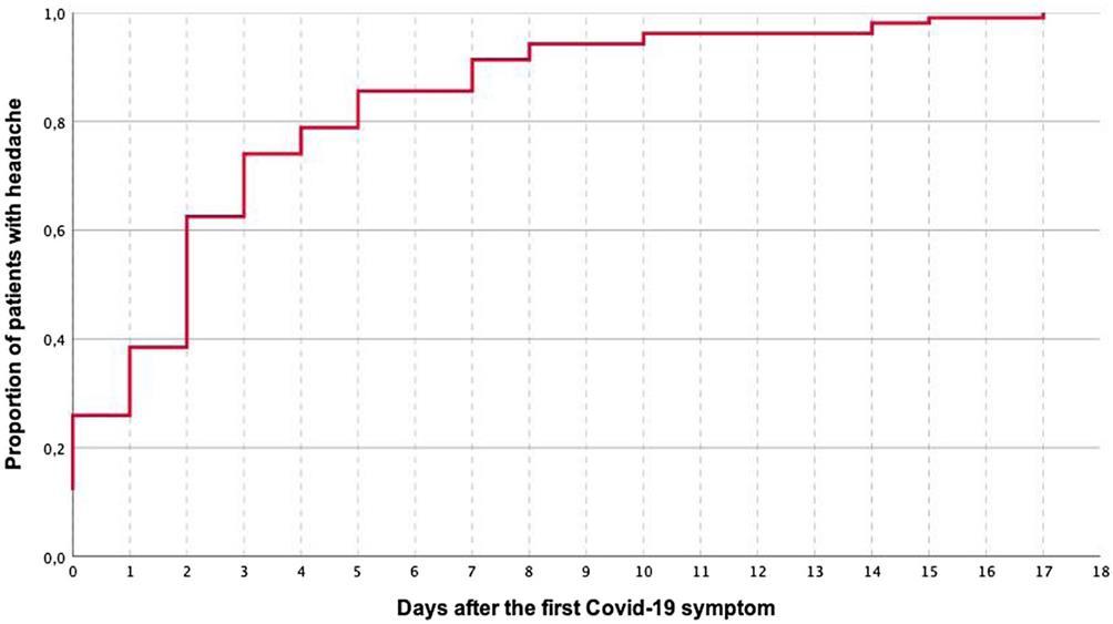
після COVID-19 зменшується після гострої фази та залишається стабільною в
різніперіодиспостереженняпісля COVID19 протягомперших 6 місяців (рис. 2) [7]. Така закономірність спостерігалась і була
Series «Medicine». Issue 45 79
The Journal of V.N. Karazin Kharkiv National University. 2022
подібною у госпіталізованих і негоспіталізованих пацієнтів з COVID-19, підтверджуючиприпущення,щоголовний біль є поширеним симптомом після
COVID-19 у важких пацієнтів (госпіталізованих), а також у пацієнтів середньої та легкої тяжкості (негоспіталізованих).
Рис.2. Тривалість постковідного головного болю Fig.2. Duration of postcovid cephalalgia ВИСНОВКИ
Вірус SARS-CoV-2 призводить до інфікування, використовуючи АСЕ2рецептори, частина, з яких локалізована у різних відділах ЦНС, що зумовлює виникнення неврологічної симптоматики. Головний біль є одним із найбільш виражених симптомів COVID-19, його
СПИСОК ЛІТЕРАТУРИ
характеристики та еволюція можуть відображати різні патофізіологічні механізми, що робить їх вивчення необхідним для ґрунтовнішого розуміння головного болю в перед- та постковідному періоді та пошук варіантів для йогоусунення.
1. The Human Protein Atlas. Available from: https://www.proteinatlas.org/ENSG00000130234-ACE2/brain.
2. Varga Z, Flammer AJ, Steiger P, et al. Endothelial cell infection and endotheliitis in COVID-19 // Lancet 2020. Vol. 395 (10234). P. 1417 - 1418. DOI: 10.1016/S0140-6736(20)30937-5
3. Edoardo Caronna, Alejandro Ballvé, Arnau Llauradó, et al. Headache: A striking prodromal and persistent symptom, predictive of COVID-19 clinical evolution. Cephalalgia 2020 November; 40(13):1410-1421, DOI: 10.1177/0333102420965157
4. Migraine and Post-COVID Headache, American Migraine Foundation, February, 2022, Available from: https://americanmigrainefoundation.org/resource-library/migraine-post-covid-headache/
5. Bolay H, GuЁ l A and Baykan B. COVID-19 is a Real Headache! Headache 2020; 60: 1415–1421. DOI: 10.1111/head.13856
6. David García-Azorín MD, Javier Trigo MD, Blanca Talavera MD, et al. Frequency and Type of Red Flags in Patients With Covid-19 and Headache: A Series of 104 Hospitalized Patients. Headache, September 2020. Vol.60, P. 1664-1672 DOI: 10.1111/head.13927
7. César Fernández-de-las-Peñas, Marcos Navarro-Santana, Víctor Gómez-Mayordomo, et al. Headache as an acute and post-COVID-19 symptom in COVID-19 survivors: A meta-analysis of the current literature. European Journal of Neurology, November 2021, Volume28, P. 3820-3825, DOI: 10.1111/ene.15040

80
HEADACHE ON THE BACKGROUND OF CORONAVIRUS INFECTION: FEATURES OF THE CLINICAL PICTURE
Feshchenko D. I. B,C,D, Malyk S. L. A,E,F, Shevnia M. B.C,E
A
research concept and design; B – collection and/or assembly of data; C – data analysis and interpretation; D
writing the article; E – critical revision of the article; F – final approval of the
Introduction. Covid-19 has become a new problem of our time. Incidence in the world is rapidly increasing, and with it, attention to the study of the mechanisms of virus pathogenesis is increasing. New symptoms of SARS-CoV-2 persistence in the human body appear. It has been established that not only respiratory manifestations are observed in infected person, but neurological symptoms are increasingly coming to the fore. The main and common manifestation of Covid-19 infection is a headache in the pre- and post-Covid period. Determining the possible routes of entry of the pathogen into the nerve cell, establishing the specifics of Covid-associated headache remains an urgent problem in medicine.
The aim of the study. To analyze the data of the world scientific literature regarding the possible mechanisms of invasion of the SARS-CoV-2 virus into the nervous system and their influence on the development of headache in the pre- and post-covid period.
Materials and methods. A retrospective analysis of the literature of scientific databases Scopus, PubMed, MedLines, Web of Science for 2020-2022 was carried out.
Results. The latest data of the researchers indicate the neurovirulence of the pathogen. SARS-CoV-2 infects humans using the angiotensin-converting enzyme 2 (ACE2) receptor, which is present in the olfactory region, cerebral cortex, basal ganglia, hypothalamus, brain stem, and endothelial cells. The detailed mechanisms of penetration into the central nervous system (CNS) have not yet been fully understood, but the following transmission routes are considered: through the olfactory nerve, the brain is subsequently affected, the second route is cellular invasion, infected monocytes and macrophages, which are a reservoir for the virus for further distribution to other tissues, the next mechanism is infection of endothelial cells of the blood-brain and epithelial blood-liquid barrier. The researchers explain the mechanism of the appearance of headache due to the direct invasion of the endings of the trigeminal nerve in the nasopharyngeal cavity, which leads to the activation of the trigeminal vascular system. Headache is observed at the beginning of the disease and can last more than 180 days after recovery. It is characterized as bilateral, compressive in nature, lasts for hours, resistant to conventional painkillers, intensifies in the morning, with coughing and physical activity.
Conclusions. The SARS-CoV-2 virus causes infection using ACE2 receptors, some of which are localized in different parts of the central nervous system, causing neurological symptoms. Headache is one of the most pronounced symptoms of COVID-19, its characteristics and evolution may reflect various pathophysiological mechanisms, which makes their study necessary for a more thorough understanding of headache in the preand post-сovid period and the search for options for its elimination.
KEY WORDS: Covid-19, headache, neurological symptoms, pre-covid period, post-covid period
INFORMATION ABOUT AUTHORS
Feshchenko Daryna, 5th –year student, medical faculty № 2, National Pirogov Memorial Medical University, 56, Pirogov Street, Vinnytsya, Ukraine, 21018; е-mail: dashafeshchenko2001@gmail.com
Malyk Svitlana, PhD, associate professor, Department of nervous diseases, National Pirogov Memorial Medical University, 56, Pirogov Street, Vinnytsya, Ukraine, 21018; е-mail: Malyksvitlana79@gmail.com
Shevnia Mykola, 5th -year student, medical faculty № 2, National Pirogov Memorial Medical University, 56, Pirogov Street, Vinnytsya, Ukraine, 21018; е-mail: nickshevnya@gmail.com
For citation:
Feshchenko DI, Malyk SL, Shevnia MB. HEADACHE ON THE BACKGROUND OF CORONAVIRUS INFECTION: FEATURES OF THE CLINICAL PICTURE. The Journal of V. N. Karazin. Kharkiv National University. Series «Medicine». 2022;45:74–81 DOI: 10.26565/2313-6693-2022-45-08
Conflicts of interest: author has no conflict of interest to declare.
Конфлікт інтересів: відсутній.
Series «Medicine». Issue 45 81
–
–
Отримано:
09.2022 Прийнято до друку:
03
20.11.2022 Received: 09.03.2022
Accepted: 11 20.2022
УДК 616.12-008.331.1(048.8)
АРТЕРІАЛЬНОГО ТИСКУ ПРИ
Каніщева О. В.
DOI: 10.26565/2313-6693-2022-45-09
Артеріальний тиск є одним з найбільш динамічних фізіологічних параметрів, який характеризується безперервними та значними змінами протягом різних проміжків часу. У фізіологічних умовах ці варіації артеріального тиску здебільшого є відповіддю на стимуляцію навколишнього середовища, спрямованою на підтримку серцево-судинного гомеостазу. Однак стійке підвищення варіабельності артеріального тиску може бути результатом патологічних змін механізмів серцево-судинної регуляції, що призводить до порушення роботи серцево-судинної системи з подальшим розвитком серцево-судинних захворювань та ускладнень.
Клінічне значення та прогностичні наслідки порушення варіабельності артеріального тиску були продемонстровані серією клінічних і популяційних досліджень, проведених в останні роки, в яких підвищення варіабельності артеріального тиску було пов’язане з вищим ризиком ураження органівмішеней, серцево-судинних подій, серцево-судинної та загальної смертності незалежно від рівня артеріального тиску
В огляді представлено сучасні уявлення про варіабельність артеріального тиску, її класифікацію, методи оцінки. Розглядається патогенетичне значення короткострокової варіабельності артеріального тиску у пацієнтів з артеріальною гіпертензією з обговоренням наявних доказів щодо значущості короткострокової варіабельності артеріального тиску в стратифікації серцево-судинного ризику.
КЛЮЧОВІ СЛОВА: короткострокова варіабельність артеріального тиску, добове моніторування артеріального тиску, серцево-судинний ризик, артеріальна гіпертензія
ІНФОРМАЦІЯ ПРО АВТОРА Каніщева Олена Володимирівна, асистент кафедри внутрішньої медицини, Харківський національний університет імені В. Н. Каразіна, майдан Свободи, 6, Харків, Україна, 61022 , e-mail: elena.petrenko@karazin.ua, ORCID: http://orcid.org/0000-0002-2955-5478
Для цитування: Каніщева ОВ. СУЧАСНІ АСПЕКТИ КОРОТКОСТРОКОВОЇ ВАРІАБЕЛЬНОСТІ АРТЕРІАЛЬНОГО ТИСКУ ПРИ АРТЕРІАЛЬНІЙ ГІПЕРТЕНЗІЇ. Вісник Харківського національного університету імені В. Н. Каразіна. Серія «Медицина». 2022;45:82–96 DOI: 0.26565/2313-6693-2022-45-09
Артеріальна гіпертензія (АГ) є важливою проблемою охорони здоров'я в усьому світі через її високу частоту та супутні ризики серцево-судинних (СС) та ниркових захворювань, такі як ішемічна хвороба серця, порушення мозкового кровообігу,хронічнахворобанирок [1] За даними Європейського товариства з АГ, поширеність цього захворювання у Центральній та Східній Європі складає близько 150 млн випадків [2] АГ є причиноюблизько 7,8 млнсмертейусвіті щорічно та посідає третє місце серед причинінвалідності [3].
Варіабельність артеріального тиску –визначення, класифікація, механізми формування
При веденні пацієнтів з АГ дослідники та клініцисти зазвичай покладаються на
Каніщева О В , 2022
рівень артеріального тиску (АТ) як значущий фактор СС ризику та маркер ефективності лікування [4]. Однак існує суттєва варіативність показників АТ протягом різних часових проміжків – від одного серцевого скорочення до іншого, відодноговимірюванняАТдоіншогопри домашньому або добовому моніторуванні
АТ, при вимірювання АТ під час різних візитів до лікаря. Рівень АТ, безумовно, є домінуючим фактором ризику СС ускладнень і смертності, але в останні десятиріччяз’являєтьсявсебільшедоказів того, що мінливість АТ – не менш значущий показник, що впливає на прогноз АГ та є незалежним предиктором ССзахворюваньтасмертності [5].
Феномен варіабельності (ВАР) АТ
відомий досить давно. Вперше його було
The Journal of V. N. Karazin Kharkiv National University. 2022 82
СУЧАСНІ АСПЕКТИ КОРОТКОСТРОКОВОЇ ВАРІАБЕЛЬНОСТІ
АРТЕРІАЛЬНІЙ ГІПЕРТЕНЗІЇ (ОГЛЯД ЛІТЕРАТУРИ)
описано англійським фізіологом Стівеном
Гейлсом у 1733р., а оцінка в клінічних
умовахсталаможливоюнаприкінці XIX –
початку XX століття, після винаходу
сфігмоманометра та впровадження
неінвазійного вимірювання АТ за
методами Ріва-Роччі та Короткова [6].
Історично ВАР АТ розглядалася як
випадковий чинник, обумовлений
похибкою вимірювання, що перешкоджає
точному вимірюванню АТ. ВАР АТ
вважали так званим «фоновим шумом»,
якому не надавали будь-якого значення і
намагалися подолати за допомогою
вдосконалення моніторингу. Але вже у
кінці минулого століття починається
обговорення клінічного значення цього
показника у пацієнтів з АГ. Концепція
ВАРАТбулаокресленанапочатку1970-х
років, у редакційній статті Джеймса
Конвея, який зазначив, що «діагнозу
гіпертензії завжди заважали коливання
артеріального тиску» [7], а у 1979 році
Rosner B. та Polk BF. звернули увагу на
важливість ВАР АТ щодо діагностики та
скринінгуАГвклінічнійпрактиці [8] ВосновусучасноїкласифікаціїВАРАТ
покладено тривалість інтервалів між
вимірюваннями [9].Розрізняютьультракороткострокову ВАР – між двома послідовними скороченнями серця; амбулаторну – ВАР АТ від вимірювання
до вимірювання протягом хвилин, годин, основних періодів добової активності (день-ніч), що відображає, в тому числі, й
циркадні коливання АТ; офісну, або внутрішньовізитну – коливання АТ від вимірювання до вимірювання під час одноговізитудолікаря;середньострокову – протягом декількох послідовних днів та довгострокову ВАР – зміни АТ протягом тижнів, місяців, річних сезонів та років, що також визначають як міжвізитну ВАР АТ. Зазначені типи ВАР АТ умовно розділяють на короткострокову та довгострокову ВАР АТ. До короткострокової відносять ультракороткострокову, амбулаторну та офісну, до довгострокової – середньострокову та міжвізитну ВАР [10,11]
АТ – динамічний показник, що змінюється залежно від багатьох зовнішніх і внутрішніх факторів, як то фізична активність, емоційний стан, прийом їжі та лікарських препаратів, нічний сон тощо, що у свою чергу визначається збалансованістю нейрогуморального контролю СС системи. Фактично ВАР АТ відображає стан процесів, що відповідають за контроль його рівня, а типи ВАР можна розглядати як результат функціонування механізмів регуляції АТ, що діють в надкороткі, короткі, середньотривалі та тривалі терміни(табл. 1,2) [12] Протеостаточний механізм взаємодії факторів, що впливаютьнарегуляціюАТіроблятьйого надзвичайно динамічною змінною, залишаєтьсяневідомим.
Таблиця 1 Table 1
Механізми регуляції АТ, що діють в надкороткі та короткі терміни
Mechanisms of blood pressure regulation acting in ultrashort and short terms
Нервові
активність симпатичної нервової системи
судинні та кардіопульмональні рефлекси Гуморальні
катехоламіни
інсулін та інсулінорезистентність
ангіотензин II, брадикінін
ендотелін-1, оксид азоту та ендотеліальна дисфункція Серцево-судинні
еластичні властивості великих артерій
вазомоторні модуляції периферичних судин
зміни ударного об’єму та серцевого викиду
Реологічні
в’язкість крові (анемія, гемоділюція)
Series «Medicine». Issue 45 83
Метаболічні
гіперкапнія / гіпоксія
ацидоз / алкалоз
дисбаланс електролітів
Ниркові
індивідуальна чутливість до NaCl
екскреція натрію з сечею
Фактори навколишнього середовища
сезонні зміни
висотні зміни (гіпобарична гіпоксія) Поведінкові
напруження на роботі
рівень фізичної активності
режим, якість та тривалість сну
зміни положення тіла
споживання солі Емоційні
психологічний стрес Генетичні
Фактори, пов’язані з лікуванням і прийомом антигіпертензивних та інших препаратів
Таблиця 2 Table 2 Механізми регуляції АТ, що діють у середньострокові та довгострокові терміни
Mechanisms of blood pressure regulation acting in the medium and long terms
Поведінкові
напруження на роботі
рівень фізичної активності
режим, якість та тривалість сну
зміни положення тіла
споживання солі
шкідливі звички (паління, вживання алкоголю) Фактори, пов’язані з антигіпертензивних лікуванням
невідповідний контроль АТ
погана прихильність пацієнта до лікування
неправильне дозування / титрування антигіпертензивних препаратів
пропуск дози або затримка прийому препарату Недотримання умов вимірювання АТ (медичними працівниками, пацієнтом при домашньому моніторування АТ)
Фактори навколишнього середовища
сезонні, пов’язані зі зміною температури навколишнього середовища
метеорологічні, пов’язані зі зміною барометричного тиску
зміни висоти над рівнем моря
Методи оцінки варіабельності артеріального тиску
Оцінка ВАР АТ протягом різних часових проміжків може бути виконана шляхом аналізу вимірювань АТ за допомогою різних методів моніторингу, як то безперервний запис АТ між серцевими скороченнями, звичайні офісні вимірювання АТ, амбулаторне добове моніторуваннятадомашнємоніторуванняАТ.
Точна оцінка коливань АТ, що відбуваються в дуже короткі терміни, проводиться в дослідних медичних
установах або поза ними, в амбулаторних
умовах, і вимагає проведення постійного
запису АТ між скороченнями серця
протягомрізнихперіодівчасу–відхвилин догодин.Длябезперервноговимірювання АТ використовують два принципово
різних підходи, а саме інвазійний та
неінвазійний. Перший вимагає введення
The Journal of V. N. Karazin Kharkiv National University. 2022 84 Продовження
1
табл.
внутрішньоартеріального катетеру в одну
з центральних артерій, другий базується
на методиці артеріальної апланаційної
тонометрії [13].Задопомогоюцихметодів
можна визначити стандартне відхилення
(SD)середніхзначеньАТ,атакожоцінити
різні частотні компоненти спектру ВАР
АТ. Вважається, що це дає можливість
провести оцінку стану механізмів, що
відповідають за регуляцію АТ, зокрема, автономної НС [14,15]. Однак технічні
проблеми, що виникають при без перервному записі АТ, як то нестабільність
неінвазійних вимірювань поза медичною
установою та труднощі з проведенням
інвазійнихзаписіввамбулаторнихумовах, завадили цьому методу набути широкого
застосовуваннявклінічнійпрактиці.
Альтернативним методом для оцінки короткострокової ВАР АТ є проведення
неінвазійногоцілодобовогоамбулаторного
моніторування з вимірюванням АТ кожні
15–30 хвилин [16,17]. Цей метод дозволяє
оцінитикороткостроковуВАРАТшляхом
обчислення стандартного відхилення (SD) такоефіцієнтаваріації(CV) –індексів,які традиційно використовуються для оцінки
короткострокової ВАР АТ [18]. Але SD відображає лише глобальні коливання АТ
навколосередньогозначенняіневраховує часову послідовність вимірювань, а значення індексу CV залежать від середнього рівня АТ, враховуючи методикурозрахункуцьогопараметру.До того ж індекси SD та CV є чутливими до циркадного ритму та змін АТ у відповідь на конкретні стресові фактори, як то біль, емоційний стрес, зміна положення тіла тощо. З метою вдосконалення методів оцінки короткострокової ВАР АТ були запропоновані альтернативні індекси, що позбавленіцихнедоліків:
ARV – середня істинна варіабельність, «averagerealvariability»; SDw –SDзважене,«SDweighted»;
VIM – варіабельність, що не залежить від середнього значення, «variation independentofmean»;
RSD – залишкове стандартне відхилення,«residualSD»;
SV – коефіцієнт послідовної варіації, «successivevariation»;
BPVR – коефіцієнт ВАР АТ, «blood pressurevariabilityratio»(табл.3).
Таблиця 3
Table 3
Індекси короткострокової варіабельності артеріального тиску Indices of short-term blood pressure variability
Формула
CV
Характеристика
Класичне стандартне відхилення. Є відображенням глобальних коливань значень АТ навколо середнього, не враховуючи часову послідовність вимірювань. Чутливий до кількості вимірювань, аутлаерів та екстремальних значень [18]
CV=100 ���� �� Коефіцієнт варіації. Коригує з середнім АТ, але ігнорує послідовність вимірювань [18]
SDw SDw= SDдень ×деннігодини+SDніч×нічнігодини деннігодини+нічнігодини
SD, скориговане з урахуванням кількості годин у кожному з основних часових періодів моніторування –денному та нічному – усереднюючи значення SD цих двох часових періодів. Таким чином вдається уникнути впливу циркадного ритму АТ на показники короткострокової ВАР АТ [19]
Series «Medicine». Issue 45 85
Індекс ВАР АТ
SD SD=√ 1 ��
1∑ (���� ��) �� �� 1 2
ARV ������ = 1 n 1∑|����+1 ��i|
VIM=SD×(��(��)
BPVR
BPVR= ����САТ ����ДАТ
Продовження табл. 3
Continued of table 3
Середнє значення абсолютних різниць
між послідовними вимірю-ваннями АТ. Враховує послідовність вимірювань, але не інтервал між ними. Зосереджується на коротко-часних змінах АТ та позбавлений впливу його
циркадного ритму. Чутливий до послідовності вимірювань та меншою мірою – до кількості вимірювань [20].
Ступінь зведення k розраховують за допомогою нелінійної регресійної моделі усієї когорти пацієнтів, що увійшли у дослідження, де SD = a×��k Не корелює з середнім рівнем АТ. Розраховується з розподілу значень АТ в кожній конкретній досліджуваній
когорті, отже, отримані значення не можна порівнювати між популяціями [21]
SD залишків лінійної регресії АТ відносно часу
АТ = a + b×час. Визначають як квадратний корінь із суми квадратів відхилень від лінії регресії АТ відносно часу. Не враховує послідовність вимірювань. Є відображенням швидких коливань, що залишаються після виключення повільніших компонентів циркадного ритму АТ [22]
Подібний до ARV. Розраховується як квадратний корінь середньої квадратичної (а не абсолютної, як у ARV) різниці між послідовними валідними вимірюваннями АТ. Так само, як і ARV, базується на часовій послідовності вимірювань і позбавлений впливу рівня АТ, але не враховує відносний інтервал між вимірюваннями, який може відрізнятися від пацієнта до пацієнта, особливо в ретроспективних наборах даних [22]
Є відношенням денної ВАР АТ до нічної, визначеною за допомогою індексу SD [23]
n - кількість вимірювань АТ в окремого пацієнта; і – порядковий номер вимірювання АТ в ряду послідовних вимірювань;
xi - окреме вимірювання АТ; �� – середнє значення вимірювань АТ в окремого пацієнта; ��(��) – середнє усіх середніх у вибірці; ���� ̂ – підігнані значення лінійної регресії АТ відносно часу
Разом з тим, до цього часу не існує консенсусу щодо того, який із цих показників найдоцільніше використовуватиприпрогнозуванніперебігуАГ.До тогож,хочадлятакоївисокоадаптивноїта динамічноїзмінної,якрівеньАТ,маєбути притаманнийпевнийступіньфізіологічної
варіабельності, для більшості індексів короткострокової ВАР АТ досі не розроблено рекомендованих цільових
значень [6]. На сьогоднішній день тільки для індексу, що традиційно використовується для оцінки ВАР АТ – SD –запропоновано порогові значення для
The Journal of V. N. Karazin Kharkiv National University. 2022 86
�� 1
��=1
VIM
�� )��
RSD RSD=√∑ (���� ���� ̂)2 �� ��=2 �� 2
√ 1 �� 1∑(����+1 ����)2 �� 1 ��=1
SV ���� =
встановлення підвищеної ВАР АТ, але й вониєпредметомдискусій [24].
Патогенетичне значення варіабельності
артеріального тиску у пацієнтів з
артеріальною гіпертензією
ВАР АТ є важливим фізіологічним
механізмом для підтримання адекватної
перфузії органів у відповідь на зміну метаболічних потреб або умов навколишньогосередовища.Однакстійкепідвищення ВАР АТ призводить до змін серцевосудинних регуляторних механізмів, що викликає несприятливі гемодинамічні, метаболічні ефекти та має негативний впливнанирки,серцеісудини [25,26]
Експерименти на тваринних моделях АГ з підвищеною ВАР АТ дозволили
виявити деякі основні молекулярні
механізми, які можуть пояснити
прогностичний вплив ВАР АТ щодо
пошкодження органів-мішеней. ПідвищенаВАРАТущурівізАГпризводитьдо
індукції локального хронічного запалення
міокарду через активацію ангіотензинперетворюючого ферменту. Зазначене
спричиняє інфільтрацію міокарда макрофагами, гіпертрофію міоцитів та периваскулярний фіброз, і, таким чином, призводить до гіпертрофії міокарда, ремоделювання та дисфункції серця [27] Такожбулопродемонстровано,щоподібні ангіотензинзалежні прозапальні зміни в нирках призводять до активізації атеросклеротичних процесів і посилюють
прегломерулярний артеріолосклероз, який лежитьвосновіураженнянироквнаслідок підвищеної ВАР АТ [28]. Так само, у щурів з АГ та підвищеною ВАР АТ було продемонстровано збільшення активності мінералокортикоїдних рецепторів коронарнихартерійтаміоцитів,щопризводить дозапальнихзмінуміокардізподальшим розвитком його гіпертрофії та фіброзу [29]
Клінічна значимість короткострокової
ВАРАТ впершебулапродемонстрованав
ході досліджень із застосуванням методів внутрішньоартеріального запису АТ між серцевимискороченнямиупацієнтівзАГ.
Результатицихдослідженьпродемонстрували прямий кореляційний зв’язок між ВАТ і ступенем ушкодження органівмішеней незалежно від середнього рівня АТ. [12]. Parati G. та співавт. в одномо-
ментному дослідженні, до якого увійшли 182 пацієнти з АГ, що не приймали гіпотензивних препаратів, проводили внутрішньоартеріальний добовий моніторинг АТ в умовах стаціонару і вперше надали докази тісного зв’язку між ураженняморганів-мішенейтамінливістю АТ протягом доби у пацієнтів з АГ [30]
Пізніше Frattola та співавт. опублікували результати обсерваційного дослідження із залученням 73 пацієнтів з АГ різного ступеню тяжкості (середня тривалість спостереження7,4±2,1років).Напочатку спостереження пацієнтам проводили добове моніторування АТ внутрішньоартеріальним методом та оцінку ступеню ушкодження органів-мішеней. Під час останнього візиту проводили повторну оцінкуступенюураженняорганів-мішеней відповідно до того ж протоколу, що й на початку спостереження. Це дослідження сталопершим,вякомубулодоведено,що СС ускладнення у майбутньому залежать від стану короткострокової ВАР АТ протягомдоби [31].
У
довготривалому обсерваційному дослідженні (середня тривалість спостереження 8,5±2,4 років) з використанням методу добового моніторування АТ (ДМАТ) Kikuya М. та співавт. дійшли висновку, що короткострокова ВАР АТ є незалежним предиктором смертності від СС захворювань у загальній популяції [32]. Подальші клінічні дослідження з використанням ДМАТ дозволили отримати докази, що підтверджують прогностичне значення підвищеної ВАР АТ як маркера несприятливого перебігу АГ. Було встановлено, що підвищення короткострокової ВАР АТ – від вимірювання до вимірювання – асоціюється з ураженням органів-мішеней незалежно від рівня АТ та є предиктором СС і загальної смертності [33–35] El Mokadem М.таспівавт.виявилинаявність прямого статистично значущого зв’язку між ВАР АТ та гіпертрофією лівого шлуночку (ГЛЖ), товщиною інтими-медії та розвитком ретинопатії у пацієнтів з контрольованою АГ [33]. У популяційному дослідженні за участю пацієнтів молодого та середнього віку з АГ підтверджено прогностичне значення короткострокової ВАР АТ для несприятливих серцево-судинних та ниркових
Series «Medicine». Issue 45 87
подій, незалежно від рівня АТ [34]. У
мета-аналізі 4-х досліджень підвищення ВАР систолічного АТ (САТ) було пов’язане зі збільшенням смертності від усіхпричин [35]
Прогностична значущість індексів короткострокової варіабельності
артеріального тиску в стратифікації серцево-судинного ризику
Натепер вивченорізнііндексиВАР АТ та підтверджено їх значення як предикторів серцево-судинних ускладнень,такихякінсульти,ішемічнахвороба серця, хронічна хвороба нирок [36–40]. У великому мета-аналізі 26-ти досліджень підвищення ВАР АТ асоціювалося зі збільшенням частоти хвороби дрібних судин головного мозку (ХДСГМ) як у пацієнтів з АГ, так і без неї. У дослідженнях, що увійшли у даний метааналіз, короткострокову ВАР АТ вимірювалирізнимиметодами,зокрема,за результатами ДМАТ визначали індекси SD, CV, SDw, ARV, VIM та SV. Варто зауважити, що не всі вони продемонстрували статистично значущі результати щодо ризиків ХДСГМ. Так, у пацієнтів з підвищеноюВАР за індексами SD та CV відношення шансів щодо розвитку ХДСГМ виявилося статистично значущим як для САТ, так і для діастолічного АТ (ДАТ), тоді як для підвищеної ВАР АТ за індексами VIM та SV статистично значущих зв’язків із ризикомрозвиткуХДСГМневстановлено ні для САТ, ні для ДАТ. Водночас для підвищеноїВАРзаіндексамиSDw таARV статистичну значущість щодо розвитку ХДСГМ було виявлено тільки для САТ [36]
У ретроспективному дослідженні Wang, Xi etal. серед різних параметрів ДМАТдобоваВАРСАТ,щовизначализа індексом SD, була єдиною незалежною змінною,пов’язаноюзізниженнямфункції нирок у пацієнтів похилого та старечого віку з добре контрольованою АГ. Слід зауважити,щовцьомудослідженніоцінку ВАРСАТтаДАТпроводилитількизацим індексом, та аналізували його разом з іншими параметрами ДМАТ, такими як середній рівень АТ, ступінь нічного зниженнятаін. [37]
Проспективне дослідження з аналізу
взаємозв’язків короткострокової ВАР АТ та розвитку ниркових подій у пацієнтів з хронічною хворобою нирок (ХХН) показало, що короткострокова ВАР САТ асоціюється з ризиком ниркових подій, а саме переведення пацієнта на діаліз або пересадка нирки, незалежно від рівнів добового, денного та нічного САТ. Необхідно наголосити, що в даному дослідженні вивчали тільки ВАР САТ, визначенузаіндексом SDw[38].
А. Hassan та співавт. провели аналіз короткострокової ВАР АТ у пацієнтів з гострим коронарним синдромом (ГКС). У дослідженняувійшли200пацієнтівзГКС, яким було проведено ДМАТ протягом
перших 2-х днів після госпіталізації. Для оцінкиВАРАТрозраховувалиіндекс SDw для САТ та, відповідно до отриманих результатів, відносили пацієнтів до групи низької або високої ВАР. Період спостереженнясклав усередньому7днів. Кінцевими точками вважали реінфаркт, розвиток шоку, інсульту, серцевої недостатності, гіпертонічного кризу або життєзагрозливої аритмії та смерть пацієнта від будь-якої причини. Слід зауважити, що усі параметри ДМАТ, що вивчалися, виявилися значно вищими в групі пацієнтів з підвищеною ВАР АТ. За результатами дослідження, авторами зроблено висновок, що мінливість АТ від вимірювання до вимірювання є не залежнимпредикторомрозвиткунесприятливих серцевих подій протягом періоду госпіталізаціїупацієнтівзГКСнезалежно віднаявностіАГ [39]
У багатоцентровому довготривалому проспективному дослідженні HARVEST, доякогоувійшли1206пацієнтіввікомвід 18 до 45років з АГ 1стадії, вивчали прогностичну значимість ВАР АТ у цієї когорти пацієнтів. Період спостереження склав15,4±7,4років.Пацієнтіввідносили догрупипідвищеноїВАРзарезультатами обчислення індексу SDw САТ, водночас індекси SD та CV САТ та ДАТ також вивчалися у цьому дослідженні. За результатами цього дослідження було встановлено, що ВАР САТ є незалежним
фактором СС ризику у пацієнтів з АГ 1
стадії – частота коронарних подій була значно вищою серед пацієнтів з підвищеною ВАР (10,2%) порівняно з
пацієнтами з нижчою ВАР АТ (3,1 %)
2022 88
The Journal of V. N. Karazin Kharkiv National University.
(р < 0,001). Аналіз отриманих даних показав, що для пацієнтів з ВАР САТ, вищою
за 12,8ммрт.ст., СС ризик є більшим
майже вчетверо (на 384%), порівняно з
пацієнтами з нижчою ВАР САТ, і навіть
застосування відповідного лікування не
зменшуєсилицієїасоціації [40].
Короткострокова варіабельність
артеріального тиску – невирішені
питання
Низкоюдослідниківнаголошується,що
клініцисти приділяють недостатньо уваги
мінливості АТ. Між тим, з точки зору
зазначених авторів, ВАР АТ може бути
важливою в якості терапевтичної цілі у
пацієнтів з АГ [41–44]. Втім, незважаючи
на встановлений зв’язок між короткостроковоюВАРАТтасерцево-судинними
подіями, залишається незрозумілим її
значення в оцінці СС ризику та
прогнозуванні
перебігу АГ [35,45]
Певною мірою це пов’язано із поганою
відтворюваністю короткострокової ВАР
АТ. Разом з тим, до цього часу
залишається нез’ясованим, чи підвищення
ВАР АТ є причиною або наслідком
гіпертензивного ушкодження органівмішеней.Такожнеіснуєконсенсусущодо того,чивартовважатипідвищенуВАРАТ
одноюзтерапевтичнихцілейупацієнтівз
АГ [46]. У деяких дослідженнях
амбулаторна ВАР АТ продемонструвала
лише помірну значимість у прогнозуванні
серцево-судинних подій, підтверджуючи
переважну роль середнього рівня АТ та довгострокової ВАР АТ в якості предикторівсерцево-судиннихускладнень [47–50]. За даними KikuyaМ., амбулаторна ВАР АТ пов’язана із ушкодженням органів-мішеней та несприятливим перебігом АГ, але її значущість у загальній оцінці серцевосудинного ризику складає лише 1% і менше [47]. У дослідженні за участі пацієнтів з хронічною хворобою нирок
(ХХН) та різним ступенем порушення
функції нирок не було виявлено доказів
прямого зв’язку амбулаторної ВАР АТ з
ризиком СС подій та смертності в когорті
такихпацієнтів [48].Водночас,результати
іншого дослідження із залученням
пацієнтів з АГ та ХХН показали, що у хворих на ХХН амбулаторна ВАР АТ має
тісніасоціаціїзрівнемАТтанекорелюєзі швидкістю клубочкової фільтрації (ШКФ)
таступенемпротеїнуріїі,такимчином,не має прогностичного значення щодо прогресуванняХХН [50]
ВАРякогосаметиску–САТабоДАТ–
єкращимпрогностичниммаркером,також залишається предметом дискусій.
Результати мета-аналізів вказують на те, щоіВАРСАТ,іВАРДАТєнезалежними факторамиризикусерцево-судиннихподій
з майже однаковими відношеннями
ризику, але їх не можна вважати взаємозамінними [51]. Дослідження вказують на те, що ВАР САТ має більш тісну кореляцію із розвитком
несприятливихССподійумайбутньомуу пацієнтівпохилоговіку,тодіякВАРДАТ асоціюється з ураженням органів-мішеней та розвитком серцево-судинних ускладненьупацієнтівмолодоговіку [11]
Найімовірніше, підвищення ВАР САТ пов’язане насамперед із ригідністю судин та старінням, а підвищенняВАР ДАТ має інші патофізіологічні механізми, до
прикладу,порушеннявегетативноїфункції з посиленою симпатичною активністю та дисфункцієюендотелію [52]
ВИСНОВОК
Таким чином, враховуючи вищенаведене, можна стверджувати, підвищення ВАР АТ є доведеним незалежним предиктором СС захворюваності та смертності у пацієнтів з АГ, але механізм впливу ВАР АТ на ураження органів мішеней та розвиток СС ускладнень достеменно не встановлений і потребує подальшого вивчення. Найбільш уживанимиіндексамиВАРАТєSD, CV та SDw, але й інші запропоновані індекси ВАР АТ, такі як ARV, VIM, SV та ін. використовуються при вивченні короткостроковоїВАРАТ,хочаостаточно діагностична та прогностична значущість кожного з них в оцінці перебігу АГ не встановлена. Зазначене обґрунтовує проведення подальших досліджень з пошукуновихметодівоцінкиВАРАТ,які б були позбавлені недоліків існуючих та могли б широко використовуватися у повсякденнійклінічнійпрактиці.
Series «Medicine». Issue 45 89
СПИСОК
ЛІТЕРАТУРИ
1. Zhou B, Bentham J, Di Cesare M, Bixby H, Danaei G, Cowan MJ, et al Worldwide trends in blood pressure from 1975 to 2015: a pooled analysis of 1479 population-based measurement studies with 19·1 million participants. Lancet. 2017;389(10064):37–55.
2. Williams B, Mancia G, Spiering W, Rosei EA, Azizi M, Burnier M, et al. 2018 ESC/ESH Guidelines for the management of arterial hypertension. Vol. 39, European Heart Journal. 2018. 3021–3104 p.
3. Forouzanfar MH, Liu P, Roth GA, Ng M, Biryukov S, Marczak L, et al. Global burden of hypertension and systolic blood pressure of at least 110 to 115mmHg, 1990-2015. JAMA – J Am Med Assoc. 2017;317(2):165
82.
4. Flack JM, Adekola B. Blood pressure and the new ACC/AHA hypertension guidelines. Vol. 30, Trends in Cardiovascular Medicine. Elsevier Inc.; 2020. p. 160–4.
5. Barnett MP, Bangalore S. Cardiovascular Risk Factors: It’s Time to Focus on Variability! J Lipid Atheroscler [Internet]. 2020 [cited 2021 Jan 9];9(2):255. Available from: https://pubmed.ncbi.nlm.nih.gov/32821735/
6. Chi XL, Guo ZR, Xu SL, Bi JZ, Ju WP, Zhang DQ, et al. Doubts and difficulties in studying blood pressure variability. Eur Rev Med Pharmacol Sci. 2016;20(9):1819–28.
7. Sogunuru GP, Kario K, Shin J, Chen CH, Buranakitjaroen P, Chia YC, et al. Morning surge in blood pressure and blood pressure variability in Asia: Evidence and statement from the HOPE Asia Network. J Clin Hypertens [Internet]. 2019 Feb 1 [cited 2022 Oct 12];21(2):324. Available from: /pmc/articles/PMC8030409/
8. Rosner B, Polk BF. The implications of blood pressure variability for clinical and screening purposes. J Chronic Dis [Internet]. 1979 [cited 2022 Oct 26];32(6):451–61. Available from: https://pubmed.ncbi.nlm.nih.gov/457829/
9. Боєв СС, Доценко МЯ, Герасименко ЛВ, Шехунова ІО. Актуальні аспекти варіабельності артеріального тиску при артеріальній гіпертензії. Артериальная гипертензия. 2018;2(58):44–51.
10.Parati G, Ochoa JE, Lombardi C, Bilo G. Assessment and management of blood-pressure variability. Nat Rev Cardiol [Internet]. 2013;10(3):143–55. Available from: http://dx.doi.org/10.1038/nrcardio.2013.1
11.Parati G, Stergiou GS, Dolan E, Bilo G. Blood pressure variability: clinical relevance and application. In: Journal of Clinical Hypertension [Internet]. Blackwell Publishing Inc.; 2018 [cited 2021 Mar 24]. p. 1133–
7. Available from: https://pubmed.ncbi.nlm.nih.gov/30003704/
12.Parati G, Torlasco C, Pengo M, Bilo G, Ochoa JE. Blood pressure variability: its relevance for cardiovascular homeostasis and cardiovascular diseases [Internet]. Vol. 43, Hypertension Research. Springer Nature; 2020 [cited 2021 Feb 4]. p. 609–20. Available from: https://pubmed.ncbi.nlm.nih.gov/32203448/
13.Roach JK, Thiele RH. Perioperative blood pressure monitoring. Best Pract Res Clin Anaesthesiol [Internet]. 2019 Jun 1 [cited 2022 Oct 30]; 33 (2): 127–38. Available from: https://pubmed.ncbi.nlm.nih.gov/31582093/
14.Olbers J, Gille A, Ljungman P, Rosenqvist M, Östergren J, Witt N. High beat-to-beat blood pressure variability in atrial fibrillation compared to sinus rhythm. Blood Press [Internet]. 2018 Sep 3 [cited 2021 Mar 30];27(5):249–55. Available from: https://pubmed.ncbi.nlm.nih.gov/29415571/
15.Tian G, Xiong L, Leung H, Soo Y, Leung T, Wong LK sing. Beat-to-beat blood pressure variability and heart rate variability in relation to autonomic dysregulation in patients with acute mild-moderate ischemic stroke. J Clin Neurosci [Internet]. 2019 Jun 1 [cited 2021 Mar 30];64:187–93. Available from: https://pubmed.ncbi.nlm.nih.gov/30876936/
16.Zeng WW, Chan SW, Tomlinson B. Patient preferences for ambulatory blood pressure monitoring devices: Wrist-type or arm-type? PLoS One [Internet]. 2021 Aug 1 [cited 2022 Oct 27];16(8). Available from: https://pubmed.ncbi.nlm.nih.gov/34370782/
17.Kario K, Hoshide S, Chia YC, Buranakitjaroen P, Siddique S, Shin J, et al. Guidance on ambulatory blood pressure monitoring: A statement from the HOPE Asia Network [Internet]. Vol. 23, Journal of Clinical Hypertension. Blackwell Publishing Inc.; 2021 [cited 2021 May 9]. p. 411–21. Available from: https://pubmed.ncbi.nlm.nih.gov/33319412/
18.Guo ZN, Qu Y, Reziya H, Liu J, Yan XL, Zhang P, et al. Which Parameters of Beat-to-Beat Blood Pressure Best Predict Poor In-Hospital Outcome in Spontaneous Intracerebral Hemorrhage? Front Aging Neurosci [Internet]. 2020 Nov 19 [cited 2021 Mar 30];12. Available from: https://pubmed.ncbi.nlm.nih.gov/33328978/
19.Fan Y, Hou C, Peng L, Gao X, Xu Y. Twenty-Four-Hour Ambulatory Blood Pressure Variability Associated With Cerebral Small Vessel Disease MRI Burden and Its Progression in Inpatients With Cerebrovascular Disease. Front Neurol [Internet]. 2020 Sep 30 [cited 2021 Apr 2];11. Available from: https://pubmed.ncbi.nlm.nih.gov/33117252/
The Journal of V. N. Karazin Kharkiv National University. 2022 90
–
20.Mena LJ, Felix VG, Melgarejo JD, Maestre GE. 24-Hour blood pressure variability assessed by average real variability: A systematic review and meta-analysis. J Am Heart Assoc [Internet]. 2017 Oct 1 [cited 2020 Dec 3];6(10):1–10. Available from: https://pubmed.ncbi.nlm.nih.gov/29051214/
21.Ebinger JE, Driver M, Ouyang D, Botting P, Ji H, Rashid MA, et al. Variability independent of mean blood pressure as a real-world measure of cardiovascular risk. eClinicalMedicine [Internet]. 2022 Jun 1 [cited 2022 Oct 30];48:101442. Available from: http://www.thelancet.com/article/S2589537022001729/fulltext
22.Xia Y, Wu D, Gao Z, Liu X, Chen Q, Ren L, et al. Association between beat-to-beat blood pressure variability and vascular elasticity in normal young adults during the cold pressor test. Med (United States) [Internet]. 2017 [cited 2021 Mar 30];96(8). Available from: https://pubmed.ncbi.nlm.nih.gov/28225488/
23.Wang J, Jiang B, Song L, Yang C, Wu Y, Chen S, et al. Correlation between visit-to-visit and short-term blood pressure variability calculated using different methods and glomerular filtration rate. J Hum Hypertens [Internet]. 2017 Feb 1 [cited 2021 Mar 25];31(2):132–7. Available from: https://pubmed.ncbi.nlm.nih.gov/27488611/
24.Німцович ТІ, Кравченко АМ. Варіабельність артеріального тиску: клінічне значення та можливості корекції. Kardiol vid Nauk do Prakt [Internet]. 2018 Jun 11;2(30)(31):70–8. Available from: http://old.kardiolog.in.ua/31-2018/702-n31s5
25.Malik EZ, Abdulhadi B, Mezue KN, Lerma E V., Rangaswami J. Clinical hypertension: Blood pressure variability. Disease-a-Month [Internet]. 2018 Jan 1 [cited 2021 Feb 4];64(1):5–13. Available from: https://pubmed.ncbi.nlm.nih.gov/28939280/
26.DeLalio LJ, Sved AF, Stocker SD. Sympathetic Nervous System Contributions to Hypertension: Updates and Therapeutic Relevance [Internet]. Vol. 36, The Canadian journal of cardiology. NLM (Medline); 2020 [cited 2021 Apr 2]. p. 712–20. Available from: https://pubmed.ncbi.nlm.nih.gov/32389344/
27.Kai H, Kudo H, Takayama N, Yasuoka S, Aoki Y, Imaizumi T. Molecular Mechanism of Aggravation of Hypertensive Organ Damages by Short-Term Blood Pressure Variability. Curr Hypertens Rev. 2015 Jan 30;10(3):125–33.
28.Wang Q, Wang Y, Wang J, Zhang L, Zhao MH. Short-Term Systolic Blood Pressure Variability and Kidney Disease Progression in Patients With Chronic Kidney Disease: Results From C-STRIDE. J Am Heart Assoc [Internet]. 2020 Jun 16 [cited 2021 Mar 23];9(12):e015359. Available from: https://pubmed.ncbi.nlm.nih.gov/32508195/
29.Yasuoka S, Kai H, Kajimoto H, Kudo H, Takayama N, Anegawa T, et al. Blood pressure variability activates cardiac mineralocorticoid receptor and induces cardiac remodeling in hypertensive rats. Circ J [Internet]. 2013 [cited 2021 Mar 23];77(6):1474–81. Available from: https://pubmed.ncbi.nlm.nih.gov/23470864/
30.Parati G, Pomidossi G, Albini F, Malaspina D, Mancia G. Relationship of 24-hour blood pressure mean and variability to severity of target-organ damage in hypertension. J Hypertens [Internet]. 1987 [cited 2022 Oct 27];5(1):93–8. Available from: https://pubmed.ncbi.nlm.nih.gov/3584967/
31.Frattola A, Parati G, Cuspidi C, Albini F, Mancia G. Prognostic value of 24-hour blood pressure variability. J Hypertens [Internet]. 1993 [cited 2022 Oct 27];11(10):1133–7. Available from: https://pubmed.ncbi.nlm.nih.gov/8258679/
32.Kikuya M, Hozawa A, Ohokubo T, Tsuji I, Michimata M, Matsubara M, et al. Prognostic Significance of Blood Pressure and Heart Rate Variabilities. Hypertension [Internet]. 2000 [cited 2021 Dec 4];36(5):901–6. Available from: https://www.ahajournals.org/doi/abs/10.1161/01.hyp.36.5.901
33.El Mokadem M, Boshra H, Abd el Hady Y, Kasla A, Gouda A. Correlation between blood pressure variability and subclinical target organ damage in patients with essential hypertension. J Hum Hypertens [Internet]. 2020 Sep 1 [cited 2021 Apr 7];34(9):641–7. Available from: https://pubmed.ncbi.nlm.nih.gov/31712711/
34.Saladini F, Fania C, Mos L, Vriz O, Mazzer A, Spinella P, et al. Short-Term but not long-term blood pressure variability is a predictor of adverse cardiovascular outcomes in young untreated hypertensives. Am J Hypertens [Internet]. 2020 Nov 1 [cited 2021 Apr 7];33(11):1030–7. Available from: https://pubmed.ncbi.nlm.nih.gov/32710778/
35.Stevens SL, Wood S, Koshiaris C, Law K, Glasziou P, Stevens RJ, et al. Blood pressure variability and cardiovascular disease: Systematic review and meta-analysis. BMJ. 2016;354(i4098):14–6.
36.Tully PJ, Yano Y, Launer LJ, Kario K, Nagai M, Mooijaart SP, et al. Association Between Blood Pressure Variability and Cerebral Small-Vessel Disease: A Systematic Review and Meta-Analysis [Internet]. Vol. 9, Journal of the American Heart Association. American Heart Association Inc.; 2020 [cited 2021 Feb 4]. Available from: https://pubmed.ncbi.nlm.nih.gov/31870233/
37.Wang X, Wang F, Chen M, Wang X, Zheng J, Qin A. Twenty-four-hour systolic blood pressure variability and renal function decline in elderly male hypertensive patients with well-controlled blood
Series «Medicine». Issue 45 91
The Journal of V. N. Karazin Kharkiv National University. 2022
pressure. Clin Interv Aging [Internet]. 2018 Apr 4 [cited 2021 May 5];13:533–40. Available from: https://pubmed.ncbi.nlm.nih.gov/29670339/
38.Wang Q, Wang Y, Wang J, Zhang L, Zhao MH. Short-Term Systolic Blood Pressure Variability and Kidney Disease Progression in Patients With Chronic Kidney Disease: Results From C-STRIDE. J Am Heart Assoc [Internet]. 2020 Jun 16 [cited 2021 Apr 7];9(12):e015359. Available from: https://pubmed.ncbi.nlm.nih.gov/32508195/
39.Hassan AKM, Abd–El Rahman H, Mohsen K, Dimitry SR. Impact of in-hospital blood pressure variability on cardiovascular outcomes in patients with acute coronary syndrome. J Clin Hypertens [Internet]. 2017 Dec 1 [cited 2021 May 5];19 (12): 1252–9. Available from: https://pubmed.ncbi.nlm.nih.gov/29105946/
40.Palatini P, Saladini F, Mos L, Fania C, Mazzer A, Cozzio S, et al. Short-term blood pressure variability outweighs average 24-h blood pressure in the prediction of cardiovascular events in hypertension of the young. J Hypertens [Internet]. 2019 Jul 1 [cited 2020 Dec 3];37(7):1419–26. Available from: https://pubmed.ncbi.nlm.nih.gov/30882599/
41.Chadachan V, Ye M, Tay J, Subramaniam K, Setia S. Understanding short-term blood-pressurevariability phenotypes : from concept to clinical practice. Int J Gen Med [Internet]. 2018;11:241–54. Available from: https://www.ncbi.nlm.nih.gov/pmc/articles/PMC6018855/
42.Omboni S, Kario K, Bakris G, Parati G. Effect of antihypertensive treatment on 24-h blood pressure variability: Pooled individual data analysis of ambulatory blood pressuremonitoring studies based on olmesartan mono or combination treatment [Internet]. Vol. 36, Journal of Hypertension. Lippincott Williams and Wilkins; 2018 [cited 2021 Apr 7]. p. 720–33. Available from: https://pubmed.ncbi.nlm.nih.gov/29045341/
43.De La Sierra A, Mateu A, Gorostidi M, Vinyoles E, Segura J, Ruilope LM. Antihypertensive therapy and short-termblood pressure variability. J Hypertens [Internet]. 2021 Feb 1 [cited 2022 Oct 27];39(2):349–55. Available from: https://journals.lww.com/jhypertension/Fulltext/2021/02000/Antihypertensive_therapy_and_short_term_b lood.22.aspx
44.Nardin C, Rattazzi M, Pauletto P. Blood Pressure Variability and Therapeutic Implications in Hypertension and Cardiovascular Diseases. High Blood Press Cardiovasc Prev [Internet]. 2019 Oct 1 [cited 2020 Dec 3];26(5):353–9. Available from: https://pubmed.ncbi.nlm.nih.gov/31559570/
45.Muntner P, Shimbo D, Carey RM, Charleston JB, Gaillard T, Misra S, et al. Measurement of blood pressure in humans: A scientific statement from the american heart association. Hypertension. 2019 May 1;73(5):E35–66.
46.Mulè’ G, Sorce A, Vario MG, Giambrone M, Cottone S. Should reduction of increased short‐term blood pressure variability be a target of antihypertensive therapy? J Clin Hypertens [Internet]. 2021 Jun 1 [cited 2022 Oct 27];23(6):1162. Available from: /pmc/articles/PMC8678723/
47.Kikuya M, Asayama K, Ohkubo T. Blood pressure variability and arterial stiffness parameters derived from ambulatory blood pressure monitoring. Kardiol Pol (Polish Hear Journal) [Internet]. 2019 [cited 2022 Sep 22];77(5):509–14. Available from: https://journals.viamedica.pl/kardiologia_polska/article/view/82322
48.Mallamaci F, Tripepi G, D’Arrigo G, Borrelli S, Garofalo C, Stanzione G, et al. Blood pressure variability, mortality, and cardiovascular outcomes in CKD patients. Clin J Am Soc Nephrol [Internet]. 2019 Feb 7 [cited 2021 Apr 7];14(2):233–40. Available from: https://pubmed.ncbi.nlm.nih.gov/30602461/
49.Rosei EA, Chiarini G, Rizzoni D. How important is blood pressure variability? Eur Hear Journal, Suppl [Internet]. 2020 [cited 2021 Apr 2];22(Suppl E):E1–6. Available from: https://pubmed.ncbi.nlm.nih.gov/32523429/
50.Borrelli S, Garofalo C, Mallamaci F, Tripepi G, Stanzione G, Provenzano M, et al. Short-term blood pressure variability in nondialysis chronic kidney disease patients: Correlates and prognostic role on the progression of renal disease. J Hypertens [Internet]. 2018 [cited 2021 May 5];36(12):2398–405. Available from: https://pubmed.ncbi.nlm.nih.gov/29995698/
51.Stevens SL, Wood S, Koshiaris C, Law K, Glasziou P, Stevens RJ, et al. Blood pressure variability and cardiovascular disease: Systematic review and meta-analysis [Internet]. Vol. 354, BMJ (Online). BMJ Publishing Group; 2016 [cited 2021 Apr 7]. Available from: https://pubmed.ncbi.nlm.nih.gov/27511067/
52.Bilo G, Parati G. Blood pressure variability and kidney disease: Another vicious circle? [Internet]. Vol. 36, Journal of Hypertension. Lippincott Williams and Wilkins; 2018 [cited 2021 Apr 21]. p. 1019–21. Available from: https://pubmed.ncbi.nlm.nih.gov/29578957/
92
MODERN ASPECTS OF SHORT-TERM BLOOD PRESSSURE VARIABILITY IN ARTERIAL HYPERTENSION
Kanishcheva Olena
Abstract Arterial hypertension is an important public health problem worldwide due to its high incidence and associated risks of cardiovascular and renal diseases such as coronary heart disease, cerebrovascular disease, chronic kidney disease. The level of blood pressure is the main indicator of hypertension treatment effectiveness and leading cardiovascular risk factor.
Blood pressure is one of the most dynamic physiological parameters, which is characterized by continuous and significant changes beat-to-beat, over 24 h, day-to-day, and visit-to-visit. Under physiological conditions, these blood pressure variations are mostly represent a response to environmental stimulation aimed at maintaining cardiovascular homeostasis. However, a persistent increase in the blood pressure variability may be the result of pathological changes in the mechanisms of cardiovascular regulation, which leads to alterations in cardiovascular system functioning with the subsequent development of cardiovascular diseases and complications.
The phenomenon of blood pressure variability has been known for a long time. It was first described by the English physiologist Stephen Gales in 1733, and its assessment in clinical conditions became possible at the end of the 19th – beginning of the 20th century, after the invention of the sphygmomanometer and the introduction of non-invasive blood pressure measurement using the Riva-Rocci and Korotkov methods. Historically, blood pressure variability has been viewed as a random factor caused by measurement error, which prevents accurate blood pressure measurement. Вlood pressure variability was considered the so-called «background noise», which was not given any importance and tried to overcome with the help of improving monitoring. However, already at the end of the last century, the discussion of the clinical significance of this indicator in patients with hypertension began.
The clinical significance and prognostic implications of blood pressure variability have been demonstrated by a series of clinical and population-based studies conducted in recent years, in which elevated blood pressure variability was associated with a higher risk of target organ damage, cardiovascular events, cardiovascular and total mortality independently of blood pressure level.
The review presents modern concepts of blood pressure variability, its classification, and assessment methods. The pathogenetic significance of short-term BP variability in patients with arterial hypertension is reviewed, with a discussion of the available evidence regarding the significance of short-term blood pressure variability in cardiovascular risk stratification.
KEY WORDS: short-term blood pressure variability, ambulatory blood pressure monitoring, cardiovascular risk, hypertension
INFORMATION ABOUT AUTHORS
Kanishcheva Olena, Assistant professor, Internal Medicine Department, V.N. Karazin Kharkiv National University, Svobody sq., 4, Kharkiv, Ukraine, 61022. e-mail: elena.petrenko@karazin.ua. ORCID: http://orcid.org/0000-0002-29555478
For citation:
Kanishcheva O. MODERN ASPECTS OF SHORT-TERM BLOOD PRESSSURE VARIABILITY IN ARTERIAL HYPERTENSION (REVIEW). The Journal of V. N. Karazin Kharkiv National University. Series «Medicine». 2022;45:82–96 DOI: 10.26565/2313-6693-2022-45-09
REFERENCE
1. Zhou B, Bentham J, Di Cesare M, Bixby H, Danaei G, Cowan MJ, et al. Worldwide trends in blood pressure from 1975 to 2015: a pooled analysis of 1479 population-based measurement studies with 19·1 million participants. Lancet. 2017;389(10064):37–55.
2. Williams B, Mancia G, Spiering W, Rosei EA, Azizi M, Burnier M, et al. 2018 ESC/ESH Guidelines for the management of arterial hypertension. Vol. 39, European Heart Journal. 2018. 3021–3104 p.
3. Forouzanfar MH, Liu P, Roth GA, Ng M, Biryukov S, Marczak L, et al. Global burden of hypertension and systolic blood pressure of at least 110 to 115mmHg, 1990-2015. JAMA – J Am Med Assoc. 2017;317(2):165–82.
4. Flack JM, Adekola B. Blood pressure and the new ACC/AHA hypertension guidelines. Vol. 30, Trends in Cardiovascular Medicine. Elsevier Inc.; 2020. p. 160–4.
5. Barnett MP, Bangalore S. Cardiovascular Risk Factors: It’s Time to Focus on Variability! J Lipid Atheroscler [Internet]. 2020 [cited 2021 Jan 9];9(2):255. Available from: https://pubmed.ncbi.nlm.nih.gov/32821735/
Series «Medicine». Issue 45 93
The Journal of V. N. Karazin Kharkiv National University. 2022
6. Chi XL, Guo ZR, Xu SL, Bi JZ, Ju WP, Zhang DQ, et al. Doubts and difficulties in studying blood pressure variability. Eur Rev Med Pharmacol Sci. 2016;20(9):1819–28.
7. Sogunuru GP, Kario K, Shin J, Chen CH, Buranakitjaroen P, Chia YC, et al. Morning surge in blood pressure and blood pressure variability in Asia: Evidence and statement from the HOPE Asia Network. J Clin Hypertens [Internet]. 2019 Feb 1 [cited 2022 Oct 12];21(2):324. Available from: /pmc/articles/PMC8030409/
8. Rosner B, Polk BF. The implications of blood pressure variability for clinical and screening purposes. J Chronic Dis [Internet]. 1979 [cited 2022 Oct 26];32(6):451–61. Available from: https://pubmed.ncbi.nlm.nih.gov/457829/
9. Boiev SS, Dotsenko MIa, Herasymenko LV, Shekhunova IO. Aktualni aspekty variabelnosti arterialnoho tysku pry arterialnii hipertenzii. Arteryalnaia hypertenzyia. 2018;2(58):44–51. [in Ukrainian]
10.Parati G, Ochoa JE, Lombardi C, Bilo G. Assessment and management of blood-pressure variability. Nat Rev Cardiol [Internet]. 2013;10(3):143–55. Available from: http://dx.doi.org/10.1038/nrcardio.2013.1
11.Parati G, Stergiou GS, Dolan E, Bilo G. Blood pressure variability: clinical relevance and application. In: Journal of Clinical Hypertension [Internet]. Blackwell Publishing Inc.; 2018 [cited 2021 Mar 24]. p. 1133–
7. Available from: https://pubmed.ncbi.nlm.nih.gov/30003704/
12.Parati G, Torlasco C, Pengo M, Bilo G, Ochoa JE. Blood pressure variability: its relevance for cardiovascular homeostasis and cardiovascular diseases [Internet]. Vol. 43, Hypertension Research. Springer Nature; 2020 [cited 2021 Feb 4]. p. 609–20. Available from: https://pubmed.ncbi.nlm.nih.gov/32203448/
13.Roach JK, Thiele RH. Perioperative blood pressure monitoring. Best Pract Res Clin Anaesthesiol [Internet]. 2019 Jun 1 [cited 2022 Oct 30];33(2):127–38. Available from: https://pubmed.ncbi.nlm.nih.gov/31582093/
14.Olbers J, Gille A, Ljungman P, Rosenqvist M, Östergren J, Witt N. High beat-to-beat blood pressure variability in atrial fibrillation compared to sinus rhythm. Blood Press [Internet]. 2018 Sep 3 [cited 2021 Mar 30];27(5):249–55. Available from: https://pubmed.ncbi.nlm.nih.gov/29415571/
15.Tian G, Xiong L, Leung H, Soo Y, Leung T, Wong LK sing. Beat-to-beat blood pressure variability and heart rate variability in relation to autonomic dysregulation in patients with acute mild-moderate ischemic stroke. J Clin Neurosci [Internet]. 2019 Jun 1 [cited 2021 Mar 30];64:187–93. Available from: https://pubmed.ncbi.nlm.nih.gov/30876936/
16.Zeng WW, Chan SW, Tomlinson B. Patient preferences for ambulatory blood pressure monitoring devices: Wrist-type or arm-type? PLoS One [Internet]. 2021 Aug 1 [cited 2022 Oct 27];16(8). Available from: https://pubmed.ncbi.nlm.nih.gov/34370782/
17.Kario K, Hoshide S, Chia YC, Buranakitjaroen P, Siddique S, Shin J, et al. Guidance on ambulatory blood pressure monitoring: A statement from the HOPE Asia Network [Internet]. Vol. 23, Journal of Clinical Hypertension. Blackwell Publishing Inc.; 2021 [cited 2021 May 9]. p. 411–21. Available from: https://pubmed.ncbi.nlm.nih.gov/33319412/
18.Guo ZN, Qu Y, Reziya H, Liu J, Yan XL, Zhang P, et al. Which Parameters of Beat-to-Beat Blood Pressure Best Predict Poor In-Hospital Outcome in Spontaneous Intracerebral Hemorrhage? Front Aging Neurosci [Internet]. 2020 Nov 19 [cited 2021 Mar 30];12. Available from: https://pubmed.ncbi.nlm.nih.gov/33328978/
19.Fan Y, Hou C, Peng L, Gao X, Xu Y. Twenty-Four-Hour Ambulatory Blood Pressure Variability Associated With Cerebral Small Vessel Disease MRI Burden and Its Progression in Inpatients With Cerebrovascular Disease. Front Neurol [Internet]. 2020 Sep 30 [cited 2021 Apr 2];11. Available from: https://pubmed.ncbi.nlm.nih.gov/33117252/
20.Mena LJ, Felix VG, Melgarejo JD, Maestre GE. 24-Hour blood pressure variability assessed by average real variability: A systematic review and meta-analysis. J Am Heart Assoc [Internet]. 2017 Oct 1 [cited 2020 Dec 3];6(10):1–10. Available from: https://pubmed.ncbi.nlm.nih.gov/29051214/
21.Ebinger JE, Driver M, Ouyang D, Botting P, Ji H, Rashid MA, et al. Variability independent of mean blood pressure as a real-world measure of cardiovascular risk. eClinicalMedicine [Internet]. 2022 Jun 1 [cited 2022 Oct 30];48:101442. Available from: http://www.thelancet.com/article/S2589537022001729/fulltext
22.Xia Y, Wu D, Gao Z, Liu X, Chen Q, Ren L, et al. Association between beat-to-beat blood pressure variability and vascular elasticity in normal young adults during the cold pressor test. Med (United States) [Internet]. 2017 [cited 2021 Mar 30];96(8). Available from: https://pubmed.ncbi.nlm.nih.gov/28225488/
23.Wang J, Jiang B, Song L, Yang C, Wu Y, Chen S, et al. Correlation between visit-to-visit and short-term blood pressure variability calculated using different methods and glomerular filtration rate. J Hum Hypertens [Internet]. 2017 Feb 1 [cited 2021 Mar 25];31(2):132–7. Available from: https://pubmed.ncbi.nlm.nih.gov/27488611/
94
24.Nimtsovych TI, Kravchenko AM. Variabelnist arterialnoho tysku: klinichne znachennia ta mozhlyvosti korektsii. Kardiol vid Nauk do Prakt [Internet]. 2018 Jun 11;2(30)(31):70–8. Available from: http://old.kardiolog.in.ua/31-2018/702-n31s5 [in Ukrainian]
25.Malik EZ, Abdulhadi B, Mezue KN, Lerma E V., Rangaswami J. Clinical hypertension: Blood pressure variability. Disease-a-Month [Internet]. 2018 Jan 1 [cited 2021 Feb 4];64(1):5–13. Available from: https://pubmed.ncbi.nlm.nih.gov/28939280/
26.DeLalio LJ, Sved AF, Stocker SD. Sympathetic Nervous System Contributions to Hypertension: Updates and Therapeutic Relevance [Internet]. Vol. 36, The Canadian journal of cardiology. NLM (Medline); 2020 [cited 2021 Apr 2]. p. 712–20. Available from: https://pubmed.ncbi.nlm.nih.gov/32389344/
27.Kai H, Kudo H, Takayama N, Yasuoka S, Aoki Y, Imaizumi T. Molecular Mechanism of Aggravation of Hypertensive Organ Damages by Short-Term Blood Pressure Variability. Curr Hypertens Rev. 2015 Jan 30;10(3):125
33.
28.Wang Q, Wang Y, Wang J, Zhang L, Zhao MH. Short-Term Systolic Blood Pressure Variability and Kidney Disease Progression in Patients With Chronic Kidney Disease: Results From C-STRIDE. J Am Heart Assoc [Internet]. 2020 Jun 16 [cited 2021 Mar 23];9(12):e015359. Available from: https://pubmed.ncbi.nlm.nih.gov/32508195/
29.Yasuoka S, Kai H, Kajimoto H, Kudo H, Takayama N, Anegawa T, et al. Blood pressure variability activates cardiac mineralocorticoid receptor and induces cardiac remodeling in hypertensive rats. Circ J [Internet]. 2013 [cited 2021 Mar 23];77(6):1474–81. Available from: https://pubmed.ncbi.nlm.nih.gov/23470864/
30.Parati G, Pomidossi G, Albini F, Malaspina D, Mancia G. Relationship of 24-hour blood pressure mean and variability to severity of target-organ damage in hypertension. J Hypertens [Internet]. 1987 [cited 2022 Oct 27];5(1):93–8. Available from: https://pubmed.ncbi.nlm.nih.gov/3584967/
31.Frattola A, Parati G, Cuspidi C, Albini F, Mancia G. Prognostic value of 24-hour blood pressure variability. J Hypertens [Internet]. 1993 [cited 2022 Oct 27];11(10):1133
7. Available from: https://pubmed.ncbi.nlm.nih.gov/8258679/
32.Kikuya M, Hozawa A, Ohokubo T, Tsuji I, Michimata M, Matsubara M, et al. Prognostic Significance of Blood Pressure and Heart Rate Variabilities. Hypertension [Internet]. 2000 [cited 2021 Dec 4];36(5):901
6. Available from: https://www.ahajournals.org/doi/abs/10.1161/01.hyp.36.5.901
33.El Mokadem M, Boshra H, Abd el Hady Y, Kasla A, Gouda A. Correlation between blood pressure variability and subclinical target organ damage in patients with essential hypertension. J Hum Hypertens [Internet]. 2020 Sep 1 [cited 2021 Apr 7];34(9):641–7. Available from: https://pubmed.ncbi.nlm.nih.gov/31712711/
34.Saladini F, Fania C, Mos L, Vriz O, Mazzer A, Spinella P, et al. Short-Term but not long-term blood pressure variability is a predictor of adverse cardiovascular outcomes in young untreated hypertensives. Am J Hypertens [Internet]. 2020 Nov 1 [cited 2021 Apr 7];33(11):1030–7. Available from: https://pubmed.ncbi.nlm.nih.gov/32710778/
35.Stevens SL, Wood S, Koshiaris C, Law K, Glasziou P, Stevens RJ, et al. Blood pressure variability and cardiovascular disease: Systematic review and meta-analysis. BMJ. 2016;354(i4098):14–6.
36.Tully PJ, Yano Y, Launer LJ, Kario K, Nagai M, Mooijaart SP, et al. Association Between Blood Pressure Variability and Cerebral Small-Vessel Disease: A Systematic Review and Meta-Analysis [Internet]. Vol. 9, Journal of the American Heart Association. American Heart Association Inc.; 2020 [cited 2021 Feb 4]. Available from: https://pubmed.ncbi.nlm.nih.gov/31870233/
37.Wang X, Wang F, Chen M, Wang X, Zheng J, Qin A. Twenty-four-hour systolic blood pressure variability and renal function decline in elderly male hypertensive patients with well-controlled blood pressure. Clin Interv Aging [Internet]. 2018 Apr 4 [cited 2021 May 5];13:533–40. Available from: https://pubmed.ncbi.nlm.nih.gov/29670339/
38.Wang Q, Wang Y, Wang J, Zhang L, Zhao MH. Short-Term Systolic Blood Pressure Variability and Kidney Disease Progression in Patients With Chronic Kidney Disease: Results From C-STRIDE. J Am Heart Assoc [Internet]. 2020 Jun 16 [cited 2021 Apr 7];9(12):e015359. Available from: https://pubmed.ncbi.nlm.nih.gov/32508195/
39.Hassan AKM, Abd
El Rahman H, Mohsen K, Dimitry SR. Impact of in-hospital blood pressure variability on cardiovascular outcomes in patients with acute coronary syndrome. J Clin Hypertens [Internet]. 2017 Dec 1 [cited 2021 May 5]; 19 (12): 1252–9. Available from: https://pubmed.ncbi.nlm.nih.gov/29105946/
40.Palatini P, Saladini F, Mos L, Fania C, Mazzer A, Cozzio S, et al. Short-term blood pressure variability outweighs average 24-h blood pressure in the prediction of cardiovascular events in hypertension of the young. J Hypertens [Internet]. 2019 Jul 1 [cited 2020 Dec 3];37(7):1419–26. Available from: https://pubmed.ncbi.nlm.nih.gov/30882599/
Series «Medicine». Issue 45 95
–
–
–
–
41.Chadachan V, Ye M, Tay J, Subramaniam K, Setia S. Understanding short-term blood-pressurevariability phenotypes : from concept to clinical practice. Int J Gen Med [Internet]. 2018;11:241–54. Available from: https://www.ncbi.nlm.nih.gov/pmc/articles/PMC6018855/
42.Omboni S, Kario K, Bakris G, Parati G. Effect of antihypertensive treatment on 24-h blood pressure variability: Pooled individual data analysis of ambulatory blood pressuremonitoring studies based on olmesartan mono or combination treatment [Internet]. Vol. 36, Journal of Hypertension. Lippincott Williams and Wilkins; 2018 [cited 2021 Apr 7]. p. 720–33. Available from: https://pubmed.ncbi.nlm.nih.gov/29045341/
43.De La Sierra A, Mateu A, Gorostidi M, Vinyoles E, Segura J, Ruilope LM. Antihypertensive therapy and short-termblood pressure variability. J Hypertens [Internet]. 2021 Feb 1 [cited 2022 Oct 27];39(2):349–55. Available from: https://journals.lww.com/jhypertension/Fulltext/2021/02000/Antihypertensive_therapy_and_short_term_b lood.22.aspx
44.Nardin C, Rattazzi M, Pauletto P. Blood Pressure Variability and Therapeutic Implications in Hypertension and Cardiovascular Diseases. High Blood Press Cardiovasc Prev [Internet]. 2019 Oct 1 [cited 2020 Dec 3];26(5):353–9. Available from: https://pubmed.ncbi.nlm.nih.gov/31559570/
45.Muntner P, Shimbo D, Carey RM, Charleston JB, Gaillard T, Misra S, et al. Measurement of blood pressure in humans: A scientific statement from the american heart association. Hypertension. 2019 May 1;73(5):E35–66.
46.Mulè’ G, Sorce A, Vario MG, Giambrone M, Cottone S. Should reduction of increased short‐term blood pressure variability be a target of antihypertensive therapy? J Clin Hypertens [Internet]. 2021 Jun 1 [cited 2022 Oct 27];23(6):1162. Available from: /pmc/articles/PMC8678723/
47.Kikuya M, Asayama K, Ohkubo T. Blood pressure variability and arterial stiffness parameters derived from ambulatory blood pressure monitoring. Kardiol Pol (Polish Hear Journal) [Internet]. 2019 [cited 2022 Sep 22];77(5):509–14. Available from: https://journals.viamedica.pl/kardiologia_polska/article/view/82322
48.Mallamaci F, Tripepi G, D’Arrigo G, Borrelli S, Garofalo C, Stanzione G, et al. Blood pressure variability, mortality, and cardiovascular outcomes in CKD patients. Clin J Am Soc Nephrol [Internet]. 2019 Feb 7 [cited 2021 Apr 7];14(2):233–40. Available from: https://pubmed.ncbi.nlm.nih.gov/30602461/
49.Rosei EA, Chiarini G, Rizzoni D. How important is blood pressure variability? Eur Hear Journal, Suppl [Internet]. 2020 [cited 2021 Apr 2];22(Suppl E):E1–6. Available from: https://pubmed.ncbi.nlm.nih.gov/32523429/
50.Borrelli S, Garofalo C, Mallamaci F, Tripepi G, Stanzione G, Provenzano M, et al. Short-term blood pressure variability in nondialysis chronic kidney disease patients: Correlates and prognostic role on the progression of renal disease. J Hypertens [Internet]. 2018 [cited 2021 May 5];36(12):2398–405. Available from: https://pubmed.ncbi.nlm.nih.gov/29995698/
51.Stevens SL, Wood S, Koshiaris C, Law K, Glasziou P, Stevens RJ, et al. Blood pressure variability and cardiovascular disease: Systematic review and meta-analysis [Internet]. Vol. 354, BMJ (Online). BMJ Publishing Group; 2016 [cited 2021 Apr 7]. Available from: https://pubmed.ncbi.nlm.nih.gov/27511067/
52.Bilo G, Parati G. Blood pressure variability and kidney disease: Another vicious circle? [Internet]. Vol. 36, Journal of Hypertension. Lippincott Williams and Wilkins; 2018 [cited 2021 Apr 21]. p. 1019–21. Available from: https://pubmed.ncbi.nlm.nih.gov/29578957/
Conflicts of interest: author has no conflict of interest to declare. Конфлікт інтересів: відсутній.
Отримано: 03.10.2022
Прийнято до друку: 20.11.2022
Received: 10.03.2021
Accepted: 11 20.2022
National
2022 96
The Journal of V. N. Karazin Kharkiv
University.
Scientific edition
The Journal of V. N. Karazin Kharkiv National University
series «Medicine»
Issue 45
Computertypesetting: BlinkovaO.V.
The journal provides easy and free access to the catalog, metadata and full-text articles on the following Internet sites: Scientific PeriodicalsofV.N.KarazinKharkivNational University,TheVernadskyNationalLibraryof Ukraine,ScientificElectronicLibrary(RSCI), Polish Scholarly Bibliography, Index Copernicus, CyberLeninka, ISSUU, Open Academic Journals Index, CiteFactor, ResearchBib, Google Scholar, Eurasian Scientific Journal Index, WorldCat, Cosmos Impact Factor, General Impact Factor, Infobase Index, Bielefeld Academic Search Engine, The European Reference Index for the Humanities and the Social Sciences, Directory of Open Access Journals (DOAJ, DOI).
Випуск 45
Комп’ютерневерстання: Блінкова О. В.
Журнал забезпечує вільний і безкоштовний доступ до каталогу, метаданих і повних текстів статей на наступних веб-ресурсах: Наукова періодика Каразінського університету, Бібліотека імені В. І. Вернадського, Наукова єлектронна бібліотека (РІНЦ), Polish Scholarly Bibliography, Index Copernicus, КіберЛенінка, ISSUU, Open Academic Journals Index, CiteFactor, ResearchBib, Google Академія, Eurasian Scientific Journal Index, WorldCat, Cosmos Impact Factor, General Impact Factor, Infobase Index, Bielefeld Academic Search Engine, The European Reference Index for the Humanities and the Social Sciences, Directory of Open Access Journals (DOAJ, DOI).
Підп. до друку 25 11.2022 р. Формат 60×84/8 Папір офсетний. Друк цифровий.
Ум. друк. арк. 10,35. Обл.-вид. арк. 12,04 Наклад 50 пр. 61022, м. Харків, майдан Свободи, 4 Харківський національний університет імені В. Н. Каразіна Надруковано ХНУ імені В. Н. Каразіна Видавництво 61022, м. Харків, майдан Свободи, 4 Тел.: 705-24-32
Свідоцтво суб’єкта видавничої справи ДК № 3367 від 13.01.09 р.
Наукове видання
Вісник Харківського
національного
університету імені В. Н. Каразіна
серія «Медицина»
