Endoscopic Transcanal Ear Anatomy and Dissection Manual
21
Observe 1. The tensor tympani tendon attaching to the neck of the malleus. 2. The broken off vertical segment of the tensor tympani fold. 3. The course of the chorda tympani nerve (Fig. 37) 4. The anterior aperture of the chorda tympani nerve running in its bony canal. 5. the topographical relationship of the chorda tympani nerve to the anterior mallear ligament. 6. The anterior tympanic spine and the attachment of the anterior mallear ligament
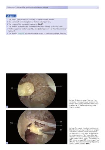
AS
SL
Left ear: Endoscopic view of the attic after removal of the incus. Articular surface of the head of malleus (AS); remnant of the superior ligament (SL) of the incus attaching to the tegmen tympani.
37
AML
CT TT TF
38
Left ear: The handle of malleus has been mobilized anteriorly to expose the tensor tympani fold which is released from its attachments; the starting point of the white arrows indicate the original position of the fold and the tips show the current point following lateralization of the malleus handle. Tensor tympani tendon (TT); chorda tympani (CT); tensor tympani fold (TF) after dislocation from its original position; anterior mallear ligament (AML).