14
Endoscopic Transcanal Ear Anatomy and Dissection Manual
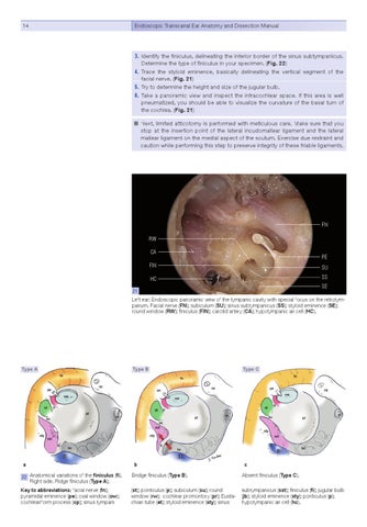
3. Identify the finiculus, delineating the inferior border of the sinus subtympanicus. Determine the type of finiculus in your specimen. (Fig. 22) 4. Trace the styloid eminence, basically delineating the vertical segment of the facial nerve. (Fig. 21) 5. Try to determine the height and size of the jugular bulb. 6. Take a panoramic view and inspect the infracochlear space. If this area is well pneumatized, you should be able to visualize the curvature of the basal turn of the cochlea. (Fig. 21) Next, limited atticotomy is performed with meticulous care. Make sure that you stop at the insertion point of the lateral incudomallear ligament and the lateral mallear ligament on the medial aspect of the scutum. Exercise due restraint and caution while performing this step to preserve integrity of these friable ligaments.
FN RW CA
PE
FIN
SU
HC
SS SE
21
Left ear: Endoscopic panoramic view of the tympanic cavity with special focus on the retrotympanum. Facial nerve (FN); subiculum (SU); sinus subtympanicus (SS); styloid eminence (SE); round window (RW); finiculus (FIN); carotid artery (CA); hypotympanic air cell (HC).
Type A
a
Type B
b
Type C
c
22 Anatomical variations of the finiculus (fi). Right side. Ridge finiculus (Type A);
Bridge finiculus (Type B).
Absent finiculus (Type C).
Key to abbreviations: facial nerve (fn); pyramidal eminence (pe); oval window (ow); cochleariform process (cp); sinus tympani
(st); ponticulus (p); subiculum (su); round window (rw); cochlear promontory (pr); Eustachian tube (et); styloid eminence (sty); sinus
subtympanicus (sst); finiculus (fi); jugular bulb (jb); styloid eminence (sty); ponticulus (p); hypotympanic air cell (hc).