7 minute read
3D Printing in Spine Surgery

Is the Hype Real?
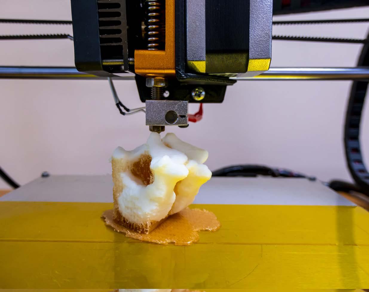
Although 3-dimensional (3D) printing, or additive manufacturing (AM), was first introduced in the 1980s, it really gained traction in spine surgery over the past 2 decades.[1] In its infancy, 3D printing was used as a means to develop custom visual aids for preoperative planning. However, with the spread of popular terms, such as “customized,” “patient-specific,” and “personalized medicine,” 3D-printed osteotomy templates, cutting guides, pedicle screw guides, and even implants have begun to flood the market.
Customized surgical implants make sense—we can forgo many unnecessary costs such as sterilizing and providing excessive items that do not match the needs of each patient. Furthermore, preoperatively planned implants may allow for more expeditious surgery, as surgeons can minimize the time it takes to template and trial sizing options.[2] On the other hand, off-the-shelf (OTS) implants come in a variety of sizes, and one must question whether there are any significant differences in outcomes that would justify the increased cost of customized implants.
Current Evidence
On one end of the spectrum, 3D printing in tumor surgery seems to make the most sense, but the literature is limited mostly to case reports.[3,4] For example, AM has been used to address kyphotic deformities involved in excision of a hemangioendothelioma by matching a cage to the patient’s cranial and caudal endplates.[5] Similarly, Chin et al used a 3D-printed implant to not only fill the gap following an en bloc spondylectomy for a giant cell tumor but also to fit into the patient’s prior instrumented construct.[6] Increased costs can be justified to match the unique anatomy, often involving the increased removal of native soft tissue and bone, or in cases of bone loss from osteoporosis or pathologic fractures.[7] A more detailed review of 13 spine tumor cases in which a 3D-printed implant was successfully used highlights the potential utility in a specific cohort of patients with unique anatomic variants.[8,9]
Anatomic anomalies exist outside of tumor surgery, however. Even asymmetric anatomic wear of an endplate can change contact pressures of a surgical implant, directly affecting fusion rates.[10] As such, limited bony work and reduced risk of endplate violation can contribute to postoperative outcomes. Similarly, pelvic incidence–lumbar lordosis (PI-LL) mismatch has been linked to postoperative outcomes and adjacent segment degeneration,[11-13] increasing the potential utility of 3D-printed implants directed toward achieving improved deformity correction.
Advantages of 3D-Printing in Spine Surgery
3D-printed spine implants, like OTS implants, are made of alloys, including titanium, Polyether Ether Ketone (PEEK), cobalt-chromium, and ceramics. The benefit of 3D printing with these alloys involves the freedom of design. As in tumor surgery, most metal implants are made via an AM process called powder bed fusion, which allows for the building of complex structures that would be otherwise cost-prohibitive to build using conventional manufacturing techniques.
The AM process also helps with osseointegration, as it allows for a more simplified addition of porous surfaces. This offers developers an easier way to manage load transfer, surface area coverage, and mechanical properties while optimizing bone integration.[14] In conventional methods, more significant post-processing steps to add such porosity contribute to increased costs.
The Federal Drug Administration 510(k) mechanism of implant approval also offers companies a unique and useful way to more readily introduce new, yet similar, products to the market. In AM, manufacturing methods may differ, but as long as the fundamental geometry remains the same as existing devices, there may be an accelerated approval process. This allows manufacturing companies to attempt various design features to an existing device with only minimal changes in the production process. In addition to manufacturing changes, 3D-printed implants allow for detailed data collection and computing analysis to evaluate how microscopic changes can have macroscopic effects—potentially slowing the course of pathology such as adjacent segment degeneration.[15]
Challenges of 3D-Printing in Spine Surgery
The most relevant limitation surrounding 3D printing in spine surgery involves regulation. The powder bed fusion process involves various parameters which may be intentionally or unintentionally varied. These parameters include printing strategies, powder particle size, purity of the alloy powder, laser beam versus electron beam usage, beam power, and subsequent heat treatments. Because there are only minimal requirements for the alloy microstructure in the 3D printing process, further variations may be seen in the mechanical and electrochemical properties of each utilized alloy.
Furthermore, studies have demonstrated that 3D-printed devices have an inferior implant-alloy microstructure when compared to conventional wrought or cast alloys.[16] This undeniably has a direct effect on the corrosion and fatigue properties of each implant. Hot cracks, build defects and gas pores, and even alterations in implant brittleness, can result in catastrophic failure and fracture. Fortunately, most impurities can be removed with post-processing and heating, though the lack of regulation and standardization limits the number of companies likely implementing this.
Final Thoughts
The enthusiasm surrounding 3D implants and their ability to address in vivo variations in bony and soft tissue anatomy, size, and pathology is warranted. The benefits have been demonstrated in the literature to support its use. However, there is far less attention paid to the quality of manufacturing with no checks-and-balances in place evaluating the alloy use of 3D implants.
References
1. Sheha ED, Gandhi SD, Colman MW. 3D printing in spine surgery. Ann Transl Med. 2019;7(Suppl 5):S164.
2. Wallace N, Schaffer NE, Aleem IS, Patel R. 3D-printed patient-specific spine Implants: a systematic review. Clin Spine Surg. 2020;33(10):400-407.
3. Burnard JL, Parr WCH, Choy WJ, Walsh WR, Mobbs RJ. 3D-printed spine surgery implants: a systematic review of the efficacy and clinical safety profile of patient-specific and off-the-shelf devices. Eur Spine J. 2020;29(6):1248-1260.
4. Wilcox B, Mobbs RJ, Wu AM, Phan K. Systematic review of 3D printing in spinal surgery: the current state of play. J Spine Surg. 2017;3(3):433-443.
5. Choy WJ, Mobbs RJ, Wilcox B, Phan S, Phan K, Sutterlin CE 3rd. Reconstruction of thoracic spine using a personalized 3D-printed vertebral body in adolescent with T9 primary bone tumor. World Neurosurg. 2017;105:1032.e13-e1032.e17.
6. Chin BZ, Ji T, Tang X, Yang R, Guo W. Three-level lumbar en bloc spondylectomy with three-dimensional-printed vertebrae reconstruction for recurrent giant cell tumor. World Neurosurg. 2019;129:531-537.e1.
7. Siu TL, Rogers JM, Lin K, Thompson R, Owbridge M. Custom-made titanium 3-dimensional printed interbody cages for treatment of osteoporotic fracture-related spinal deformity. World Neurosurg. 2018;111:1-5.
8. Girolami M, Boriani S, Bandiera S, et al. Biomimetic 3D-printed custom-made prosthesis for anterior column reconstruction in the thoracolumbar spine: a tailored option following en bloc resection for spinal tumors: preliminary results on a case-series of 13 patients. Eur Spine J. 2018;27(12):3073-3083.
9. Li X, Wang Y, Zhao Y, Liu J, Xiao S, Mao K. Multilevel 3D printing implant for reconstructing cervical spine with metastatic papillary thyroid carcinoma. Spine. 2017;42(22):E1326-E1330.
10. Mobbs RJ, Parr WCH, Choy WJ, McEvoy A, Walsh WR, Phan K. Anterior lumbar interbody fusion using a personalized approach: is custom the future of implants for anterior lumbar interbody fusion surgery? World Neurosurg. Published online January 8, 2019. doi:10.1016/j.wneu.2018.12.144
11. Rothenfluh DA, Mueller DA, Rothenfluh E, Min K. Pelvic incidence-lumbar lordosis mismatch predisposes to adjacent segment disease after lumbar spinal fusion. Eur Spine J. 2015;24(6):1251-1258.
12. Tempel ZJ, Gandhoke GS, Bolinger BD, et al. The influence of pelvic incidence and lumbar lordosis mismatch on development of symptomatic adjacent level disease following single-level transforaminal lumbar interbody fusion. Neurosurgery. 2017;80(6):880-886.
13. Senteler M, Weisse B, Snedeker JG, Rothenfluh DA. Pelvic incidence-lumbar lordosis mismatch results in increased segmental joint loads in the unfused and fused lumbar spine. Eur Spine J. 2014;23(7):1384-1393.
14. Kelly CN, Wang T, Crowley J, et al. High-strength, porous additively manufactured implants with optimized mechanical osseointegration. Biomaterials. 2021;279:121206.
15. Serra T, Capelli C, Toumpaniari R, et al. Design and fabrication of 3D-printed anatomically shaped lumbar cage for intervertebral disc (IVD) degeneration treatment. Biofabrication. 2016;8(3):035001.
16. Neto MQ, Radice S, Hall DJ, Mathew MT, Mercuri LG, Pourzal R. Alloys used in different temporomandibular joint reconstruction replacement prostheses exhibit variable microstructures and electrochemical properties. J Oral Maxillofac Surg. 2022;80(5):798-813.
Arash J. Sayari, MD