6 minute read
Virtual Reality for Preoperative Planning in Spine Surgery

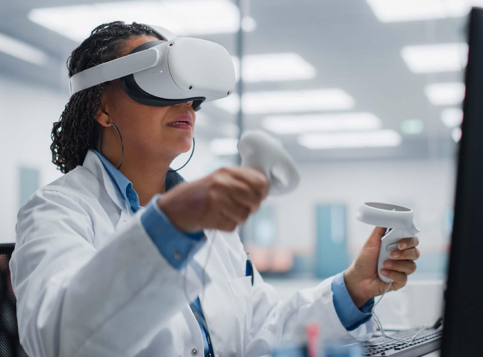
Since the 1970s, preoperative planning in spine surgery has been reliant on computed tomography (CT) and magnetic resonance imaging (MRI) to constructing 2-dimensional (2D) cross-sections to examine patient anatomy.[1] Using these modalities, the surgeon relies on mentally integrating a combination of these images to visualize a 3-dimensional (3D) model of the patient’s anatomy. Furthermore, these modalities do not correspond with the surgeon’s operative view.[2–4] However, the introduction of virtual reality (VR) has allowed for a commercially available method in translating these 2D cross-sections into 3D virtual models.[2] Using the head-mounted display (HMD) and controllers, the surgeon may manipulate the anatomical structures to obtain a better understanding of the patient’s anatomy and plan their surgical approach accordingly. Improving visualization and providing tactile manipulation has been demonstrated to improve identification of anatomical abnormalities not typically visualized through conventional radiographic modalities.[1,2] As such, VR preoperative planning has been increasingly utilized in decision-making in complex cranial procedures.[1,5–8]
Although VR preoperative planning has not seen as much adoption in spine surgery, some articles have published its utility in decision-making, satisfaction, and clinical outcomes. In this article, we discuss the affordability of incorporating VR preoperative planning in spine surgery as well as how VR planning may influence decisions in surgical approach in the cervical and lumbar spine.
With advancements in commercially available VR systems, forming 3D models from Digital Imaging and Communications in Medicine (DICOM) files has become increasingly affordable to implement in a surgeon’s practice. One of the most affordable VR systems is the Google Cardboard, a VR system formed by cardboard cutouts and a smartphone. Other commercially available VR headsets, such as the Oculus systems, cost approximately $300. For preoperative planning, Salvatore et al compared preoperative planning techniques using Google Cardboard and conventional CT planning in 65 patients undergoing correction of adolescent idiopathic scoliosis.[2] The authors systematically evaluated the number of fusion levels, screw direction, potential structural abnormalities, potential bone abnormalities, type of approach, and number of required osteotomies.[2] Patients who underwent VR planning had significantly lower operative times, blood loss, and hospital stay with higher surgeon satisfaction.[2] The authors reported that the improved visualization of the patient anatomy allowed for greater comprehension of delicate structures in the operative field and therefore provided greater awareness of potential risks.[2] As such, incorporating VR in preoperative planning in spine surgery is affordable and may provide significant benefits in perioperative outcomes.
The use of VR for preoperative planning may influence surgeons in the type of surgeries performed. Zawy Alsofy et al compared the impact of VR planning in minimally invasive and open single-level lumbar decompression and/or fusion.[3] The authors reported that VR significantly influenced whether the surgeons utilized decompression only or decompression and fusion, minimally invasive or open technique, and type of surgical approach.[3] The use of VR allowed for greater visualization of pars defects and arthritic changes in the facet joints, influencing surgeons to select decompression and fusion.[3] Additionally, as previous studies have demonstrated the advantages of VR for learning surgical approaches, the improved visualization may allow for greater confidence in recognizing critical structures for surgeons to become comfortable with a minimally invasive approach.[3,9,10] Furthermore, selection of a minimally invasive approach typically results in less blood loss and shorter postoperative length of stay with similar postoperative clinical outcomes, thus reducing healthcare expenditures.[3,11–13] As such, use of VR in preoperative planning may significantly influence surgical decision-making in the lumbar spine and lead to less invasive approaches when possible.
As in the lumbar spine, the use of VR planning may significantly impact surgical decision-making in the cervical spine. In a separate article by Zawy Alsofy et al, the authors examined the influence of VR on preoperative planning in patients with single-level cervical foraminal stenosis.[4] Zawy Alsofy et al reported that VR influenced the style of anterior approach instrumentation utilized (ie, cage, cage+plate, or arthroplasty), with a trend toward significance in favoring a posterior over anterior surgical approach.[4] Although Oshina et al did not utilize VR, the authors formed a 3D MRI/CT fusion image and reported that 18.1% of cervical radiculopathy cases had a change in strategy once the neuroforamen was fully visualized.[14] Specifically, the surgeons in this study opted for more extensive decompression.[14] As such, improved virtual visualization of the cervical spine may significantly impact surgical decision-making regarding approach and extent of decompression.
Preoperative surgical planning using VR allows for more intuitive visualization of patient anatomy compared to traditional radiographic evaluation. Furthermore, improved visualization and tactile manipulation may allow surgeons to determine anatomical abnormalities not typically seen on CT or MRI. With commercially available VR systems, incorporating VR in preoperative planning is affordable and may significantly influence surgical decision-making.
References
1. Lan L, Mao RQ, Qiu RY, Kay J, de Sa D. Immersive virtual reality for patient-specific preoperative planning: a systematic review. Surg Innov. 2023;30(1):109-122.
2. De Salvatore S, Vadalà G, Oggiano L, Russo F, Ambrosio L, Costici PF. Virtual reality in preoperative planning of adolescent idiopathic scoliosis surgery using Google Cardboard. Neurospine. 2021;18(1):199-205.
3. Zawy Alsofy S, Nakamura M, Ewelt C, et al. Retrospective comparison of minimally invasive and open monosegmental lumbar fusion, and impact of virtual reality on surgical planning and strategy. J Neurol Surg A Cent Eur Neurosurg. 2021;82(5):399-409.
4. Zawy Alsofy S, Stroop R, Fusek I, et al. Virtual reality-based evaluation of surgical planning and outcome of monosegmental, unilateral cervical foraminal stenosis. World Neurosurg. 2019;129:e857-e865.
5. Stadie AT, Kockro RA, Reisch R, et al. Virtual reality system for planning minimally invasive neurosurgery [technical note]. J Neurosurg. 2008;108(2):382-394.
6. Steineke TC, Barbery D. Virtual reality preoperative planning to reduce procedure time of microsurgical clipping of MCA aneurysm. Neurosurgery. 2020;67(Supplement_1).
7. Kockro RA, Serra L, Tseng-Tsai Y, et al. Planning and simulation of neurosurgery in a virtual reality environment. Neurosurgery. 2000;46(1):118-135; discussion 135-137.
8. Anthony D, Louis RG, Shekhtman Y, Steineke T, Frempong-Boadu A, Steinberg GK. Patient-specific virtual reality technology for complex neurosurgical cases: illustrative cases. J Neurosurg Case Lessons. 2021;1(23):CASE21114.
9. McCloskey K, Turlip R, Ahmad HS, Ghenbot YG, Chauhan D, Yoon JW. Virtual and augmented reality in spine surgery: a systematic review. World Neurosurg. 2023;173:96-107.
10. Lohre R, Wang JC, Lewandrowski KU, Goel DP. Virtual reality in spinal endoscopy: a paradigm shift in education to support spine surgeons. J Spine Surg. 2020;6(Suppl 1):S208-S223.
11. McClelland S 3rd, Goldstein JA. Minimally invasive versus open spine surgery: what does the best evidence tell us? J Neurosci Rural Pract. 2017;8(2):194-198.
12. Mooney J, Michalopoulos GD, Alvi MA, et al. Minimally invasive versus open lumbar spinal fusion: a matched study investigating patient-reported and surgical outcomes. J Neurosurg Spine. 2021;36(5):753-766.
13. Holy CE, Corso KA, Bowden DE, et al. Evaluation of cost, payments, healthcare utilization, and perioperative and post-operative outcomes of patients treated with posterior lumbar spinal surgery using open versus minimally invasive surgical approaches. Med Devices. 2021;14:173-183.
14. Oshina M, Oshima Y, Tanaka S, et al. Utility of oblique sagittal reformatted and three-dimensional surface reconstruction computed tomography in foraminal stenosis decompression. Sci Rep. 2018;8(1):16011.
James W. Nie, BS
Timothy J. Hartman, MD
Keith R. MacGregor, BS
Omolabake O. Oyetayo, BS
Eileen Zheng, BS
Kern Singh, MD
