10 minute read
Ocular Complications in Spine Surgery


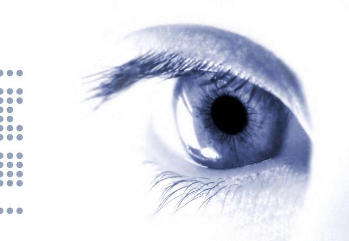
Visual perception is initiated as light reaches the cornea, a transparent structure continuous with the sclera. Incoming light is focused by the lens onto the retina, a neural tissue lined with photoreceptors that transduces visual stimuli into electrochemical signals. These signals then track posteriorly into the occipital lobe of the brain via the optic nerve to render visual perception. Although ocular complications are rare, they are increasingly being reported across several surgical fields, including orthopaedic, cardiothoracic, and general surgery.[1–4] In 1948, Slocum et al documented the first spine surgery–linked case of blindness, which occurred as a result of incorrect intraoperative head positioning.[5] Over the past few decades, the complexity and number of spine surgeries has increased in the United States.[6] According to a study from the Scoliosis Research Society, the frequency of eye complications is 1 per every 100 spinal procedures.[7] Visual complications after spinal surgery can potentially bring about additional severe adverse postoperative outcomes for patients. Within spine surgery, perioperative vision loss (POVL) rates have been reported around 0.14% for lumbar fusion procedures and 0.28% for deformity correction cases. Individual patient characteristics such as preexisting health conditions, including hypertension, diabetes mellitus, and peripheral vascular disease or lifestyle choices, such as smoking, may potentially increase the risk of experiencing POVL.[7]
Complications
POVL and central retinal artery occlusion (CRAO) are 2 of the most feared surgical complications by both patients and physicians because they often culminate in visual field loss or irreversible blindness. However, corneal abrasion is the most commonly reported ocular complication.[8,9] A corneal abrasion occurs when the epithelial layer of the cornea is separated from the underlying basement membrane. Corneal abrasions are generally classified by location, extent, and depth of the epithelial defect.[9] Other less reported ocular complications include conjunctivitis, direct trauma, or chemical injury.
Neurological origins for vision loss after surgery are also reported. Posterior reversible encephalopathy syndrome (PRES) is a neurological condition characterized by symptoms like seizures, vision disturbances, headaches, and a reduced level of alertness. While it is more commonly associated with the field of obstetrics, cases of PRES have been documented in the field of orthopedics following procedures such as lumbar fusion.[10] In these cases, causes have been associated with rapid spikes in blood pressure that surpass the brain’s self-regulating capacity, which eventually culminates in brain swelling. However, experiencing recovery from PRES is much more likely in comparison to CRAO or other complications like ischemic optic neuropathy (ION).[11]
The most significant ocular complication for patients undergoing spine surgery are those associated with blindness due to the drastic impact on the patient’s life. POVL can be attributed to various origins, including occipital lobe infarction resulting in cortical blindness, blockages in the central retinal artery or ophthalmic vein, and inadequate blood flow to the optic nerve resulting in ION.[12,13] Individuals who required postoperative recovery in the hospital or who required high acuity care in an intensive care unit are at increased risk of experiencing vision loss.[14] Of all surgical subspecialties, spine and cardiac surgery reportedly have higher than average rates of POVL.[8,14,15] In a cohort study by Hofer et al, individuals reporting perioperative ocular injury versus those without injury were associated with instrumentation procedures, decompression and fusion procedures, right lateral positioning, and surgery involving the lumbar spine.[14,16] In the United States, CRAO occurs at an approximate rate of 1 per every 100,000 people, whereas following spinal surgery, it has been reported to be significantly higher at an estimated 1 per every 1000 people.[17]
Risk Factors
Patient positioning during a surgical procedure significantly influences the risk of a patient experiencing POVL postoperatively. Prone positioning increases the risk of ocular complications by upwards of 10-fold.[4,14,18] This increase is a result of the periorbital region receiving more direct pressure in the prone position resulting in increased intraocular pressure and potential complications of physical trauma or ischemic injury due to CRAO.[18] However, prone, lateral, and Trendelenburg positioning of a patient in surgery may result in corneal contact with various items or surfaces, thereby increasing the risk for corneal abrasions.[3,19,20] Additionally, gender has been suggested to significantly influence the risk of POVL after spinal fusion with a greater risk of occurrence in men than in women. Furthermore, individuals aged 50–64 years showed a heightened risk of ocular complications, thus adding age as another contributing factor. These findings are supported by a case series published by the American Society of Anesthesiologists from a POVL registry across multiple centers.[21–23] Additional risk factors include deliberate hypotension, excessive blood loss, or an operative time exceeding 7 hours.[18]
Diagnosis, Treatment, and Prevention
The most reported symptoms of those experiencing ocular complications include complaints of blurry vision, swelling, and pain around the eye. Patients experiencing any symptoms should be urgently referred for an ophthalmologic consultation.[24] Up to 80% of patients with corneal abrasion present without evidence of trauma, leading to the conclusion that despite taping of the eyelids, approximately 59% of patients do not experience complete closure of the eyelids—an occurrence called lagophthalmos.[18,25] One cause for intraoperative lagophthalmos and consequential corneal abrasions has been suggested to be a potential result of the frequent checks of the depth of anesthesia sedation in which the patient’s eyelids are lifted and assessed with reactive pupil testing.[26] Should a corneal abrasion occur during surgery, the most reliable method to confirm the diagnosis is using fluorescein staining.[8,9] The most effective protective measure against corneal abrasions remains intraoperative eyelid taping, but alternative protective measures include the use of ointments or cushioned pads over the eyes.[27,26] ION typically presents as an abrupt, yet painless, loss of vision in one or more visual fields. The neuropathy can originate on the anterior or posterior aspect of the optic nerve based on the location of injury. ION often results in partial vision loss in both eyes or total blindness in more severe incidences. Diagnoses resulting from ION are typically detected after the patient wakes up after surgery.[29]
CRAO during spine fusion surgery can be prevented by protecting eyes from compression.[13] In the operating room, placing the surgical table in a 5° reverse Trendelenburg position helps to decreased intraocular pressure in comparison to traditional prone positioning for surgery times shorter than 120 minutes.[30] Hofer et al noted that patients with optic nerve ischemia were older, underwent longer operations, experienced more blood loss, and received more crystalloid fluids.[14] Prevention of postoperative hypotension or hypovolemia leading to POVL may be accomplished through the use of fluid replacement in which colloid solution is substituted for crystalloid solution.[17] It is recommended that high-risk patients undergo an eye examination of visual fields and pupillary reflexes as they regain consciousness after surgery.[13]
Conclusion
The intricate and crucial process of visual perception is susceptible to several perioperative complications ranging from the most reported corneal abrasions to more severe consequences such as POVL and CRAO. It is imperative that healthcare professionals in the spinal field are educated regarding the risks for ocular injury during surgery. In prioritizing this vital education, healthcare professionals can avoid serious consequences such as irreversible blindness. The most prudent recommendation when suspecting an ocular complication is the immediate referral for ophthalmologic consultation. However, the best strategy centers on prevention, such as ensuring effective intraoperative eyelid taping, reducing operative time, limiting prone positioning, and optimizing surgical table position to decrease intraocular pressure.
References
1. Su AW, Lin SC, Larson A N. Perioperative vision loss in spine surgery and other orthopaedic procedures. J Am Acad Orthop Surg. 2016;24:702–710.
2. Yu HD, Chou AH, Yang MW, Chang CJ. An analysis of perioperative eye injuries after nonocular surgery. Acta Anaesthesiol Taiwan. 2010;48:122–129.
3. Roth S, Thisted RA, Erickson JP, Black S, Schreider BD. Eye injuries after nonocular surgery. A study of 60,965 anesthetics from 1988 to 1992. Anesthesiology. 1996;85:1020–1027.
4. Li A, Swinney C, Veeravagu A, Bhatti I, Ratliff J. Postoperative visual loss following lumbar spine surgery: a review of risk factors by diagnosis. World Neurosurg. 2015;84:2010–2021.
5. Slocum HC, O’Neal KC Allen CR. Neurovascular complications from malposition on the operating table. Surg Gynecol Obstet . 1948;86:729–734.
6. Rajaee SS, Bae HW, Kanim LEA, Delamarter RB. Spinal fusion in the United States: analysis of trends from 1998 to 2008. Spine . 2012;37:67–76.
7. Myers MA, Hamilton SR, Bogosian AJ, Smith CH, Wagner TA. Visual loss as a complication of spine surgery. A review of 37 cases. Spine . 1997;22:1325–1329.
8. Baig MN, Lubow M, Immesoete P, Bergese SD, Hamdy EA, Mendel E. Vision loss after spine surgery: review of the literature and recommendations. Neurosurg Focus . 2007;23:E15.
9. Moos DD, Lind DM. Detection and treatment of perioperative corneal abrasions. J Perianesth Nurs . 2006;21:332–338.
10. Yi JH, Ha SH, Kim YK, Choi EM. Posterior reversible encephalopathy syndrome in an untreated hypertensive patient after spinal surgery under general anesthesia—a case report. Korean J Anesthesiol. 2011;60:369–372.
11. Nickels TJ, Manlapaz MR, Farag E. Perioperative visual loss after spine surgery. World J Orthop. 2014;5:100–106.
12. Grover V, Jangra K. Perioperative vision loss: a complication to watch out. J Anaesthesiol Clin Pharmacol. 2012;28:11–16.
13. Roth S, Moss HE, Vajaranant TS, Sweitzer B. Perioperative care of the patient with eye pathologies undergoing nonocular surgery. Anesthesiology. 2022;137:620–643.
14. Hofer RE, Evans KD, Warner MA. Ocular injury during spine surgery. Can J Anaesth. 2019;66:772–780.
15. Stevens WR, Glazer PA, Kelley SD, Lietman TM, Bradford DS. Ophthalmic complications after spinal surgery. Spine. 1997;22:1319–1324.
16. Ho VTG, Newman NJ, Song S, Ksiazek S, Roth S. Ischemic optic neuropathy following spine surgery. J Neurosurg Anesthesiol. 2005;17:38–44.
17. Patil CG, Lad EM, Lad SP, Ho C, Boakye M. Visual loss after spine surgery: a population-based study. Spine. 2008;33:1491–1496.
18. Stambough JL, Dolan D, Werner R, Godfrey E. Ophthalmologic complications associated with prone positioning in spine surgery. J Am Acad Orthop Surg. 2007;15:156–165.
19. Lee SH, Chung I, Choi DS, et al. Visual loss due to optic nerve infarction and central retinal artery occlusion after spine surgery in the prone position: a case report. Medicine. 2017;96:e7379.
20. Xiong J, Liang G, Hu L, et al. Transient visual acuity loss after spine surgery in the prone position: a case report and literature review. J Int Med Res. 2020;48:300060520952279.
21. Postoperative Visual Loss Study Group. Risk factors associated with ischemic optic neuropathy after spinal fusion surgery. Anesthesiology. 2012;116:15–24.
22. Lee LA, Roth S, Posner KL, et al. The American Society of Anesthesiologists Postoperative Visual Loss Registry: analysis of 93 spine surgery cases with postoperative visual loss. Anesthesiology. 2006;105:652–659.
23. Holy SE, Tsai JH, McAllister RK, Smith KH. Perioperative ischemic optic neuropathy: a case control analysis of 126,666 surgical procedures at a single institution. Anesthesiology. 2009;110:246–253.
24. Hoff JM, Varhaug P, Midelfart A, Lund-Johansen M. Acute visual loss after spinal surgery. Acta Ophthalmol. 2010;88:490–492.
25. Kaye AD, Renschler JS, Cramer KD, et al. Postoperative management of corneal abrasions and clinical implications: a comprehensive review. Curr Pain Headache Rep. 2019;23:48.
26. Yanagidate F, Dohi S. Corneal abrasion after the wake-up test in spinal surgery. J Anesth. 2003;17:211–212.
27. Grixti A, Sadri M, Watts MT. Corneal protection during general anesthesia for nonocular surgery. Ocul Surf. 2013;11:109–118.
28. Miller NR. Current concepts in the diagnosis, pathogenesis, and management of nonarteritic anterior ischemic optic neuropathy. J Neuroophthalmol. 2011;31:e1–e3.
29. Hayreh SS. Ischemic optic neuropathies—where are we now? Graefes Arch Clin Exp Ophthalmol. 2013;251:1873–1884.
30. Carey TW, Shaw KA, Weber ML, DeVine JG. Effect of the degree of reverse Trendelenburg position on intraocular pressure during prone spine surgery: a randomized controlled trial. Spine J. 2014;14:2118–2126.
Authors: Andrea M. Roca, MS
Fatima N. Anwar, BA
Alexandra C. Loya, BS
Srinath S. Medakkar, BS
Richa Singh, MD
Kern Singh, MD
From the Department of Orthopaedic Surgery, Rush University Medical Center, in Chicago, Illinois.